The use of liquid biopsy in early breast cancer: clinical evidence and future perspectives - NET
←
→
Page content transcription
If your browser does not render page correctly, please read the page content below
D’Amico et al. J Cancer Metastasis Treat 2021;7:3
DOI: 10.20517/2394-4722.2020.93
                                                                                                    Journal of Cancer
                                                                                             Metastasis and Treatment

Review                                                                                                                         Open Access

The use of liquid biopsy in early breast cancer:
clinical evidence and future perspectives
Paolo D’Amico1,2,3, Carla Corvaja4,5, Lorenzo Gerratana1,4,5,6, Carolina Reduzzi1, Giuseppe Curigliano2,3,
Massimo Cristofanilli1
1
 Division of Hematology and Oncology, Department of Medicine, Northwestern University Feinberg School of Medicine,
Chicago, IL 60611, USA.
2
  Division of Early Drug Development for Innovative Therapies, IEO, European Institute of Oncology IRCCS, Milan 20132, Italy.
3
  Department of Oncology and Hemato-Oncology, University of Milano, Milan 20122, Italy.
4
  Department of Oncology, ASUFC, University Hospital of Udine, Udine 33100, Italy.
5
  Department of Medicine, University of Udine, Udine 33100, Italy.
6
  Department of Medical Oncology, Centro di Riferimento Oncologico (CRO), IRCCS, Pordenone 33081, Italy.

Correspondence to: Dr. Paolo D’Amico, Division of Hematology and Oncology, Department of Medicine, Northwestern University
Feinberg School of Medicine, 420 E Superior St., Chicago, IL 60611, USA. E-mail: paolodam@gmail.com

How to cite this article: D’Amico P, Corvaja C, Gerratana L, Reduzzi C, Curigliano G, Cristofanilli M. The use of liquid biopsy in
early breast cancer: clinical evidence and future perspectives. J Cancer Metastasis Treat 2021;7:3.
http://dx.doi.org/10.20517/2394-4722.2020.93

Received: 1 Sep 2020 First Decision: 22 Oct 2020 Revised: 23 Oct 2020 Accepted: 15 Dec 2020 Published: 7 Jan 2021

Academic Editor: Wei Zhang Copy Editor: Miao Zhang Production Editor: Jing Yu

Abstract
Liquid biopsy, including both circulating tumor cells and circulating tumor DNA, is gaining momentum as a
diagnostic modality adopted in the clinical management of breast cancer. Prospective studies testing several
technologies demonstrated clinical validity and, in some cases, achieved the United States Food and Drug
Administration approval. The initial testing and clinical application of liquid biopsy focused primarily on the
diagnosis, while molecular characterization and monitoring of metastatic disease, with larger data from prospective
studies, came in the last two decades. Although its role in metastatic setting is thus widely recognized, the current
evidence does not provide support for the routine clinical use of liquid biopsy methods for the earlier stage of this
disease. Considering the relevance of early detection, characterization, and management of breast cancer in the
early-stage, this clinical setting is the most suitable to increase the chances for effective treatment selection and
improved prognosis, and a better understanding of the main application of liquid biopsy tools in the earlier stage
of breast cancer is therefore crucial. The aim of this review is to provide an overview of the clinical evidence and
subsequent potential applications of liquid biopsy in early breast cancer, identifying the main existing caveats and
the possible future scenarios.

                       © The Author(s) 2021. Open Access This article is licensed under a Creative Commons Attribution 4.0
                       International License (https://creativecommons.org/licenses/by/4.0/), which permits unrestricted use,
    sharing, adaptation, distribution and reproduction in any medium or format, for any purpose, even commercially, as long
    as you give appropriate credit to the original author(s) and the source, provide a link to the Creative Commons license,
    and indicate if changes were made.

                                                                                                                        www.jcmtjournal.com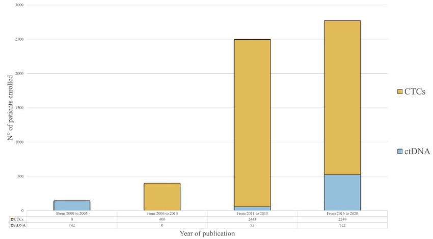
Page 2 of 16              D’Amico et al. J Cancer Metastasis Treat 2021;7:3 I http://dx.doi.org/10.20517/2394-4722.2020.93

Keywords: Liquid biopsy, early breast cancer, circulating tumor cells, circulating tumor DNA, clinical trials, screening,
early detection, minimal residual disease

INTRODUCTION
About 276,480 new cases of invasive breast cancer will be diagnosed among American women within
2020[1]. This incidence means breast cancer is the second most common cancer, showing a slightly
increasing trend in recent years (by 0.3% per year). Around 6% of these patients will present with metastatic
disease at diagnosis, while, among the remaining, approximately 40% will develop a systemic recurrence,
despite the surgery and eventual use of systemic and radiation therapy[2]. The 5-year overall survival (OS)
rate among patients with localized disease ranges between 86% and 99%. Conversely, only 27% of patients
with metastatic breast cancer (mBC) will reach the 5th year after diagnosis[3]. In consideration of these data,
it is fundamental that efforts are made to ensure that diagnosis of both primary and potential recurrence is
made as early as possible.

The current evidence suggests that mammography remains the only standard of care diagnostic imaging
for breast cancer screening (with the exception of high-risk patients, in which case the use of Magnetic
Resonance Imaging may be preferred) and, performing a biopsy of the lesion is mandatory to confirm the
diagnosis and provide further tumor pathological and molecular characteristics. The standard surveillance
methods evaluated for early detection of recurrence of disease are mostly similar to those used for the
primary diagnosis. Moreover, some serological markers (such as CA15-3 and CEA) are available for clinical
use, although none of these were demonstrated to improve the detection ability of the standard procedures
and improve outcome. Tissue biopsies are invasive, time-consuming, quite expensive and for those reasons
not often replicable within the same patient. Furthermore, the tissue biopsies appear to be too shortsighted
to capture the true spatial and temporal tumor heterogeneity.

The terms liquid biopsy mainly refers to several different diagnostic tests performed on biological fluids
(i.e., blood, saliva, urine, etc.) with the aim of investigating the presence of circulating cancer cells (CTCs)
or fragments of DNA [circulating tumor DNA (ctDNA)] that can be shed from the tumor. Despite the
development and testing of several enrichment techniques to identify, isolate and characterize CTCs in
the blood of cancer patients, the only Food and Drug Administration (FDA) approved method is the
CellSearchTM (Menarini Silicon Biosystems, Bologna, Italy), that allows the identification and enumeration
of CTCs using an immunomagnetic enrichment of epithelial cells by anti-EpCAM antibodies. With
this method, the detection of 5 CTCs or more in the peripheral blood of patients with metastatic breast
cancer is an independent indicator of worse prognosis[4]. In a retrospective study analyzing data from 18
international centers, the same cutoff identified a group of mBC patients with more aggressive disease
irrespective of disease subtype, recurrence site, type and line of therapy suggesting the need to use this
test to stratify mBC, Stage IVaggressive vs. Stage IVindolent. In addition, CTCs often bear the same genomic
abnormalities detected in metastatic tumor tissue with some additional peculiar phenotypic changes that
are responsible for systemic disease spread (epithelial-mesenchymal transition and stem cell markers),
or drug resistance (PI3KCA‑AKT signaling pathway)[5], suggesting that CTCs may have a key role in the
evolution of the disease and can be considered as a sensitive, quantitative and qualitative surrogate of the
overall systemic disease.

The median level of circulating DNA is approximately five-fold higher in plasma of breast cancer patients
compared to healthy controls[6]. In a sub-cohort of breast cancer patients, ctDNA was detected in both
metastatic (90%) and localized disease (55%), and its presence was correlated overall with each stage
of the disease and its prognosis[7]. Moreover, ctDNA levels in metastatic breast cancer patients have
D’Amico et al. J Cancer Metastasis Treat 2021;7:3 I http://dx.doi.org/10.20517/2394-4722.2020.93                           Page 3 of 16

Figure 1. Histogram representation of patients enrolled in clinical trials assessing the role of CTCs and ctDNA detection in early breast
cancer, grouped by year of publication of the results. Showing a relevant rise in the number of enrolled patients in the past 20 years,
the chart highlights an increasing interest in liquid biopsy as a tool for the management of early breast cancer patients. CTCs studies
have provided a substantial contribution, particularly in the last decade (from 2011 to 2020). A more recent increase in the number of
patients enrolled in trials analyzing ctDNA (from 2016 to 2020) suggests a growing interest to investigate the role of ctDNA in this
setting. CTCs: circulating tumor cells; ctDNA: circulating tumor DNA

been associated with tumor burden with a better performance than canonical serological biomarkers[8].
Furthermore, common subtype-specific breast cancer mutations such as phosphatidylinostitol 3-kinase
catalytic subunit (PIK3CA) and tumor protein 53 gene (TP53) and resistance genomic abnormalities
like Estrogen Receptor 1 (ESR1) and erythroblastic leukemia viral oncogene homolog 2 (ERBB2) can be
detected through ctDNA analysis[9,10], proposing it as an effective tool to assess the genomic characteristics
and follow the mutant evolution of metastatic breast cancer.

Notwithstanding the well-recognized role of liquid biopsy in the metastatic setting, the importance of these
tests for early breast cancer (eBC) patients is still under investigation. However, the increasing number of
patients enrolled in investigational trials is evidence of high interest in this matter [Figure 1]. Thus, the aim
of this review is to report the current evidence suggesting the potential clinical utility of liquid biopsy for
both screening and early diagnosis of recurrence, highlighting the existing limitations and possible future
developments.

EARLY DIAGNOSIS AND BASELINE CHARACTERIZATION
The detection of eBC at pre-clinical stages is the “holy grail” of every diagnostic modality but, also the most
challenging. Indeed, the development and clinical testing of highly sensitive diagnostic tools is urgent to
ideally reduce BC-related morbidity and mortality, tailor less morbid treatment strategies and eventually
improve clinical outcomes. Blood-based detection of circulating cell-free DNA (cfDNA) and the ctDNA
fraction holds the great promise to address the need for non-invasive early diagnosis alongside overcoming
the shortcomings of tissue biopsies. Moreover, genomic analysis of ctDNA could provide prompt and
valuable prognostic information[11]. However, cfDNA and ctDNA levels are significantly low at pre-clinical
stages and typically below the level of sensitivity of current clinical testing, affecting the accuracy of ctDNA
Page 4 of 16            D’Amico et al. J Cancer Metastasis Treat 2021;7:3 I http://dx.doi.org/10.20517/2394-4722.2020.93

extraction and analyses through high-throughput sequencing[12]. Indeed, the sensitivity of conventional
next generation sequencing (NGS) in detecting DNA alterations is limited to a relatively high fraction
of mutant to wild-type DNA (> 1%)[13]. Nonetheless, the conceptual value of ctDNA-based driver gene
mutations identification stands in tumor cells clonality, which might in fact determine a high degree
of specificity compared to other blood-based biomarkers, endorsing its clinical utility in the diagnostic
process.

Beaver et al.[14] prospectively analyzed plasma ctDNA of 29 eBC patients before and after surgery through
digital polymerase chain reaction (ddPCR) and compared it with tissue-based Sanger sequencing and
ddPCR analyses. PIK3CA mutations were identified in 15 tumor tissues and 13 of them had a matching
pre-operatory plasma sample mutation. Of note, in 5 cases PIK3CA mutations were still detectable in
post-surgical plasma samples. ddPCR high sensitivity allowed for ctDNA mutation detection with even
low levels of tumor burden, providing an initial proof-of-concept to support the feasibility of ctDNA-
based analyses in the detection of somatic DNA mutations. Phallen et al.[15] developed the targeted error
correction sequencing (TEC-Seq) approach to retrospectively detect cfDNA sequence changes through
ultra-sensitive massive genome sequencing. Overall, 58 cancer-related driver genes were analyzed in 200
patients with early-stage colorectal, breast, lung, and ovarian cancer and somatic mutations were detected
in 71%, 59%, 59%, and 68% of cases, respectively. Of note, BC ctDNA samples had the lowest mutant
allele fraction. High concordance between plasma and tumor-detected alterations was observed and the
apparent lack of concordance between some specific alterations, potentially determined by intratumoral
heterogeneity, might in fact be overcome by plasma-based analyses.

Recently, a single, multi-analyte blood-test, named CancerSEEK, was developed by Cohen et al.[16] to assess
cfDNA mutations and it was applied to 1.005 clinically diagnosed stage I-III cancer patients to screen for
eight common cancer types, including eBC. The circulating levels of eight serum protein biomarkers were
simultaneously evaluated to overcome the issue of low detectable levels of ctDNA, therefore improving
sensitivity. This approach also included machine-learning tools to accurately narrow down the location of
a tumor to a small number of anatomic sites. A relatively small yet robust and highly selective 61-amplicon
panel was designed through multiplex-polymerase chain reaction to detect ctDNA mutations with an
acceptable degree of screening sensitivity and specificity. Median sensitivity was 70% overall and 33% in
eBC patients, increasing alongside tumor stage, with 43% for stage I, 73% for stage II, and 78% for stage
III cancers. Specificity was higher than 99%, with only 7 out of 812 healthy controls scoring positive and
localization of tissue of origin (TOO) was possible in a median of 83% patients. Intriguingly, a significant
concordance between ctDNA- and tissue-detected mutations was observed in 90% of cases among all
tumors.

Other circulating biomarkers, including exosomes, miRNA, and methylated DNA sequences could
be integrated in multi-analyte blood tests to provide complementary early information through gene
expression profiling. Proof-of-concept studies have demonstrated that miRNA, packed into tumor-released
exosomes, can be detected at higher concentrations compared to ctDNA in early-stage cancer patients.
Moreover, their qualitative analyses might be informative both of DNA somatic mutations and epigenetic
alterations[17,18]. Indeed, large-scale epigenetic alterations might improve sensitivity in early cancer detection.
For instance, the enrichment of methylated cfDNA fragments, which are cancer-specific, could potentially
overcome the current technical constraints of mutation-based ctDNA detection methods, mostly
determined by the limited amount of recurrent mutations that discriminate tumor and normal cfDNA.
Aiming to profile the methylome of small amounts of cfDNA, Shen et al.[19] developed a non-invasive
sequencing approach that included cell-free methylated DNA immunoprecipitation and high throughput
sequencing (cfMeDIP-seq) for genome-wide bisulfite-free plasma DNA. An optimized Me-DIP-sequencing
protocol was first analyzed in a cohort of early-stage pancreatic cancer tissues and healthy controls and
D’Amico et al. J Cancer Metastasis Treat 2021;7:3 I http://dx.doi.org/10.20517/2394-4722.2020.93     Page 5 of 16

DNA methylation profiles were generated through reduced representation bisulfite sequencing (RRBS),
demonstrating high analytical sensitivity in detecting cancer-derived DNA. Further analyses suggested
the ability of cfMeDIP-seq in detecting tumor-specific methylation events in ctDNA and highlighted that
cfMeDIP-seq methylomes could identify active transcriptional networks in tumors or other tissues through
plasma cfDNA. CfMeDIP-seq was then explored in a discovery cohort of 189 plasma samples from healthy
controls and patients with seven tumor types. Interestingly, this approach demonstrated a high and cost-
effective performance in detecting and classifying tumors by recovering ctDNA-associated methylation
profiles and it certainly requires further investigation in larger cohorts of eBC patients.

The performance of cfDNA-based targeted methylation sequencing was also evaluated in a prospective
case-control sub-study conducted by Liu et al.[20], that assessed the accuracy of a panel of > 10,000
methylation regions to detect and localize a broad range of cancer types in 6,689 participants. In the
validation set, sensitivity was 18% among all cancer types, with detection increasing with higher stages, and
the specificity was 99.3%. TOO prediction was significantly accurate. This targeted methylation approach
allowed deeper sequencing of informative sites compared with whole genome sequencing (WGS).

Cristiano et al.[21] developed a diagnostic tool to investigate genome-wide cfDNA fragmentation patterns
of 236 breast, colorectal, lung, ovarian, pancreatic, gastric, or bile duct cancer patients and 245 healthy
controls. This study provided further proof-of-concept for plasma-based screening and early cancer
detection, as cfDNA altered fragmentation profiles were detected in cancer patients compared to
healthy subjects, whose cfDNA rather reflected nucleosomal patterns of white blood cells. In addition,
fragmentation profiles were used to confine the TOO to a limited number of sites in 75% of cases.

Besides early diagnosis, plasma-based gene expression profiling allows for the characterization of
the genomics, epigenomics, and proteomics of BC patients and ctDNA-based analyses might lead to
the identification of targetable mutations at baseline, laying the foundation for very-early precision
medicine[22,23].

Granting the value of these preliminary thrilling results, some issues need to be addressed. An accurate
blood-based cancer detection test would need a high degree of sensitivity to detect very early-stage
disease in a target, asymptomatic, but high-risk population. Notably, most of the studies have evaluated a
population of diagnosed but early-stage cancer patients so far. In fact, sensitivity can be improved by deeper
sequencing, enhanced error correction methods (including molecular barcoding technology), increased
blood amounts and longitudinal evaluation through multiple liquid biopsies. Considering that spontaneous
mutation incident rates in healthy subjects may be influenced by multiple non-cancer related biological
factors, in addition to sensitivity, specificity is crucial to minimize the risk of high-false positive rates and
subsequent unnecessary follow-up tests and to eventually guide the diagnostic workup. Given the impact of
early-stage BC diagnosis on cancer-specific survival, the implementation of cfDNA-based cancer detection
tools undoubtedly needs further validation in long-term prospective trials to establish its impact on clinical
outcomes.

ctDNA DCTECTION AT THE TIME OF PRIMARY DIAGNOSIS
The role of ctDNA detection after primary diagnosis was assessed at different timepoints with respect to
surgery, neoadjuvant, and adjuvant systemic treatments. In a pioneering prospective trial from Silva et al.[24]
(2002), a correlation between the presence of at least 1 out of 6 different chromosomal region’s loss of
heterozygosis (LOH), commonly represented among breast carcinomas, or a mutation in TP53 before
mastectomy was a predictive factor of disease free survival. Despite the significant results, only 76% of
relapsed patients were ctDNA positive. The limited number of alterations explored and the technologies
available at that time may explain the low sensitivity.
Page 6 of 16            D’Amico et al. J Cancer Metastasis Treat 2021;7:3 I http://dx.doi.org/10.20517/2394-4722.2020.93

Taking in consideration the clinical evidence produced in the following years, partly due to the deployment
of new technologies, here we address the potential prognostic or predictive role of ctDNA detection before
any primary treatment (neoadjuvant, surgical, adjuvant) and the possible implications.

Limited and contrasting evidence is available in support of ctDNA as a predictive biomarker of response
to neoadjuvant treatment. In this regard, in a translational ctDNA sub-study of the Neoadjuvant Lapatinib
and/or Trastuzumab Treatment Optimisation trial (NeoALTTO)[25], among 69 patients with human
epidermal growth factor receptor 2 (HER2) positive+ breast cancer bearing the mutation of TP53 or
PIK3CA gene in the primary tumor, the detection of the corresponding alteration in plasma before
neoadjuvant treatment was associated with failure to achieve a pathological complete response (pCR).
Interestingly, neither detection during week 2 neoadjuvant treatment nor prior surgery were associated with
pCR. In this study, ctDNA detection before any treatment was not associated with the time to recurrence
(TTR), although the analysis was underpowered to assess the correlation with both TTR and failing to
achieve a pCR. Similar results were reported from a prospective study with 55 early breast cancer patients
with different subtypes at the diagnosis[26], which demonstrated that ctDNA detection with personalized
digital polymerase chain reaction (dPCR) assays of somatic mutations is feasible, but fail to have a
prognostic value if detected before neoadjuvant treatment. Conversely, in the same timepoint, a significant
association between ctDNA detection and relapse was, instead, found in a bigger prospective cohort of 101
early-stage breast cancer patients [hazard ratio (HR), 5.8; 95% confidence interval (CI): 1.2-27.1; P = 0.01],
irrespective of hormone receptor and HER2 status[27]. Due to the conflicting results obtained from these
investigations, the prognostic role of ctDNA detection before any treatment remains a matter of debate.

Several other studies addressed the prognostic potential of ctDNA detection before surgery. For instance,
despite the modest number of patients recruited on the BRE09-146 phase II clinical trial, Chen et al.[28]
were retrospectively capable of identifying a group of triple negative breast cancer (TNBC) patients with
residual disease after neoadjuvant treatment with detected ctDNA and a very poor prognosis. Although
with high specificity (100%) but very low sensibility (31%), the patients identified by ctDNA detection had
a significantly inferior disease-free survival (DFS) if compared with relapsed patients and negative ctDNA (P
< 0.0001, median DFS: 4.6 months. vs. Not Reached (NR); HR = 12.6, 95%CI: 3.06-52.2). Considering that
only 1 ml of blood was dedicated to this analysis, this study represents the “worst-case scenario” in which
the detection of ctDNA identifies an exceptionally high-risk patient population. However, the previous
study is not the sole addressing and supporting the potential clinical role of ctDNA in this timepoint.
Indeed, to the present day, one of the strongest pieces of evidence is the preplanned analyses from the
phase II trial BRE12-158 presented by Radovich et al.[29]. DFS, distant DFS (DDFS), and OS at 24 months of
196 patients with early-stage TNBC, who had residual disease after neoadjuvant treatment, were analyzed.
Detection of ctDNA before surgery was significantly associated with an inferior DFS (HR = 2.67, 95%CI:
1.28-87 5.57; P = 0.009), DDFS (median DDFS: 32.5 months vs. Not Reached; HR = 2.99, 95%CI: 1.38-6.48;
P = 0.006) and OS (HR = 4.16, 95%CI: 1.66-10.42; P = 0.002). Notably, the distant DFS probability was 56%
in ctDNA-positive patients as compared to 81% in ctDNA-negative patients. Taken together, these results
indicate that the detection of ctDNA in early-stage TNBC after neoadjuvant treatment is an independent
predictor of disease recurrence.

Leveraging the lessons we have learned from these trials, it appears legitimate to further investigate the
potential of ctDNA, among the different breast cancer molecular subtypes, to select high risk patients that
could benefit from more demanding adjuvant treatment, sparing, in the meanwhile, unnecessary toxicity
to those at lower risk of recurrence. Similar strategies are being adopted and investigated in trials enrolling
stage II colon cancer. (“The DYNAMIC study- Circulating tumor DNA analysis informing adjuvant
chemotherapy in Stage II Colon Cancer”, registration number: ACTRN126150003815830).
D’Amico et al. J Cancer Metastasis Treat 2021;7:3 I http://dx.doi.org/10.20517/2394-4722.2020.93       Page 7 of 16

ctDNA AND DETECTION OF MINIMAL RESIDUAL DISEASE: EARLY DIAGNOSIS OF
RECURRENCE
Patients with potentially curable localized disease will receive surgical treatment and/or eventually
radiation and neoadjuvant/adjuvant systemic therapy. The choice of the systemic treatment and the
selection of patients that are candidates to receive more aggressive systemic therapies are research fields
in constant evolution. Despite the optimal multidisciplinary treatment and the use of appropriate subtype
specific systemic therapies, a portion of these patients will relapse as a result of undetected micrometastatic
disease. The minimal residual disease (MRD) is the presence of cancer cells that remain in the tissues
and/or in the blood after treatment. Considering that the amount of ctDNA, intended as clonal plasma
variant allele frequency (VAF), directly correlates to pathological tumor size[30], the detection of ctDNA
was hypothesized to be suggestive of MRD. Therefore, several prospective and retrospective trials have
addressed the potential of ctDNA to determine those patients who, even if they are considered “disease-free”
after surgery, will relapse. In a prospective, multicenter, validation study reported by Garcia-Murillas et al.[27]
(2019) after 35.5 months of median follow-up of early breast cancer patients, the detection of ctDNA after
surgery was associated with relapse-free survival (HR = 5.8; 95%CI: 1.2-27.1; P = 0.01), with a median lead
time of 10.7 months compared with clinical relapse. In this study, the detection of ctDNA trough dPCR
assays, based on somatic mutations identified from diagnostic biopsy samples, reached a sensibility of 96%
for extracranial distant metastatic relapses. In contrast, brain-only relapses were unlikely to be detected
(17%). Similar results were presented by Coombes et al.[31]. In this prospective trial, serial blood draws
were performed during a 6-month follow-up, subsequent to surgery and adjuvant treatments, showing
a correlation between ctDNA detection in the first post-surgical plasma sample and poorer prognosis
(HR = 11.8; 95%CI: 4.3-32.5). In this case, using upfront exome profiling of tumor tissue followed by a
personalized targeted multiplex plasma sequencing, ctDNA was detected ahead of metastatic relapse with
a median lead-time of 8.9 months. It is worth mentioning that a similar prognostic correlation was shown
for ctDNA detection in the successive follow-up plasma samples, giving rise to new question about the
potential additional meaning of seriate multiple testing.

Consistent results were reported by Garcia-Murillas et al.[26] (2015), with a median lead time over clinical
relapse of 7.9 months. In addition, the authors addressed the above-mentioned question if several repeated
sampling after surgery (“mutational tracking”) might outperform the single post-surgical blood draw,
showing that repeated samples achieved a higher sensitivity detecting recurrences in estrogen receptor
positive (ER+) breast cancer patients. Moreover, in the former study, the alterations highlighted in
the ctDNA were more aligned with the alterations found in metastatic biopsy than in primary tumor,
supporting the hypothesis that detecting MRD through ctDNA is both a potential prognostic factor and a
reliable tool for the molecular characterization of the upcoming metastatic disease.

Despite these promising achievements, sensibility in detecting low concentration of ctDNA in patients
apparently “disease-free”, or at least bearing a minimal tumor burden, remains an issue. New technologies
are currently being developed and others have been already tested. Among them, in a small exploratory
cohort of early breast cancer patients, the targeted digital sequencing (TARDIS) achieved up to a 100-fold
improvement beyond the current limit of ctDNA detection[32].

CLINICAL ROLE OF CTCS IN EARLY BREAST CANCER
The amount of circulating tumor cells in peripheral blood before the administration of systemic treatment
has been proven to be an independent predictor of progression-free survival (PFS) and OS in metastatic
breast cancer[4]. However, around 20% early-stage breast cancer patients have CTCs detected in blood[33]. In
contrast with the current CTCs positivity cut-off used in metastatic setting, considering the lower detection
rate in the earlier stages, a cut-off of 1 or more CTCs was proposed and subsequently adopted in the vast
majority of the investigations[34].
Page 8 of 16            D’Amico et al. J Cancer Metastasis Treat 2021;7:3 I http://dx.doi.org/10.20517/2394-4722.2020.93

The first prospective assessment of the role of CTCs in early breast cancer patients was published in 2006 by
Xenidis et al.[35], suggesting that detection of peripheral blood CK-19 mRNA-positive cells in eBC patients
were associated with early clinical relapse (P = 0.00001) and disease-related death (P = 0.008). Since then,
several big randomized prospective trials preplanned a CTCs analysis with the aim to better evaluate the
prognostic and predictive value of CTCs detection in early breast cancer patients.

The main evidences supporting the prognostic value of CTCs detection in the earlier setting of breast
cancer were provided by the translational research program of the phase 3 SUCCESS-A trial[36]. Pre- and
post-adjuvant treatment blood samples from 2026 patients were evaluated and the presence of CTCs was
associated with poor DFS (P < 0.0001), DDFS (P < 0.001), breast cancer-specific survival (P = 0.008), and
OS (P = 0.0002). Similar correlations were demonstrated by several other trials, across different subtypes
and clinical features[37-41]. Interestingly, the large sized cohort allowed both correlation of CTCs and clinical
characteristics (such as node-positivity), and to show an additional negative influence on DFS and OS in
case of persistence of CTCs after therapy.

From the same group, a more recent analysis of correlation between CTC counts at the 2-year visit after
treatment and outcome provided evidences about follow-up CTCs detection. The presence of CTCs at
2 years after chemotherapy significantly predicted both shorter OS (HR = 4.24, 95%CI: 2.32-7.73; log-rank
test, P < 0.001) and shorter DFS (HR = 2.37, 95%CI: 1.57-3.58; log-rank test, P < 0.001) in the univariate
analyses. Of note, a subgroup analysis was not able to show a correlation in HER-2 positive breast cancer[42].

The importance of follow-up CTCs detection was more recently assessed in a per-protocol secondary
analysis of a double-blinded phase III trial, whose important results were published by Sparano et al.[43]
in 2018. Among 353 estrogen receptor positive eBC patients without any evidence of recurrence between
4.5 years and 7.5 years after primary surgery, a positive CTC assay result was associated with a 13.1-fold
higher risk of recurrence (hazard ratio point estimate, 13.1; 95%CI: 4.7-36.3), reinforcing the value of
CTCs detection during follow-up and the possible future implication in the decisional process on adjuvant
endocrine therapy duration of treatment. In the CTCs analysis of REMAGUS 02 trial, Pierga et al.[37] found
that circulating tumor cell detection before and/or after neoadjuvant chemotherapy was significantly
associated with early metastatic relapse (P = 0.013; median follow-up, 18 months). An updated analysis of
this cohort from Bidard et al.[44] confirmed the results with higher statistical significance. However, neither
CTC detection before or after neoadjuvant chemotherapy nor changes in CTC count during neoadjuvant
chemotherapy was predictive of pathologic complete response.

Patient and treatment selection for neoadjuvant therapy has gained greater momentum in breast
cancer since a correlation between tumor response and long-term outcome was observed. However, in
contrast with the metastatic setting, the utility of the CTC number change to monitor the efficacy of
neoadjuvant treatment has not been demonstrated. In the sub-study of the NeoALTTO phase III trial,
a numerically lower pCR rate was observed in patients with detectable CTCs, although no statistically
significant association was found[45]. A lack of correlation was highlighted also in the CTCs analyses of
the inflammatory breast cancer (IBC) patients enrolled in both BEVERLY 1 and 2 trials, even though the
association of achieved pCR and negative baseline CTCs identified a very good prognosis subgroup of
HER2+ IBC (95% 3-years DFS)[39,40].

In summary, notwithstanding the potential of CTCs in monitoring treatment response, there is still no
solid evidence about their role in driving the treatment intensity as, on the other hand, has been recently
suggested in the metastatic setting[46].

Despite the growing evidence with regards to the potential clinical relevance of pre-treatment detection,
several investigations have called into question the value of CTC count after neoadjuvant treatment.
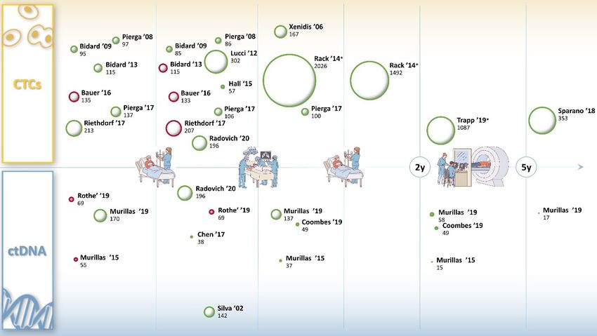
D’Amico et al. J Cancer Metastasis Treat 2021;7:3 I http://dx.doi.org/10.20517/2394-4722.2020.93                                     Page 9 of 16

Figure 2. Bubble representation of the most important clinical trials (name of first author and year of publication) assessing the
association between CTCs, ctDNA detection and prognosis. The data are retrieved from original manuscripts and shown in respect
of the 5 main management timepoints of early breast cancer (neoadjuvant treatment, surgery, adjuvant treatment, 2 years follow-
up, 5 years follow-up). Bubble size represents the number of analyzed patients. Bubble color represents a statistical association
between detection and either worse disease-free survival or worse overall survival (green = significant association, red = not significant
association). X-axis represents the treatment time frame for which data from the study refers to. Y-axis represents the year of
publication: closer to the x axis are the most recent trials. CTCs: circulating tumor cells; ctDNA: circulating tumor DNA. *Bubbles
dimension resized by a factor of 3. Figure references: Silva ’02[24], Rothe ’19[25], Murillas ’15[26], Murillas ’19[27], Chen ’17[28], Radovich
’20[29], Coombes ’19[31], Xenidis ’06[35], Rack ’14[36], Pierga ’08[37], Lucci ’12[38], Pierga ’17[40], Trapp ’19[42], Bidard ’09[44], Hall ’15[48],
Riethdorf ’17[74], Sparano ’18[76], Bidard ’13[73], Bauer ’16[78]

The CTCs analysis from the Geparquattro trial[41], after a median follow-up of 67.1 months, showed a
correlation between CTCs detection and reduced DFS and OS (P = 0.031 and P = 0.0057) only before
neoadjuvant treatment. The lack of prognostic value of post-neoadjuvant detection of CTCs was confirmed
in several studies[39,40,47]. However, Hall et al.[48] specifically addressed this question. In a cohort of 57 TNBC,
detection of at least 1 CTC after NAT predicted decreased DFS (log-rank P = 0.03, HR = 5.25, 95%CI: 1.34-
20.56) and OS (log-rank P = 0.03, HR = 7.04, 95%CI: 1.26-39.35), suggesting a potential different biological
behavior across subtypes. For a comprehensive graphic representation of trials assessing the role of CTCs
and ctDNA detection as a prognostic biomarker among different timepoints of eBC treatment, see Figure 2.

A combined comparison of the data from the main prospective trials that occurred in the past years has
partially answered the above-mentioned questions. In a pooled analysis of 3,173 eBC patients, the presence
of CTCs at the time of primary diagnosis was significantly associated with shorter DFS and DDFS, with
multivariate HRs of 1.822 (95%CI: 1.470-2.258; P < 0.001) for DFS and 1.888 (95%CI: 1.485-2.401; P < 0.001)
for DDFS. The prognostic value of CTCs was significant for all CTC cut-off values ranging from 1 to 20
both for DFS (all P < 0.01) and for DDFS (all P < 0.01) and was confirmed across all subtypes. However,
CTC detection did not significantly predict OS in the subgroup of patients with nodal stage N0 disease,
showing indeed, higher effect on OS in high risk patients, defined as those with primary tumors larger than
2 cm and lymph node involvement (HR = 2.460; 95%CI: 1.784-3.390; P = 0.001)[49].

Furthermore, in a comprehensive international meta-analysis of 21 trials (the IMENEO study), the
number of CTCs detected before NAT administration had no detrimental impact on pathological complete
Page 10 of 16           D’Amico et al. J Cancer Metastasis Treat 2021;7:3 I http://dx.doi.org/10.20517/2394-4722.2020.93

response. Although only a few trials showed that increasing number of CTCs enumerated was correlated
with a higher hazard ratio for disease recurrence[29,38], in the former meta-analysis each CTC detected
added a quantum of poor prognosis, suggesting that CTCs can be considered as a quantitative marker even
in eBC[50].

CAVEATS AND PITFALLS OF ctDNA IN EARLY-STAGE BREAST CANCER
Notwithstanding the potential of liquid biopsy in the metastatic setting, its deployment for early disease
diagnosis, MRD monitoring and characterization still poses multiple challenges[51].

As a matter of fact, ctDNA is directly associated with tumor burden, with consequently lower MAF
in early-stage, which is moreover characterized by a significantly low number of ctDNA-detectable
aberrations. In addition, low levels of ctDNA could result in low reproducibility, as high pre-analytical and
analytical variables could influence the characterization of alterations in situations with a near to the limit
of detection MAF[52,53].

Besides tumor burden, low ctDNA levels could also be linked to the differential shedding across metastatic
sites. In a study of Serpas et al.[54], knocking-down the DNase1L3 in mice resulted in a change in DNA
fragment length, suggesting that different nuclease mechanisms among different tissues could provide more
information about the origin of the ctDNA, allowing increased specificity. However, Garcia-Murillas et al.[27]
showed that single site relapses were characterized by low, undetectable, ctDNA levels and in particular
brain-only relapses were unlikely to be detected with rates of ctDNA detection similar to those of primary
brain tumors, probably due to the role of the blood-brain-barrier in hindering ctDNA shedding into the
plasma.

All these assumptions translate in the need to develop more sensible techniques, capable of detecting lower
and lower ctDNA concentrations.

A first layer of selection could be made by fragment size-based enrichment and selective sequencing that
can increase ctDNA detection and could therefore enhance downstream characterizations[55].

Additional mutation-agnostic ctDNA features, such as epigenetics, could potentially increase not only
sensitivity, but also specificity[56,57], which is of pivotal importance in the early setting to avoid false
negatives with their consequent impact on healthcare costs and patients’ quality of life.

As a matter of fact, the increasingly high sensitivity of sequencing technologies can detect, as a side-effect,
somatic mutations that are present across normal tissues. The onset of such confounding somatic mutations
may depend on tissue-specific factors, exposure to mutagens as chemotherapy and age[58]. These clones may
consequently result in a genetic drift that could potentially cause false-positive results in ctDNA analysis[59].

In particular, the clonal hematopoiesis of indeterminate potential (CHIP) is common with increasing age[60-62].
Garcia-Murillas et al.[27] prospectively assessed the detection of CHIP during MRD monitoring, showing
how persistently high levels of truncal mutations such as TP53 or PIK3CA could be detected also in the
Buffy coat as a result of CHIP and would otherwise generate false-positive ctDNA results.

Thus, breaking down the barrier of low tumor burden detection through ctDNA analysis, an increasing
need to develop highly sensitive and specific techniques has arisen. Several approaches have been
previously attempted to combine the two philosophies by applying wide targeted NGS panels or exome
sequencing to the primary tumor and consequently screen the resulting mutations on longitudinal ctDNA
samples through personalized ddPCR primers[27,32]. Such techniques include BEAMing, SafeSeqS, and
TAmSeq[63-65].
D’Amico et al. J Cancer Metastasis Treat 2021;7:3 I http://dx.doi.org/10.20517/2394-4722.2020.93    Page 11 of 16

Although offering a reasonable balance, these approaches have a potential downside. In fact, adjuvant and
neoadjuvant treatments may select new mutant clones that would be therefore missed, with consequently
false-negative results.

To overcome the mentioned drawbacks, hybrid capture-based NGS was developed to improve the detection
without prior knowledge of primary tumor alterations. In this approach, biotinylated probes, designed
against recurrently mutated genomic regions in breast cancer, select relevant DNA sequences for NGS
analysis. These technologies allowed the implementation of error-correction strategies (such as integrated
digital error suppression) that pushed the ctDNA detection limit to ~ 0.001% (CAPP-Seq[66], TEC-Seq[15]).
Commercial assays that utilizes hybrid capture-based NGS technology, as Guardant360®, PlasmaSELECT®
and FoundationOne® Liquid, are available for clinical use. However, despite the high self-reported accuracy,
specificity and sensitivity, a comparative study of two of these platforms demonstrated a low concordance
between the two assays[67].

CAVEATS AND PIRFALLS OF CTCs IN EARLY-STAGE BREAST CANCER
CTC deployment in early-stage breast cancer has encountered several analytical and pre-analytical
obstacles. The most challenging limitation encountered by CTC detection in early disease is that CTCs
are slightly less represented in blood, compared to physiological cellular components (approximately
one cancer cell among a billion normal blood cells, in the metastatic setting). Therefore, considering this
setting, even highly sensitive technologies could require a huge amount of blood to detect few CTCs,
threatening the clinical feasibility.

Thus, to overcome blood volume limitations, new detection approaches were developed. Inserting a 2 cm
long steel wire with a hydrogel functional tip coated with EpCAM antibody, the GILUPI CellCollector is a
method that allows to expose the collection tube to approximately 1 L of blood, instead of the canonical 7.5 ml
analyzed by CellSearch[68]. However, even if this promising approach has shown a higher CTC capture rate
from stage IV and III lung cancer patients compared to the FDA cleared CellSearch, these results have not
yet been achieved in a breast cancer patients’ cohort. Furthermore, this technology requires the insertion of
a noodle in the cubic vein of the patient for 30 min, resulting to be slightly more invasive than blood draw-
based analysis.

CTCs are highly heterogenous and dynamically change their shape and morphology. For instance,
downregulation of EpCAM during epithelial-mesenchymal transition (EMT) is a known phenomenon[69].

Accordingly, the inability to select CTCs that underwent to mesenchymal transition represents a further
limitation of the EpCAM based CTCs detection approaches. Since the transition of adherent epithelial
cells to a migratory mesenchymal state has been implicated in tumor metastasis, as well as in the process
in which CTCs acquire greater ability to metastasize[70], new approaches that allow the detection of not-
epithelial CTCs are being developed[71].

Among them, is worth of mention the use of carbon nanotubes for adherence-based capture and isolation
of CTCs of different phenotypes. This technology, called nanotube-CTCchip, has shown the potential to
overcome both selective phenotype detection and size-dependent loss of cells, proposing itself as a versatile
platform to capture CTCs of different phenotype[72]. Nevertheless, considering that it has only been tested
in a very small cohort of breast cancer patients (7 pts with different clinical staging and 2 healthy controls),
this technique is far from clinical deployment.

An additional point of weakness of the literature produced so far in this setting of BC is the arbitrary
nature of the positivity cut-off adopted among the different trials. Indeed, in contrast with the well-
Page 12 of 16            D’Amico et al. J Cancer Metastasis Treat 2021;7:3 I http://dx.doi.org/10.20517/2394-4722.2020.93

established prognostic cut-off used in the metastatic setting of 5 CTCs detected in 7.5 ml of blood through
the CellSearch, discretionary cut-off of either 1 CTCs, 5 CTCs or both have been used. However, since
the number of CTCs appeared to be directly correlated with the prognosis[36] and considering the lower
number of CTCs presents in the earlier stages, the lowest cut-off (1 CTCs) has been empirically adopted by
most of the investigators.

CONCLUSION AND FUTURE DIRECTIONS
Improving the management of early disease patients represents one of the most pressing challenges in
breast cancer care. Thus, in order to enhance the chances of early diagnosis, early detection of recurrence
and personalized treatment, several liquid biopsy tools have been deployed in this setting in the last 20 years.
Among them, CTCs and ctDNA detection and characterization have played a fundamental role, showing a
potential prognostic and predictive value in numerous prospective trials.

As a matter of fact, the sensitivity and specificity of the current methods are still suboptimal to candidate
ctDNA detection as a tool of early diagnosis. In this context, the assessment of epigenetic alterations (e.g.,
methylomes analysis) has shown promising preliminary results, demonstrating the potential utility of
ctDNA methylation profiles as a basis for non-invasive, sensitive and accurate early tumor detection[19].

Despite the highlighted current analytical and technical limitations of ctDNA detection in early setting,
encouraging results are shown by investigations addressing the potential prognostic role of ctDNA
detection after primary treatment[27,31], which suggest considering the detection as a good indicator of MRD.
Furthermore, the molecular characterization of ctDNA could act as a versatile, dynamic and not invasive
tool of disease characterization that might lead clinicians in the process of treatment choice, anticipating or,
in a foreseeable future, even partially replacing the tissue biopsy[46].

Combined analysis of ctDNA and CTCs could improve the prognostic value and clinical utility. In fact,
under certain circumstances, even with undetectable amount of ctDNA, CTCs can be isolated from the
circulation. Indeed, in a cohort of 196 TNBC patients that received a blood draw after NAT (results recently
published by Radovich et al.[29]), the combination of ctDNA and CTCs provided additional information
to identify patients with worse prognosis, such that the sensitivity to detect recurrences within 24 months
went from 79% with ctDNA detection alone to 90% when combined.

These results suggest that these methods have, at least in part, a complementary sensitivity and that the
combined use could improve the performances. Another successful combinatorial approach is represented
by the PCR-based NGS assay called CancerSEEK, that provided high performance in diagnosis and
assessment of primary tumor combining ctDNA detection with different circulating protein biomarkers[16].

However, although the findings provided by several investigations currently suggest a potential clinical
impact of ctDNA mutation tracking, clinical utility is still far from being demonstrated, as the highlighted
lead time among several studies has not yet been translated in a ctDNA based eBC management that has
prospectively shown a significant increase of overall survival.

CTC detection in early-stage breast cancer is associated with prognostic information in patients with
locally advanced or high-risk primary breast cancer at different time points in their treatment history[42,47,50].
In particular, the number of CTCs detected before any treatment has been associated with prognosis in
several investigations[37,40,44,73,74], suggesting a potential role of CTCs in stratifying patients for neoadjuvant
treatment selection. Furthermore, large data analysis including patients with early low-risk luminal breast
cancer seem to indicate that CTCs detection could serve as a predictive biomarker for loco regional
radiotherapy (XRT)[75]. In that respect, there is currently a plan for a prospective trial to test de-escalating or
D’Amico et al. J Cancer Metastasis Treat 2021;7:3 I http://dx.doi.org/10.20517/2394-4722.2020.93                               Page 13 of 16

omitting XRT in this low-risk CTC-negative disease, essentially focusing for the first time on this “indolent”
luminal cohort. On the other hand, much of the attention has been focused on patients with detectable
CTCs. Studies aimed at using CTC enumeration as selection criteria for additional “adjuvant” therapy have
been planned, particularly in hormone-receptor positive disease where CTCs appear associated with late-
recurrence and, possibly endocrine resistance[76]. One of such potential, example has been the study testing
HER2+ therapies in patients with detectable cells, TREAT-CTC represented an intriguing approach with an
ambitious goal but it was stopped for futility due to slow accrual and no indication of benefit[77]. In fact, the
study enrolled sixty-three patients with non-metastatic HER2-negative breast cancer and detectable CTCs (>
1/7.5 mL blood) after neoadjuvant chemotherapy and surgery randomized to trastuzumab (Herceptin) or
observation. There was no difference in CTCs at week 18, the trial’s primary endpoint, maybe due to CTCs-
HER2 definition or true lack of efficacy of the intervention.

In summary, this review showed that in spite of emerging data on the prognostic role of CTCs, the lack
of consensus about the “positive” detection cut-off (0, > 1, or > 5), the evaluation timing (at completion of
surgery or adjuvant treatment), and the lack of biomarker assessment along with missing clinical utility
prospective data may have limited the clinical application of CTC enumeration in early breast cancer.
Future studies will evaluate therapeutic strategies for both, de-escalation for CTC-negative disease and,
secondary adjuvant for potential late-recurrence in CTC-positive disease. It is likely that combining
molecular information such as CTCs, biomarkers, or integration with sensitive and complementary ctDNA
technologies may be the key for the practice-changing implementation of liquid biopsy in early disease.

DECLARATIONS
Authors’ contributions
Made substantial contributions to conception and design of the work: D’Amico P
Performed acquisition and analysis of data and have drafted the manuscript: D’Amico P, Corvaja C,
Gerratana L, Reduzzi C
Revised the manuscript critically and added important intellectual content: Cristofanilli M, Curigliano G
Availability of data and materials
Not applicable.
Financial support and sponsorship
PD is supported by the American-Italian Cancer Foundation Post-Doctoral Research Fellowship, year
2019-2020.
Conflicts of interest
All authors declared that there are no conflicts of interest.
Ethical approval and consent to participate
Not applicable.
Consent for publication
Not applicable.
Copyright
© The Author(s) 2021.

REFERENCES
1.   Society A cancer. Cancer Facts and Figures 2020. Available from: https://www.cancer.org/research/cancer-facts-statistics/all-cancer-facts-
Page 14 of 16                   D’Amico et al. J Cancer Metastasis Treat 2021;7:3 I http://dx.doi.org/10.20517/2394-4722.2020.93

      figures/cancer-facts-figures-2020.html. [Last accessed on 21 Sep 2020]
2.    Colleoni M, Sun Z, Price KN, et al. Annual hazard rates of recurrence for breast cancer during 24 years of follow-up: results from the
      international breast cancer study group trials I to V. J Clin Oncol 2016;34:927-35.
3.    Howlader N, Noone AM, Krapcho M, et al. SEER cancer statistics review, 1975-2016 National Cancer Institute. Bethesda MD. Posted to
      the SEER web site, April 2019. Available from: https://seer.cancer.gov/csr/1975_2016. [Last accessed on 21 Sep 2020]
4.    Cristofanilli M, Budd GT, Ellis MJ, et al. Circulating tumor cells, disease progression, and survival in metastatic breast cancer. N Engl J
      Med 2004;351:781-91.
5.    Bredemeier M, Kasimir-Bauer S, Kolberg HC, et al. Comparison of the PI3KCA pathway in circulating tumor cells and corresponding
      tumor tissue of patients with metastatic breast cancer. Mol Med Rep 2017;15:2957-68.
6.    Huang ZH, Li LH, Hua D. Quantitative analysis of plasma circulating DNA at diagnosis and during follow-up of breast cancer patients.
      Cancer Lett 2006;243:64-70.
7.    Bettegowda C, Sausen M, Leary RJ, et al. Detection of circulating tumor DNA in early- and late-stage human malignancies. Sci Transl
      Med 2014;6:224ra24.
8.    Dawson SJ, Tsui DW, Murtaza M, et al. Analysis of circulating tumor DNA to monitor metastatic breast cancer. N Engl J Med
      2013;368:1199-209.
9.    Madic J, Kiialainen A, Bidard FC, et al. Circulating tumor DNA and circulating tumor cells in metastatic triple negative breast cancer
      patients. Int J Cancer 2015;136:2158-65.
10.   Rodriguez BJ, Córdoba GD, Aranda AG, et al. Detection of TP53 and PIK3CA Mutations in Circulating Tumor DNA Using Next-
      Generation Sequencing in the Screening Process for Early Breast Cancer Diagnosis. J Clin Med 2019;8:1183.
11.   Alimirzaie S, Bagherzadeh M, Akbari MR. Liquid biopsy in breast cancer: A comprehensive review. Clin Genet 2019;95:643-60.
12.   Chen KZ, Lou F, Yang F, et al. Circulating tumor DNA detection in early-stage non-small cell lung cancer patients by targeted sequencing.
      Sci Rep 2016;6:31985.
13.   Zhang XF, Ju SQ, Wang XD, Cong H. Advances in liquid biopsy using circulating tumor cells and circulating cell-free tumor DNA for
      detection and monitoring of breast cancer. Clin Exp Med 2019;19:271-9.
14.   Beaver JA, Jelovac D, Balukrishna S, et al. Detection of cancer DNA in plasma of early stage breast cancer patients. Clin Cancer Res
      2014;20:2643-50.
15.   Phallen J, Sausen M, Adleff V, et al. Direct detection of early-stage cancers using circulating tumor DNA. Sci Transl Med
      2017;9:eaan2415.
16.   Cohen JD, Li L, Wang Y, et al. Detection and localization of surgically resectable cancers with a multi-analyte blood test. Science
      2018;359:926-30.
17.   Salido-Guadarrama I, Romero-Cordoba S, Peralta-Zaragoza O, Hidalgo-Miranda A, Rodríguez-Dorantes M. MicroRNAs transported by
      exosomes in body fluids as mediators of intercellular communication in cancer. Onco Targets Ther 2014;7:1327-38.
18.   Gold B, Cankovic M, Furtado LV, Meier F, Gocke CD. Do circulating tumor cells, exosomes, and circulating tumor nucleic acids have
      clinical utility? A report of the association for molecular pathology. J Mol Diagn 2015;17:209-24.
19.   Shen SY, Singhania R, Fehringer G, et al. Sensitive tumour detection and classification using plasma cell-free DNA methylomes. Nature
      2018;563:579-83.
20.   Liu M, Oxnard G, Klein E, et al. Sensitive and specific multi-cancer detection and localization using methylation signatures in cell-free
      DNA. Annals of Oncology 2020;31:745-59.
21.   Cristiano S, Leal A, Phallen J, et al. Genome-wide cell-free DNA fragmentation in patients with cancer. Nature 2019;570:385-9.
22.   Gerratana L, Davis AA, Shah AN, Lin C, Corvaja C, Cristofanilli M. Emerging Role of Genomics and Cell-Free DNA in Breast Cancer.
      Curr Treat Options Oncol 2019;20:68.
23.   Koboldt DC, Fulton RS, McLellan MD, et al. Comprehensive molecular portraits of human breast tumors. Nature 2013;490:61-70.
24.   Silva JM, Silva J, Sanchez A, et al. Tumor DNA in plasma at diagnosis of breast cancer patients is a valuable predictor of disease-free
      survival. Clin Cancer Res 2002;8:3761-6.
25.   Rothé F, Silva MJ, Venet D, et al. Circulating tumor DNA in HER2-amplified breast cancer: a translational research substudy of the
      NeoALTTO phase III trial. Clin Cancer Res 2019;25:3581-8.
26.   Garcia-Murillas I, Schiavon G, Weigelt B, et al. Mutation tracking in circulating tumor DNA predicts relapse in early breast cancer. Sci
      Transl Med 2015;7:302ra133.
27.   Garcia-Murillas I, Chopra N, Comino-Méndez I, et al. Assessment of molecular relapse detection in early-stage breast cancer. JAMA
      Oncol 2019;5:1473-8.
28.   Chen YH, Hancock BA, Solzak JP, et al. Next-generation sequencing of circulating tumor DNA to predict recurrence in triple-negative
      breast cancer patients with residual disease after neoadjuvant chemotherapy. NPJ Breast Cancer 2017;3:24.
29.   Radovich M, Jiang G, Hancock BA, et al. Association of circulating tumor DNA and circulating tumor cells after neoadjuvant
      chemotherapy with disease recurrence in patients with triple-negative breast cancer: preplanned secondary analysis of the BRE12-158
      randomized clinical trial. JAMA Oncol 2020;6:1410-5.
30.   Swanton C, Jamal-Hanjani M, Abbosh C, et al. Phylogenetic ctDNA analysis depicts early stage lung cancer evolution. Nature
      2017;545:446-51.
31.   Coombes RC, Page K, Salari R, et al. Personalized detection of circulating tumor DNA antedates breast cancer metastatic recurrence. Clin
      Cancer Res 2019;25:4255-63.
32.   McDonald BR, Contente-Cuomo T, Sammut SJ, et al. Detection of residual disease after neoadjuvant therapy in breast cancer using
You can also read