Recapitulation of HIV-1 Env-antibody coevolution in macaques leading to neutralization breadth
←
→
Page content transcription
If your browser does not render page correctly, please read the page content below
RESEARCH ARTICLES

                                                                                                                               Cite as: R. S. Roark et al., Science
                                                                                                                                10.1126/science.abd2638 (2020).

Recapitulation of HIV-1 Env-antibody coevolution in
macaques leading to neutralization breadth
Ryan S. Roark1*, Hui Li1*, Wilton B. Williams2,3*, Hema Chug4*, Rosemarie D. Mason5*, Jason Gorman5*,
Shuyi Wang1, Fang-Hua Lee1, Juliette Rando1, Mattia Bonsignori2,3, Kwan-Ki Hwang2, Kevin O. Saunders2,6,
Kevin Wiehe2,3, M. Anthony Moody2,7, Peter T. Hraber8, Kshitij Wagh8, Elena E. Giorgi8, Ronnie M. Russell1,
Frederic Bibollet-Ruche1, Weimin Liu1, Jesse Connell1, Andrew G. Smith1, Julia DeVoto1, Alexander I. Murphy1,
Jessica Smith1, Wenge Ding1, Chengyan Zhao1, Neha Chohan1, Maho Okumura1, Christina Rosario1, Yu Ding1,
Emily Lindemuth1, Anya M. Bauer1, Katharine J. Bar1, David Ambrozak5, Cara W. Chao5, Gwo-Yu Chuang5,
Hui Geng5, Bob C. Lin5, Mark K. Louder5, Richard Nguyen5, Baoshan Zhang5, Mark G. Lewis9, Donald
Raymond4, Nicole A. Doria-Rose5, Chaim A. Schramm5, Daniel C. Douek5, Mario Roederer5, Thomas B.
Kepler10,11, Garnett Kelsoe2,6, John R. Mascola5, Peter D. Kwong5, Bette T. Korber8, Stephen C. Harrison4,12,

                                                                                                                                                                           Downloaded from http://science.sciencemag.org/ on January 2, 2021
Barton F. Haynes2,3, Beatrice H. Hahn1, George M. Shaw1†
1
 Departments of Medicine and Microbiology, Perelman School of Medicine, University of Pennsylvania, Philadelphia, PA 19104, USA. 2Duke Human Vaccine Institute, Duke
University School of Medicine, Durham, NC 27710, USA. 3Department of Medicine, Duke University School of Medicine, Durham, NC 27710, USA. 4Laboratory of Molecular
Medicine, Boston Children’s Hospital and Harvard Medical School, Boston, MA 02115, USA. 5Vaccine Research Center, National Institute of Allergy and Infectious Diseases,
National Institutes of Health, Bethesda, MD 20892, USA. 6Departments of Immunology and Surgery, Duke University School of Medicine, Durham, NC 27710, USA.
7
  Departments of Pediatrics and Immunology, Duke University School of Medicine, Durham, NC 27710, USA. 8Theoretical Biology and Biophysics, Los Alamos National
Laboratory, Los Alamos, NM 87545, USA. 9Bioqual, Inc., Rockville, MD 20850, USA. 10Department of Microbiology, Boston University School of Medicine, Boston, MA 02118,
USA. 11Department of Mathematics and Statistics, Boston University, Boston, MA 02215, USA. 12Howard Hughes Medical Institute, Harvard Medical School, Boston, MA
02115, USA.
*These authors contributed equally to this work.
†Corresponding author. Email: shawg@pennmedicine.upenn.edu

Neutralizing antibodies elicited by HIV-1 coevolve with viral envelope proteins (Env) in distinctive patterns,
in some cases acquiring substantial breadth. We report that primary HIV-1 envelope proteins—when
expressed by simian-human immunodeficiency viruses in rhesus macaques—elicited patterns of Env-
antibody coevolution strikingly similar to those in humans. This included conserved immunogenetic,
structural and chemical solutions to epitope recognition and precise Env-am ino acid substitutions,
insertions and deletions leading to virus persistence. The structure of one rhesus antibody, capable of
neutralizing 49% of a 208-strain panel, revealed a V2-apex mode of recognition like that of human bNAbs
PGT145/PCT64-35S. Another rhesus antibody bound the CD4-binding site by CD4 mimicry mirroring
human bNAbs 8ANC131/CH235/VRC01. Virus-antibody coevolution in macaques can thus recapitulate
developmental features of human bNAbs, thereby guiding HIV-1 immunogen design.

A major roadblock to rational HIV-1 vaccine design is the lack                         Envs to elicit bNAbs and to characterize the co-evolutionary
of a suitable primate model in which broadly neutralizing an-                          pathways of bNAb lineages and the cognate Env intermedi-
tibodies (bNAbs) can be commonly induced, and the molecu-                              ates that elicited them (2–8), thereby serving as a molecular
lar, biological and immunological mechanisms responsible                               guide for rational vaccine design. Recent innovations in SHIV
for such responses studied in a reproducible and iterative                             design (9) now make this experimental strategy testable.
fashion. Since most HIV-1 bNAbs identified to date have                                   HIV-1 bNAbs target one of several conserved regions on
come from humans chronically infected by HIV-1, we hypoth-                             the native Env trimer, including the CD4-binding site
esized that one means to elicit such antibodies in primates                            (CD4bs), V2 apex, V3 high mannose patch, gp120/gp41 inter-
might be by infecting Indian rhesus macaques (RMs) with                                face, fusion peptide, glycosylated silent face, and membrane-
simian-human immunodeficiency virus (SHIV) strains that                                proximal external region (MPER) (10, 11). Such antibodies
bear primary or transmitted/founder (T/F) HIV-1 Envs that                              generally share certain features such as exceptional HCDR3
induced bNAbs in humans (1–7). SHIV infected RMs could                                 length, extensive somatic hypermutation, autoreactivity or
then be employed to assess the potential of particular HIV-1                           unusual mechanisms for binding glycans or glycopeptides
                                                                                       (10–13). Long HCDR3s result from initial germline

First release: 19 November 2020                                         www.sciencemag.org                (Page numbers not final at time of first release) 1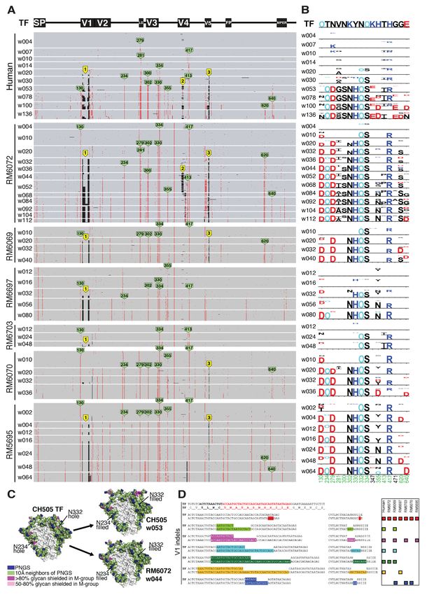
immunoglobulin gene rearrangements whereas the charac-              superinfected by a second genetically divergent virus
teristic high frequencies of V(D)J mutations that lead to af-       (CAP256SU, superinfection) (22). The CAP256SU variant was
finity maturation and neutralizing breadth result from              thought to trigger the V2 bNAb lineage in this individual (5),
persistent virus replication, epitope variation, and continued      so we constructed and analyzed a SHIV containing this Env
Env-Ab coevolution. In some subjects, cooperating antibody          (fig. S1). SHIVs containing CH505, CH848 and CAP256SU
lineages that target the same epitope have been identified,         Envs were modified at gp120 residue 375 to enhance binding
and together they contribute to Env diversification and bNAb        and entry into rhesus CD4 T cells. The Env375 substitutions
development (4, 6). A critical question in the HIV-1 vaccine        resulted in no discernible change in antigenicity or sensitivity
field is whether the relatively rare examples of high titer,        to anti-HIV-1 NAbs compared with wild-type virus, but they
bNAbs that have been identified in a subset of chronically in-      were essential for replication in primary RM CD4+ T cells [fig.
fected humans represent stochastic accidents of nature, not         S1 and (9)]. We inoculated 22 RMs (table S1) with
likely to be replicated by a vaccine, or if there are special       SHIV.CH505 (n = 10), SHIV.CH848 (n = 6) or
properties of particular HIV-1 Envs that along with determin-       SHIV.CAP256SU (n = 6) by the intravenous route and fol-
istic features of Env-Ab coevolution make HIV-1 vaccine de-         lowed them for an average period of two years (mean of 103
velopment more plausible.                                           weeks, range 36 to 184) for viral replication kinetics, develop-

                                                                                                                                         Downloaded from http://science.sciencemag.org/ on January 2, 2021
    The current study is based on the premise that while Env        ment of strain-specific (autologous) and broadly reactive
diversity within HIV-1 group M is extraordinarily high, pri-        (heterologous) NAbs, and patterns of Env sequence evolution.
mary HIV-1 Envs [i.e., native Env trimers on infectious viri-       Eight of the animals were treated with anti-CD8 mAb at or
ons that allow for persistent replication and clinical              near the time of SHIV inoculation in order to transiently de-
transmission in humans (1)] are nonetheless constrained in          plete CD8+ cells and increase peak and set point plasma virus
their immediate evolutionary potential (14–16). This para-          titers. One animal (RM5695), which was repurposed from an
dox—extreme Env diversity globally but constraints on imme-         earlier study of HIV-1 gp120 protein immunization (23), was
diate or near-term evolution in an individual—can be                inoculated with plasma from three animals recently infected
explained by competing evolutionary forces: positive selec-         by SHIV.CH505 to increase early virus diversity.
tion imposed by immune evasion balanced by purifying (neg-
ative) selection acting to retain viral fitness. At least four      SHIV replication in vivo and elicitation of neutralizing
interrelated Env properties, acting in concert, facilitate im-      antibodies
mune escape—occlusion of trimer-interface epitopes (17), gly-       Figure S2 depicts the kinetics of virus replication and the de-
can shielding (18), epitope variation (19), and conformational      velopment of autologous, strain-specific tier 2 NAbs as meas-
masking (20)—while conservation of structure and cell-entry         ured by plasma vRNA and inhibition of virus entry into TZM-
properties and retention of protein stability constrain each        bl cells (18). Acute virus replication kinetics were consistent
primary Env’s immediate and short-term evolutionary trajec-         for all SHIVs in all animals with peak viremia occurring
tory. When introduced into a human as HIV-1 infection or            about 14 days post-inoculation and reaching titers in the 105
into RMs as SHIV infections, each Env will have unique re-          to 108 vRNA/ml range [geometric mean (GM) = 4.2 × 10⁶].
strictions in its exposure of antigenic sites, thus favoring dis-   Peak vRNA titers were higher in anti-CD8 treated animals
tinct molecular pathways of escape from neutralizing                than in untreated animals [geometric mean titers (GMT) of
antibodies with each pathway exacting a different fitness cost      2.6 × 10⁷ versus 1.5 × 10⁶, respectively; P < 0.0001]. Plasma
from the virus. These considerations led us to hypothesize          virus load set points at 24 weeks post-infection varied widely
that SHIV infection of RMs will recapitulate HIV-1 infection        between 100 and 988,110 vRNA/ml with a mean for anti-CD8
of humans with respect to molecular patterns of Env-Ab co-          treated animals of 248,313 ± 136,660 vRNA/ml compared
evolution, within bounds set by any particular Env sequence         with 8313 ± 3509 vRNA/ml for untreated animals (P = 0.002).
and the respective rhesus and human immunoglobulin gene             Set point vRNA levels in human subjects infected with the
repertoires that respond to it (21).                                corresponding HIV-1 CH505, CH848 or CAP256 strains were
    Some of the best studied examples of HIV-1 Env-Ab co-           81,968, 132,481 and 750,000 vRNA/ml, respectively. Com-
evolution have come from the analysis of HIV-1 infected hu-         pared with two human cohort studies of acute and early HIV-
man subjects CH505, CH848 and CAP256, who developed                 1 infection (24, 25), peak and setpoint vRNA titers in SHIV
bNAb responses targeting the CD4bs, V3 high mannose patch           infected animals were comparable, although several ma-
and V2 apex of HIV-1 Env, respectively (2–7). Subjects CH505        caques developed viral loads near the lower limit of detection
and CH848 were each productively infected by a single T/F           (100 vRNA/ml), which is rare in humans. Six macaques de-
virus, and previously we constructed SHIVs containing each          veloped AIDS-defining events and were euthanized 36, 40,
of these T/F Envs (9). CAP256 was infected by a single virus        60, 65, 89 or 136 weeks post-SHIV infection; four of these an-
(CAP256PI, primary infection) and then became                       imals had been treated with anti-CD8 mAb (table S1).

First release: 19 November 2020                         www.sciencemag.org       (Page numbers not final at time of first release)   2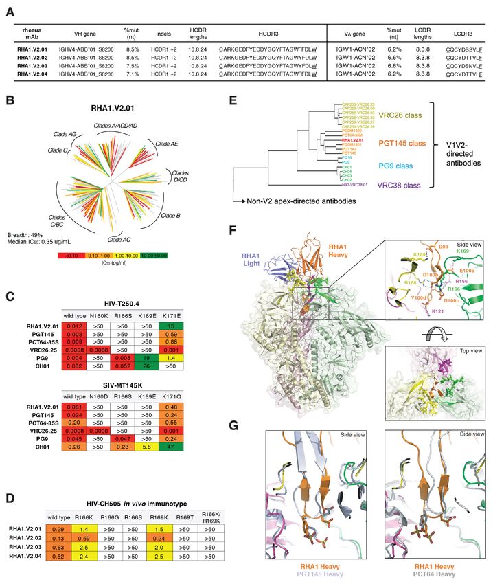
Autologous tier 2 NAb responses to the three SHIVs were          elicitation that are characteristic of HIV-1 infection in hu-
detected as early as 8 weeks post-infection and peaked be-           mans, including the potential to elicit bNAbs.
tween 24 and 80 weeks with 50% inhibitory dilutions (ID50)               Figure 1B highlights the kinetics, potency and breadth of
between 0.05 (1:20 dilution) and 0.000125 (1:8,000 dilution)         neutralization by plasma from the SHIV.CH505 infected ani-
(fig. S2). The kinetics of appearance and titers of autologous       mal RM5695 and identifies immunoglobulin G (IgG) as the
tier 2 Nabs that developed in response to SHIV infections            active component. By 16 weeks post-infection, an autologous
were generally comparable to those observed in humans in-            NAb response to SHIV.CH505 was detectable at an ID50 titer
fected by viruses bearing the homologous Envs (2, 7, 22).            of 0.02 along with heterologous responses to viruses bearing
Among all animals, there was a significant association be-           HIV-1 25710, X1632, Q23, ZM233, T250 and WITO Envs at ti-
tween higher setpoint virus titers and higher autologous tier        ters of 0.05–0.01. NAbs increased rapidly in breadth and titer
2 NAb titers (Spearman correlation rank correlation coeffi-          thereafter. By week 48, bNAbs targeting Q23 and T250
cient rs = 0.74, P < 0.0001). Heterologous plasma neutralizing       jumped in titer to 0.0002–0.0005 and against X1632, 246F3,
activity was assessed against a diverse 22-member global             ZM233 and WITO to titers of 0.005. By week 56, bNAbs in the
panel of tier 1a (n = 3) or tier 2 (n = 19) HIV-1 strains (26–29)    plasma of RM5695 neutralized 17 of 18 viruses in the heterol-
(table S2). All animals developed potent neutralizing re-            ogous HIV-1 test panel at titers ranging from 0.05–0.0002,

                                                                                                                                          Downloaded from http://science.sciencemag.org/ on January 2, 2021
sponses to the three tier 1a viruses (GMT ID50 = 0.0004). Tier       along with a divergent tier 2 simian immunodeficiency virus
1a viruses have “open” Env trimers that spontaneously expose         strain (SIVcpz.MT145.Q171K) that shares selective antigenic
linear V3 and conformational CD4 induced (CD4i) bridging             cross-reactivity with HIV-1 in the V2 apex bNAb epitope clus-
sheet epitopes, thus explaining their extreme sensitivity to         ter (35) at a titer of 0.006. IgG was purified from RM5695
what are otherwise non-neutralizing antibodies. Such “non-           week 56 plasma and assayed for neutralizing activity against
neutralizing” antibodies that target linear V3 and CD4i              the same 19 heterologous viruses: IgG concentrations be-
epitopes are elicited in virtually all HIV-1 infected humans,        tween 0.002 mg/ml (corresponding to a ~1:10,000 dilution of
thereby selecting for Envs with an open-closed equilibrium           rhesus plasma) and 4 mg/ml neutralized 18 of the 19 viruses
that strongly favors the closed configuration (20, 30–32). Tier      in a rank order similar to the polyclonal plasma (Fig. 1B, far
2 viruses typify primary or T/F viruses that are generally re-       right panels). Neither plasma nor purified IgG neutralized
sistant to neutralization by heterologous plasma antibodies          control viruses pseudotyped with the murine leukemia virus
except for those that target one of the canonical bNAb epitope       (MLV) Env. Thus, anti-HIV-1 specific IgG accounted for all of
clusters (1, 10, 11, 26). In this study, we used a reciprocal neu-   the autologous and heterologous neutralizing activity in the
tralization titer cutoff of ID50 ≤ 0.05 against ≥33% of heterol-     RM5695 plasma. Of note, RM5695 plasma and plasma IgG
ogous tier 2 viruses as an indication of neutralization              from week 56 reached ID90 or ID95 thresholds against most
breadth. This is a conservative threshold consistent with            viruses and exhibited steep inflections at the ID50 midpoint,
other studies that have characterized bNAb prevalence in hu-         indicating potent neutralization. Neutralization breadth de-
man cohorts (28, 33, 34). Seven of 22 RMs developed antibody         tected as early as 16 weeks post-infection is unusual in HIV-1
responses that neutralized between 6 and 18 of 18 heterolo-          infection but not unprecedented (36) and occurs most often
gous HIV-1 tier 2 viruses in our test panel (Fig. 1A and table       with V2 apex bNAbs, likely because their activity depends
S2). Heterologous neutralization was first detected as early as      more on long HCDR3s than on extensive somatic hypermu-
8 to 16 weeks post-SHIV infection in two animals and as late         tation. We show below that the bNAb activity in RM5695
as 88 weeks post-SHIV infection in others. Heterologous              plasma and its isolated IgG fraction as well as monoclonal
NAbs reached ID50 titers as high as 0.0001, with GMTs in the         bNAbs derived from RM5695, all targeted a bNAb epitope in
seven animals ranging from 0.018 (1:55) to 0.004 (1:271) (Fig.       the V2 apex that included the conserved lysine rich C-strand
1A). Peak and setpoint plasma virus titers were higher in the        and N160 glycan. The kinetics of appearance, breadth, titers
seven animals that developed bNAbs (GMT = 18,150,586 and             and potency of bNAbs elicited in the six other SHIV-infected
47,471 vRNA/ml, respectively) than in animals that did not           RMs, including three animals (RM6070, RM40591 and
(GMT = 2,101,814 and 1945 vRNA/ml, respectively; P < 0.015           RM42056) whose bNAbs also targeted the V2 apex C-strand,
for both). Two animals (RM5695 and RM6070) with bNAbs                are summarized in Fig. 1A and table S2.
were infected by SHIV.CH505, two (RM6163 and RM6167) by
SHIV.CH848, and three (RM40591, RM42056 and RM6727)                  Env evolution in SHIV infected macaques and HIV-1
by SHIV.CAP256SU. The remaining 15 animals in the study              infected humans
showed either no or very limited, low titer neutralization of        To explore if SHIV Env evolution in RMs recapitulates that of
heterologous tier 2 viruses (table S2). Altogether, the findings     HIV-1 in humans in a strain-specific manner, we analyzed
show that SHIVs bearing primary T/F Envs reproduce in RMs            Env sequences in the 22 SHIV infected animals over one to
key features of virus replication dynamics and NAb                   three years of follow-up and compared them with the

First release: 19 November 2020                         www.sciencemag.org        (Page numbers not final at time of first release)   3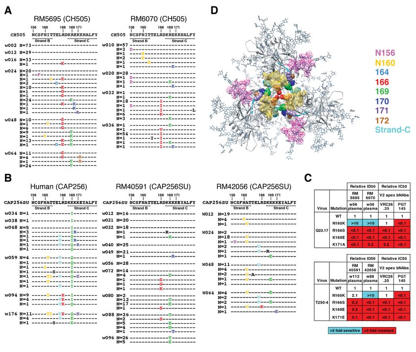
evolution of the homologous Envs in humans infected by              humans and rhesus animals infected by CH505 and CH848
HIV-1. We used single genome sequencing (SGS) for this anal-        viruses were different still from the CAP256SU infected RMs
ysis, since it allows for retention of genetic linkage across in-   in which 45 selected sites were identified, including 25 that
tact viral genes and enables precise molecular tracking of          were shared in more than one animal (figs. S7, A and B, and
sequence diversification from unambiguous T/F genomes (1,           S8). A comparable Env-wide analysis in the human subject
37–39). We analyzed cDNA sequences derived from plasma              CAP256 could not be performed because this individual was
virion RNA, since the half-life of circulating plasma virus is      infected and then superinfected by two widely divergent vi-
observed in different individuals could occur by chance and These are common contact residues of human V2 apex bNAbs
that likelihood was estimated to be vanishingly small (see (5, 8, 48). A minor fraction of sequences lost PNGs at 156 and
“Extended Discussion” in the supplementary materials). 160. By 24 weeks post-infection in RM5695 and 32 weeks
post-infection in RM6070, most circulating virus contained
NAbs select for mutations in Env mutations at residues 166 or 169; by 36 to 48 weeks post-in-
CH505 fection, all sequences were mutant at one or the other of these
To examine if NAb-mediated selection was a primary driving positions. We corroborated this single genome sequence evi-
force in the Env-specific evolutionary patterns that we ob- dence of strong virus selection in the central cavity of the V2
served, early strain-specific NAb responses and later bNAb apex of RM5695 by next generation sequencing, which re-
responses were mapped using site-directed mutagenesis to vealed that 99.3% of 10,000 sequences sampled between
introduce observed mutations into neutralization sensitive weeks 48 and 64 post-infection contained mutations at resi-
autologous and heterologous Envs. We then tested these mu- dues 166 or 169. When these residues were mutated in the
tant Envs, compared with their wild-type counterparts, for wild type versions of heterologous primary virus strains T250,
sensitivity to polyclonal plasma and mAbs isolated from the Q23, MT145K, 246F or BG505 that were otherwise neutral-
infected RMs or human subjects. Variable sites in the CH505 ized by RM5695 and RM6070 plasma, neutralization was ab-
Downloaded from http://science.sciencemag.org/ on January 2, 2021
Env (Fig. 2, A and B) were interrogated alone or in combina- rogated (Fig. 3C and fig. S12). Neutralization of these
tion for their effects on Env sensitivity to autologous antibod- heterologous strains was variably dependent on the glycan at
ies. Aside from variable residues in the leader sequence of N160 (fig. S13), similar to what has been reported for V2 apex
gp160 that generally represent CTL escape mutations (37, 43), bNAbs in subject CAP256SU (49). These findings indicated
CTL epitope reversions to global consensus at or near residue the presence of potent V2 apex C-strand targeted bNAbs in
417 (4), and mutations at residues 300, 620 and 640 that oc- RM5695 and RM6070. In summary, the mapping of autolo-
curred inconsistently and late in the course of infection, most gous and heterologous Nab responses in RMs infected by
of the variable residues were found to represent escape mu- SHIV.CH505 indicated that most mutations in Env that could
tations from autologous NAbs (fig. S11A). These included res- not be ascribed to CTL selection were the result of NAb selec-
idues 234 and 334 where mutations restored PNGs that filled tion.
T/F glycan holes (fig. S3), loop D residues 279 and 281 in-
volved in CD4 binding, CD4 contact residues in V5 (460/ΔV5), CH848
and residue 130. The temporal appearance of these NAbs co- We also examined Env mutations shared between
incided with the appearance of phenotypically demonstrable SHIV.CH848 infected RMs and the HIV-1 infected CH848 hu-
NAb escape mutations in Env (Fig. 2, A and B). These results man subject as potential sites targeted by NAbs. The earliest
were corroborated by neutralization patterns of V3 targeted Env substitution in the human subject that was also mutated
mAbs DH647 and DH648 and CD4bs targeted mAbs in sequences from all six RMs was at position R336, which is
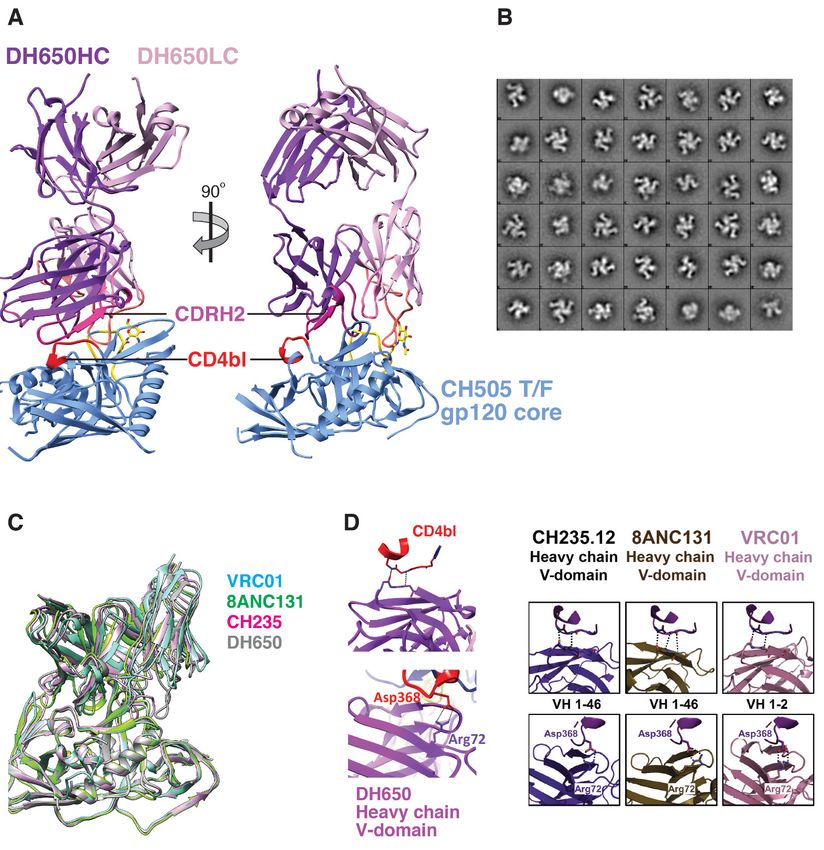
DH650.UCA and DH650 (all isolated from the SHIV.CH505 surface exposed on the HIV-1 trimer. The R366G substitution
infected RM6072) and the human CD4bs targeted mAbs in RM6167 occurred at week 10 post-SHIV infection when au-
CH235.UCA, CH235.IA3 and CH235.9 isolated from human tologous NAb titers to the T/F SHIV were 1:220. NAb titers to
subject CH505 (6) (fig. S11, B and C). Human subject CH505 a site-directed mutant of the T/F virus that contained the
developed two cooperative lineages of CD4bs antibodies that R366G substitution fell three-fold to 1:72 (fig. S14A), indica-
targeted epitopes that included loop D residues 279-281 and tive of an early immunodominant epitope-focused NAb re-
residues in V5, eventually leading to neutralization breadth sponse. Autologous Nab titers in RM6167 increased to 1:435
by both antibody lineages (4, 6). In RM6072, a mAb lineage, by week 36 and this was accompanied by strong selection on
termed DH650, was isolated that targeted these same the evolving virus quasispecies resulting in deletions in V1
epitopes but it never developed neutralization breadth, a and V5 (figs. S4 and S10) and amino acid substitutions at po-
finding for which we found a structural explanation (see be- sitions indicated in fig. S4. When these consensus mutations
low). were introduced into the CH848 T/F Env and tested for neu-
In addition to the strain-specific NAb responses that we tralization sensitivity to week 10 and 36 plasma, the mutant
identified in SHIV.CH505 infected RMs, we found in two virus showed complete escape (fig. S14A). In the human sub-
CH505 animals (RM5695 and RM6070) neutralizing antibod- ject CH848, an early strain-specific NAb response targeted
ies that targeted heterologous tier 2 viruses (Fig. 1 and table many of these same sites including V1 (7). The virus qua-
S2). These bNAbs were first detected at weeks 8 (RM6070) sispecies in subject CH848 escaped by deleting ~10 amino ac-
and 16 (RM5695) post-infection and their development was ids in V1, which was followed by the development of the V3
temporally associated with the appearance of mutations in glycan focused bNAb lineage DH270 (7, 50). Remarkably, the
the Env V2 apex, including residues 166 or 169 (Fig. 3A). same sequence of events occurred in SHIV.CH848 infected
First release: 19 November 2020 www.sciencemag.org (Page numbers not final at time of first release) 5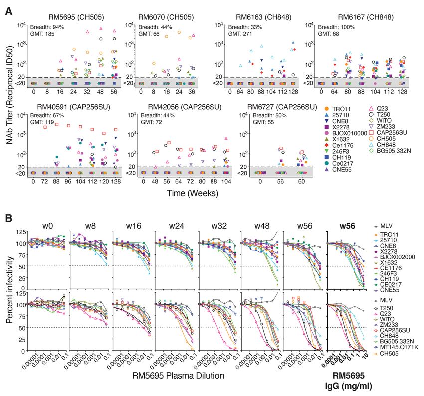
animals RM6163 and RM6167 (fig. S10). V1 was first targeted and follow similar patterns of Ab-Env coevolution. We ex-
by an early autologous NAb response (fig. S14), followed by plored the latter possibility by isolating and characterizing
deletions in V1, which was in turn followed by the develop- neutralizing mAbs from two RMs that were infected by
ment of V3 glycan targeted bNAbs (Fig. 1A and table S2). We SHIV.CH505. We selected these animals for study because
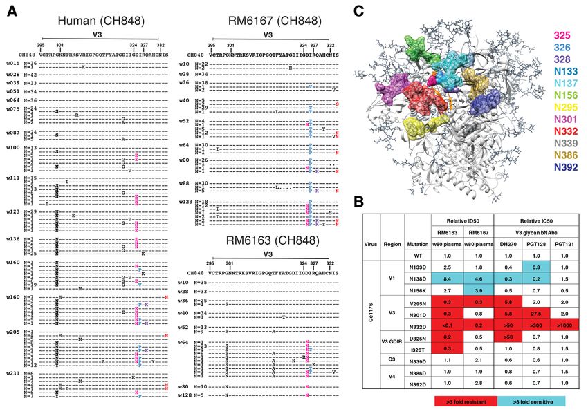
mapped the epitopes of these bNAb responses to the glycan both exhibited favorable virus replication kinetics, compara-
at residue N332 and the Gly-Asp-Ile-Arg (GDIR) motif at po- ble early NAb responses, and an overall pattern of Env evolu-
sitions 324-327 (Fig. 4B). Consistent with this epitope map- tion similar to that in the human subject CH505; nonetheless,
ping, we identified in the evolving viral quasispecies of both one animal (RM5695) developed bNAbs and the other
RMs, mutations at residues 332-334 and GDIR residues 324- (RM6072) did not. We asked what might be the genetic and
327 (Fig. 4A). Like the prototypic human V3 glycan bNAbs structural similarities and dissimilarities between the NAbs
DH270, PGT121 and PGT128, the bNAbs that we identified in elicited in these animals.
RMs 6163 and 6167 were strictly N332 dependent, and muta-
tions in spatially associated surface residues V295, N301, CD4bs-directed antibodies
N138 and N133 had similar effects on neutralization potency Two broadly neutralizing mAb lineages (CH235 and CH103)
of both rhesus and human bNAbs (Fig. 4, B and C). targeting the HIV-1 CD4bs were previously isolated from the
Downloaded from http://science.sciencemag.org/ on January 2, 2021
human subject CH505 and their evolutionary pathways from
CAP256 germline receptors to mature antibodies determined (2, 4, 6).
In the human subject CAP256, recombination between PI and In animal RM6072, serological analysis of early plasma sam-
SU lineage sequences (fig. S7C) precluded a gp160-wide Env ples suggested the presence of CD4bs targeted Abs based on
analysis. We focused instead on mutations in and near the V2 selective Ab binding to CH505 T/F gp120 and resurfaced Env
C strand, since the human subject and two of six RMs (40591 core but not to their isogenic ∆371I mutants, which do not
and 42056) developed V2 apex targeted bNAbs (Fig. 1A and bind CD4 (fig. S15A). Additional evidence of CD4bs targeted
table S2). In both human and RMs, very similar patterns of Abs in the plasma of this animal included competitive block-
Env V2 sequence evolution occurred (Fig. 3B). These muta- ing of soluble CD4 (sCD4) and CH235 and CH106 mAbs (fig.
tions included substitutions at the canonical residues 160, S15B). We sorted memory B cells from RM6072 from longitu-
166, 169 and 171 shown to be contact residues for other pro- dinal timepoints 20, 24 and 32 weeks post-SHIV infection and
totypical human V2 apex bNAbs (5, 8, 51–53). When intro- selected cells that reacted with CH505 T/F gp120 but not with
duced into heterologous neutralization-sensitive Envs, the ∆371I mutant. We isolated a 15-member B cell clonal lin-
mutations at 166, 169 and 171 abrogated neutralization by eage designated DH650 (fig. S15, C and D) that selectively
plasma from RMs 40591 and 42056 as it did for control hu- bound the autologous CH505 T/F Env gp120 but not the ∆371I
man bNAbs PGT145 and VRC26.25, the latter having been iso- mutant (fig. S15E). Some of these mAbs competed with sCD4
lated from subject CAP256 (49, 53) (Fig. 3C). In RMs 40591 and CH235 and CH106 mAbs for binding CH505 T/F gp120
and 42056, we observed that V2 apex targeted antibodies (fig. S15F). Mature DH650 lineage mAbs, but not the inferred
were variably dependent on binding to the glycan at Env res- germline UCA, bound CH505 T/F gp140 SOSIPv4.1 trimer
idue 160 for neutralizing activity, a pattern that was similar (fig. S16A). Most members of the DH650 lineage neutralized
to antibodies from animals RM5695 and RM6070 and the hu- a glycan-deficient mutant of CH505 Env (gly4) and two-thirds
man subject CAP256 (49). This variable dependence on N160 of them neutralized the wildtype CH505 T/F strain (fig. S16B).
for bNAb activity is further shown in fig. S13 and is discussed The DH650 UCA neutralized neither. None of the DH650 lin-
in supplementary materials. Overall, the molecular and tem- eage mAbs neutralized heterologous viruses (fig. S16C and ta-
poral patterns of Env sequence evolution reported here for ble S3).
SHIV-infected macaques, and previously in humans (18, 37– The immunogenetic features of the DH650 lineage mAbs
39, 43), highlight the utility of dynamic measurements of lo- suggest how they recognize HIV-1 Env. The lineage comes
calized sequence variation as a highly sensitive indicator of from V(D)J recombination of the macaque VH1-h gene (fig.
epitope-specific adaptive immune responses. S15D), which is 91% similar to an orthologous human gene
VH1-46 used by the CD4bs bNAbs CH235 and 8ANC131 (6, 54).
Homologous B cell responses in humans and macaques DH650 antibodies share key VH residues with CH235 (fig.
The observation that homologous Envs evolved in similar S17A), which were shown previously to be contact sites with
molecular patterns in humans and macaques could be ex- gp120 Env (6, 55). These included residue 57, which in both
plained by limited numbers of antigenic sites accessible for the CH235 UCA and DH650 UCA underwent affinity matura-
antibody binding and restricted pathways of virus escape. In tion to R57, which is important for CH235 bNAb activity and
addition, homologous human and rhesus germline B cell re- is shared among CD4bs bNAbs using VH1-46 (6, 55). We found
ceptors could favor binding to common HIV-1 Env epitopes this N57R substitution in DH650 to be essential for binding
First release: 19 November 2020 www.sciencemag.org (Page numbers not final at time of first release) 6to CH505 T/F Env (fig. S17B). Other DH650 VH1-h gene resi- (Fig. 6A and fig. S18). All four rhesus mAbs belonged to a sin-
dues that we found to be important for Env binding included gle lineage that we designated RHA1.V2 (Rhesus HIV Anti-
N35, Q62 and R72 (Fig. 5 and fig. S17, C and D). A distinguish- body 1 targeting a V2 epitope, with lineage members
ing feature of DH650 lineage antibodies was the IGVk2 light RHA1.V2.01-04). The IGVH and IGVL genes of the four
chain, which has an exceptionally long LCDR1 of 17 amino RHA1.V2.01-04 mAbs were closely related with maximum nu-
acids (fig. S17E) that we explored by structural studies. cleotide diversities of 5.3% and 3.9%, respectively. We em-
The crystal structure of DH650 bound to the gp120 Env ployed NextGen sequencing to characterize immunoglobulin
core of the CH505 T/F virus showed that its interactions with transcripts expressed by naïve IgD+IgM+IgG- B cells from
the gp120 CD4bs closely resembled those of the human RM5695 and used IgDiscover (57) and a recent database of
CD4bs mAbs CH235, 8ANC131 and VRC01 (Fig. 5, A to D, and RM Ig alleles (21) to identify a personalized immunoglobulin
table S4). This similarity included conserved HCDR2- gene repertoire (table S5). From this analysis, we determined
mediated CD4-mimicry and coordination of Env Asp368 by the germline origins of the mature RHA1 mAbs to include a
Arg72. An important difference between the rhesus and hu- novel IGVH4 allele, IGHV4-ABB-S*01_S8200 (fig. S18A), and
man antibody lineages was in the light chains (DH650, ma- IGλV1-ACN*02 (fig. S18B). Somatic hypermutation within the
caque IGVk2; CH235, human IGVλ3), in which the LCDR1 of lineage was modest, with nucleotide divergence from
Downloaded from http://science.sciencemag.org/ on January 2, 2021
the DH650 light chain was six residues longer than its CH235 germline of 7.1 to 8.5% for VH and 6.2 to 6.6% for VL (Fig. 6A).
counterpart (fig. S17E). The structure showed that in the Env- These values are comparable to some human V2 apex bNAbs,
Ab complex, the CH505 gp120 loop D had undergone a con- which typically have lower frequencies of VH mutations than
formational change to accommodate the longer DH650 members of other human bNAb classes. The mature rhesus
LCDR1. We inferred from the structure that this shift could bNAb heavy chains contained a two amino acid insertion
occur only because of the absence of a commonly found gly- within HCDR1 (figs. S18A and S19A) and a 24 residue long
can at gp120 position 234 in the CH505 TF virus. Moreover, HCDR3 (IMGT numbering) that was derived from rearrange-
addition of that glycan, which occurred in both the human ment of the VH4-ABB-S*01_S8200, DH3-9 and JH2-P genes
donor and RM6072 by 30 to 36 weeks post infection, con- plus six nontemplated amino acids (figs. S18A and S19B). This
ferred resistance to DH650 (fig. S11C) and likely eliminated HCDR3 was rich in aromatic and negatively charged residues,
selective pressure in the monkey to enforce deletions in like HCDR3s of human V2 apex bNAbs (fig. S19C). Despite
LCDR1. Thus, infection of RM6072 with SHIV.CH505 ex- this long HCDR3, the mature rhesus bNAb RHA1.V2.01 was
panded a B cell clone bearing an antigen receptor encoded by not auto- or polyreactive (fig. S20). The HCDR3 of the RHA1
the RM VH1-h gene segment that is orthologous to the human lineage antibodies was similar in length to the human V2
VH1-46 gene. This B cell lineage underwent affinity matura- apex bNAb PCT64-35S (24 versus 25 amino acids, respec-
tion, including selection for a critical R57 VH mutation that tively), and the two broadly neutralizing mAbs contained
is also found in the human CH235 bNAb lineage. It has been conserved motifs within their respective HCDR3s including a
reported that the maturation of VRC01-class CD4bs bNAbs negatively charged “DDY” segment (fig. S19C), which in
generally includes deletions in LCDR1 or mutations to glycine PCT64-35S was shown to be tyrosine-sulfated and to interact
that confer flexibility (56). Evolution of the DH605 lineage in with positively charged V2 apex-hole residues of Env (8, 58).
RM6072 failed to include deletions or flexibility in LCDR1 All four rhesus mAbs were tested for neutralization against
and hence neutralization breadth did not develop. the 19-member global panel of tier 2 viruses and showed sim-
ilar patterns of reactivity, neutralizing 15 to 17 strains (fig.
V2 apex-directed antibodies S21). One antibody (RHA1.V2.01) was tested for neutraliza-
RM5695, infected with an early SHIV.CH505 plasma virus tion against a 208 member global virus panel and was found
quasispecies (see supplementary materials), developed to neutralize 102 heterologous virus strains, or 49%, at a max-
broadly neutralizing antibodies which, based on Env escape imum concentration threshold of 50 μg/ml (Fig. 6B and fig.
patterns in vivo and neutralization phenotypes of site-di- S22). Neutralization of heterologous virus strains depended
rected Env mutants in vitro, targeted the V2 apex (Fig. 3). We on Env residues N160, R/K166 and R/K169, with partial de-
used an unbiased FACS strategy to isolate 20,000 individual pendence on K171 (Fig. 6C and fig. S23). This precise pattern
memory B cells from peripheral blood mononuclear cells of neutralization sensitivity to N160, R/K166 and C-strand
(PBMCs) 65 weeks post-SHIV infection and expanded these residue R/K169 and K171 mutations was shared by the human
cells in 384-well plates. Culture supernatants were screened V2 apex bNAbs PCT64-35S and PGT145 but was different
for neutralizing activity against two heterologous virus from that of PG9, VRC26.25 and CH01. CH505 Envs that
strains (T250-4 and Q23.17). Five wells scored positive and evolved in RM5695 in vivo coincident with the development
paired heavy and light chain immunoglobulin genes were and maturation of RHA1 lineage antibodies showed evidence
successfully amplified from cellular mRNA from four of them of strong selection at residues 166 and 169 (Fig. 3A).
First release: 19 November 2020 www.sciencemag.org (Page numbers not final at time of first release) 7Introduction of these mutated residues into the CH505 T/F CDRH3s adopt a needle-like antiparallel β-hairpin confor-
Env resulted in loss of neutralization sensitivity to RHA1 mation that extends from the combining surface of the Fab
mAbs (Fig. 6D). and inserts into a cationic hole at the trimer apex. The N-
terminal ends of each of the three C-strands abut the apex
Bioinformatical comparisons of human and RM V2- hole and are oriented perpendicular to the inserting HCDR3.
apex bNAbs Like PCT64-35S and PGT145, the acidic EDDY motif of
Using a neutralization “fingerprint” analysis (59), which com- RHA1.V2.01 was tyrosine-sulfated and made key contacts
pares the potency of individual bNAbs against a large set of with Env residues 121, 166 and 169. (Fig. 6F, boxed insert).
HIV-1 strains (fig. S22), we observed clustering of RHA1.V2.01 When the Env-bound structures of RHA1.V2.01, PCT64-35S
within the PGT145 class of V2 apex bNAbs that includes and PGT145 were overlaid, the respective EDDY motifs
PCT64-35M and PGDM1400 (Fig. 6E and fig. S19, D and E). aligned at the tips of their respective HCDR3 loops around
In a hierarchical clustering analysis of the neutralization pro- the β-hairpin turn (Fig. 6G). Otherwise, the overall Fab ori-
files of RHA1.V2.01 and other prototypic human V2 apex entations differed, indicating the HCDR3 tip structural mim-
bNAbs measured against the 208 virus panel, RHA1.V2.01 icry to be the main source of the neutralization similarity
grouped most closely with PCT64-35M (fig. S24A). This find- among these antibodies. The HCDR1 of RHA1.V2.01, which
Downloaded from http://science.sciencemag.org/ on January 2, 2021
ing was strongly supported statistically by the overlap of vi- contained a nontemplated two amino acid insertion in addi-
ruses that were sensitive or resistant to those antibodies tion to other strongly selected mutations, was sandwiched be-
[Fisher’s exact test P = 2 × 10⁻¹⁶; odds ratio = 13.14; accuracy tween the Env N160 glycans of two protomers and proximal
ratio (all concordant = 1, none = 0) = 0.78] (fig. S24B) and by to the C-strand of one, with buried surface area of 52 Å2 and
correlation of IC50 titers of RHA1.V2.01 and PCT64-35M (R2 = 49 Å2 for the two glycans and a key electrostatic interaction
0.4467; P = 1.4 × 10⁻¹⁰) compared with other V2 apex bNAbs between D29 and K171 (Fig. 6F and figs. S19A and S27F).
(fig. S24C). We next examined neutralization profiles across Thus, the V2 apex bNAb lineage in RM5695 exhibits genetic,
different HIV-1 group M subtypes (fig. S25). We found that chemical and structural solutions to epitope recognition that
RHA1.V2.01 was more subtype-independent than any of the are shared with human V2 apex targeted bNAbs, especially
human V2 apex bNAbs but was otherwise most similar to PCT64-35S and PGT145.
PCT64-35M and CAP256.VRC26. Finally, we performed a
neutralization signature analysis using GenSig Discussion
(www.hiv.lanl.gov/content/sequence/GENETICSIGNATURE A principal finding of this study is that SHIVs bearing pri-
S/gs.html) (fig. S26). Neutralization “signatures” identify in- mary T/F HIV-1 Envs elicit strain-specific and heterologous
dividual Env residues that contribute directly or indirectly to NAbs in RMs that can replicate to a striking degree responses
antibody binding, including those potentially involved in se- to HIV-1 in humans. This mimicry includes the frequency, ki-
lecting for affinity maturation (60). The signature analysis netics, titers, immunogenetics, structures and target epitopes
was performed using Fisher’s test with a binary IC50 threshold of elicited antibodies; structural and chemical features of
greater or less than 50 μg/ml and a Wilcoxon test that epitope recognition; and coevolutionary pathways of anti-
measures the difference in IC50 distributions with and with- body maturation and Env escape. All are key features to be
out a given amino acid residue. High-confidence signature considered in vaccine design. Our findings add substantially
sites were defined as those meeting at least two of three cri- to earlier reports of sporadic neutralization of heterologous
teria: (i) contact sites; (ii) at least one phylogenetically cor- tier 2 viruses elicited in RMs by SHIVs bearing lab-adapted
rected signature at the site; or (iii) at least one signature at or animal passaged HIV-1 Envs (61–64) or Env immunogens
the site that had a false discovery rate q < 0.1. For the rhesus (65–72). The current results, together with a recent report by
mAb RHA1.V2.01, statistically robust signatures were identi- Wang and colleagues (73), show how closely neutralizing an-
fied at residues 130, 160, 166, 167 and 169. Among all of the tibody responses in RMs can mirror responses in humans and
V2 apex bNAbs analyzed, only PCT64-35M shared all five of indicate the extent to which protective responses elicited by
these signature sites. reverse engineered or lineage-based vaccines in RMs might
be expected to predict human responses to candidate vac-
Structure of V2-apex bNAb RHA1.V2.01 cines (10, 11, 74, 75).
The structure of mAb RHA1.V2.01 in complex with the BG505 While bNAbs from animal RM5695 were exceptional, the
DS-SOSIP Env trimer, determined by cryo-EM at 3.9-Å reso- breadth and potency of bNAbs from the other six animals
lution, showed striking similarity to PGT145 and PCT64-35S were more in keeping with a large cross section of chronically
(Fig. 6, F and G, and table S6). These antibodies bind Env infected humans who were studied for neutralization breadth
with a 1:1 stoichiometry near the trimer 3-fold axis and are generally after many years of infection (28, 33, 34). In those
surrounded by the three N160 glycans. Their respective studies, “elite neutralizers” with the most broadly reactive
First release: 19 November 2020 www.sciencemag.org (Page numbers not final at time of first release) 8antibodies and from whom many of the best bNAb mAbs canonical bNAb epitope clusters and suggest a tendency for
have been isolated, actually represented a very small fraction certain Envs to preferentially elicit bNAb responses targeting
(100-fold more resistant (Fig. 6D). Thus, se-
charged Env residues at positions 121, 166, 169 and 171. To- quential Envs that varied at residues 166 and 169 in animal
gether, the conserved patterns of Env-specific sequence vari- RM5695 showed progressive phenotypic escape from V2 apex
ation and the homologous and orthologous B cell responses bNAb antibodies, closely resembling the viral Env-bNAb co-
in humans and rhesus represent remarkable examples of con- evolution observed in humans CAP256 and PCT64. Cryo-EM
vergent evolution (14) that may aid in the design and testing analysis of the RHA1.V2.01 mAb provided a structural expla-
of novel HIV-1 vaccines. nation for this loss of antibody recognition by showing that
Our findings suggest that HIV-1 Envs are not equal in Env residues 166 and 169 were primary electrostatic contacts
their propensity for eliciting epitope specific bNAb responses. with the antibody. Mutations in these two residues in the V2
For example, we found that CAP256SU Env, which elicited V2 apex appear to be largely or solely responsible for driving af-
apex bNAbs in the human subject CAP256 (3, 5, 22), induced finity maturation of diverse antibody lineages to breadth in
bNAbs of the same specificity in 2 of 6 SHIV infected RMs. multiple rhesus animals and humans within a relatively short
CH848 Env, which elicited V3 glycan targeted bNAbs in a hu- time (targeted by bNAbs in HIV-1 infected humans (33) and are of were sedated for blood draws or excisional biopsies of lymph
high interest for HIV-1 vaccine development (50, 76, 77). Site- nodes, anti-CD8 mAb infusions, and SHIV inoculations. A
directed mutagenesis coupled with antibody neutralization subset of animals received an intravenous infusion of either
showed that polyclonal bNAb responses in SHIV.CH848 in- 25-50 mg/kg of anti-CD8alpha mAb (MT807R1) or anti-
fected RMs 6167 and 6163 targeted canonical N332 and CD8beta mAb (CD8beta255R1) one week prior to or at the
324
GDIR327 motifs, similar to human V3 glycan bNAbs (78, 79). time of SHIV inoculation. The intent of administering anti-
Deletions in V1 of SHIV.CH848 sequences preceded the de- CD8 mAbs was to transiently reduce CD8+ cells, thereby al-
velopment of V3 glycan bNAbs in both monkeys and in the lowing for higher peak and setpoint viral loads. This was
human subject CH848. Long, glycosylated (N133, N138) V1 done in only a subset of animals because it was not known a
segments obstruct access of V3 glycan bNAbs (7, 80), and priori if this might accelerate disease progression to an extent
germline-targeted Env immunogens with shortened V1 seg- that would preclude long term follow-up of animals for bNAb
ments depleted of glycans enhance Ab access to V3 high man- induction. In humans, higher plasma virus loads and lower
nose patch epitopes (50, 76). Because Env-Ab coevolution CD4+ T cell counts are correlated with the development of
leading to V3 glycan bNAbs generally requires more extensive bNAbs (33, 34). SHIV infections were done intravenously. All
somatic hypermutation compared with V2 apex bNAbs (10, animals in this study, with the exception of RM5695, were
Downloaded from http://science.sciencemag.org/ on January 2, 2021
11), a rhesus model in which vaccinations with germline-tar- inoculated with molecularly cloned virus (50 or 500 ng p27Ag
geted Envs (50, 76, 77) is followed by infection with SHIVs in DMEM or RPMI 1640 with 10% heat-inactivated fetal bo-
whose Envs are similarly targeted, offers a novel strategy for vine serum) containing the preferred Env375 variant or a
identifying the “finishing” or “polishing” immunogens neces- mixture of Env375 Env variants. Animals RM6069, 6070,
sary for bNAb affinity maturation and an outbred primate 6072, 6163 and 6167 were repurposed from a previous study
model system to test them. of SHIV replication dynamics and immunopathogenesis (9).
It is generally believed that the development of an effec- Animal RM5695 was repurposed from a previous study of
tive neutralizing antibody-based HIV-1 vaccine will require HIV-1 CH505 Env gp120 protein immunization (23). This an-
consistent activation of multiple germline precursor B cells imal was infected with 0.2 ml of SHIV-positive plasma from
that express immunoglobulin receptors specific for one or monkeys RM6069 (weeks 10 and 20), RM6070 (weeks 10 and
more of the canonical bNAb epitope clusters, followed by ef- 20) and RM6072 (weeks 04, 10 and 20), for a total plasma
ficient antigen-driven selection for antibody affinity matura- inoculum of 1.4 ml. The rationale for this experiment was to
tion (10, 11, 50, 74–77, 81). The present study demonstrates increase early viral diversity and potentially include antigen-
that the SHIV-infected rhesus model can inform both of these antibody immune complexes in the inoculum. The genetic
critical steps in bNAb elicitation. The fact that only a minor- composition of this plasma virus inoculum, and the particu-
ity of SHIV infected animals in the current study developed lar viral genomes that were successfully transmitted to
bNAbs is a faithful reflection of the natural prevalence of RM5695, can be seen in the Highlighter and LASSIE panels
bNAb responses in HIV-1 infected humans (28, 33, 34, 82) and in Fig. 2, A and B, and the sequences can be accessed from
further argues for the relevance of the rhesus model. A limi- GenBank (accession # MT484881-MT484977).
tation of our study is that SHIV infection, of course, is not a
viable vaccine strategy for humans, nor is CD8 depletion, Processing and storage of clinical specimens
which was employed to increase peak and setpoint virus All blood samples were collected in sterile vacutainers con-
loads. Nonetheless, it should be possible to combine estab- taining ACD-A anticoagulant. Forty ml of ACD-A anticoagu-
lished immunization platforms such as Env trimers, outer do- lated blood was combined in a sterile 50 ml polypropylene
main scaffolds, virus-like particles, or DNA/RNA expression conical tube, centrifuged at 2100 rpm (1000xg) for 10 min at
followed by SHIV infection to identify optimized priming and 20°C, and the plasma collected in a fresh 50 ml conical tube
boosting immunogens that elicit broad neutralization in ma- without disturbing the buffy coat WBC layer and large red
caques as a molecular guide for HIV-1 vaccine design in hu- cell pellet. The plasma was centrifuged again at 2500 rpm
mans. (~1500g) for 15 min at 20°C in order to remove all platelets
and cells. Plasma was collected and aliquoted into 1 ml cryo-
Materials and methods vials and stored at −80°C. The RBC/WBC pellet was resus-
Nonhuman primates and SHIV inocula pended in an equal volume of Hanks balanced salt solution
Indian RMs were housed at Bioqual, Inc., Rockville, MD, ac- (HBSS) without Ca++ or Mg++ and containing 2mM EDTA and
cording to guidelines of the Association for Assessment and then divided into four 50 ml conical tubes. Additional HBSS-
Accreditation of Laboratory Animal Care standards. Experi- EDTA (2 mM) buffer was added to bring the volume of the
ments were approved by the University of Pennsylvania and RBC/WBC mixture to 30 ml in each tube. The cell suspension
Bioqual Institutional Animal Care and Use Committees. RMs was then carefully underlayered with 14 ml 96% Ficoll-Paque
First release: 19 November 2020 www.sciencemag.org (Page numbers not final at time of first release) 10and centrifuged at 1800 rpm (725xg) for 20 min at 20°C in a added dropwise to tissue culture dishes. Media containing vi-
swinging bucket tabletop centrifuge with slow acceleration rus was harvested on day 3 and aliquoted for storage at
and braking so as not to disrupt the ficoll-cell interface. Mon- −80°C. Virus concentration was estimated by p27 antigen
onuclear cells at the ficoll interface were collected and trans- (p27Ag) ELISA (Zeptometrix) and infectious particle concen-
ferred to a new 50ml centrifuge tube containing HBSS-EDTA tration was determined by entry into TZM-bl cells in the pres-
(w/o Ca++ or Mg++) and centrifuged at 1000 rpm (~200g) for ence of DEAE–Dextran, as previously described (18).
15 min at 20°C. This pellets PBMCs and leaves most of the Typically, 293T-derived SHIV stocks contained >1000 ng/ml
platelets in the supernatant. The supernatant was removed p27Ag and >1,000,000 IU/ml on TZM-bl cells. The replication
and the cell pellet was resuspended in 40 ml HBSS (with kinetics of each of the SHIV.CAP256SU Env375 variants in
Mg++/Ca++ and without EDTA) + 1% FBS. To remove addi- primary, activated human and rhesus CD4 T cells were deter-
tional contaminating platelets, the cell suspension was cen- mined as previously described (9). 293T supernatants con-
trifuged again at 1000 rpm (~200g) for 15 min at 20°C and taining 300 ng p27Ag of each variant, were added to 2 × 10⁶
the supernatant discarded. The cell pellet was tap-resus- purified human or rhesus CD4 T cells in complete RPMI
pended in the residual 0.1 to 0.3 ml of media and then growth medium (RPMI1640 with 15% FBS (Hyclone), 100
brought to a volume of 10 ml HBSS (with Mg++/Ca++) + 1% U/ml penicillin–streptomycin (Gibco), 30 U/ml IL-2
Downloaded from http://science.sciencemag.org/ on January 2, 2021
FBS. Cells were counted and viability assessed by trypan blue (aldesleukin, Prometheus Laboratories) and 30 μg/ml DEAE-
exclusion. Cells were centrifuged again at 1200 rpm (300xg) Dextran. 300 ng p27Ag is equal to ~3 × 10⁹ virions, ~3 × 10⁵
for 10 min at 20°C, the supernatant discarded, and the cells IU on TZM cells, or ~3 × 10⁴ IU on primary CD4 T-cells, so
resuspended at a concentration of 5 to 10 × 106 cells/ml in the estimated MOI of this titration was estimated to be ≤ 0.1.
CryoStor cell cryopreservation media (Sigma Cat. C2999) and The cell and virus mixtures were incubated for 2 hours under
aliquoted into 1-ml cryovials (CryoClear cryovials; Globe Sci- constant rotation at 37C to facilitate infection, washed three
entific Inc., Cat. 3010). Cells were stored in a Mr. Frosty at times with RPMI1640, and resuspended in complete
−80°C overnight and then transferred to vapor phase liquid RPMI1640 medium lacking DEAE-Dextran. Cells were plated
N2 for long-term storage. Mononuclear cells collected from into 24-well plates at 2 million cells in 1 ml and cultured for
lymph nodes (LN) and spleen were processed similar to blood 11 days, with sampling of 0.2 ml supernatant and media re-
mononuclear cells. LN nodes and spleen were excised and placement every 2 to 3 days for 11 days. Supernatants were
placed immediately into RPMI1640 medium on wet ice. LNs assayed for p27Ag concentration by ELISA (Zeptometrix).
were diced with a sterile scalpel and spleen was homogenized
and the material passed through a sterile mesh grid. Cells Plasma vRNA quantification
were collected from the pass-through and subjected to Ficoll Plasma viral load measurements were performed by the
density gradient purification as described above. NIH/NIAID-sponsored Nonhuman Primate Virology Core La-
boratory at the Duke Human Vaccine Institute. This core fa-
SHIV construction and characterization cility is CLIA certified and operates a highly standardized,
The experimental design for constructing SHIVs bearing pri- quality-controlled Applied Biosystems Real-Time SIV and
mary or transmitted/founder Envs with allelic variation at HIV vRNA PCR assays. QIAsymphony SP and QIAgility auto-
gp120 residue 375 was previously described, including mated platforms (QIAGEN) are used for high throughput
SHIV.CH505 and SHIV.CH848 (9). For the construction of sample processing and PCR setup. Viral RNA is extracted and
SHIV.CAP256SU, we synthesized (GenScript) sequence purified from plasma, annealed to a target specific primer
KF996583.1 from GenBank (GenBank: KF996583.1) and and reverse transcribed into cDNA. The cDNA is treated with
cloned it into the pCRXL-TOPO-SIVmac766 backbone (9) by RNase and added to a custom real-time PCR master mix con-
recombinant PCR. The QuikChange II XL Site-Directed Mu- taining target specific primers and a fluorescently labeled hy-
tagenesis kit (Agilent Technologies) was used to create allelic drolysis probe. Thermal cycling is performed on a
variants (M, Y, F, W, or H) of the wild type Env375S codon. QuantStudio3 (ThermoFisher Scientific) real-time quantita-
Wild type and mutant plasmids were transformed into MAX tive PCR (qPCR) instrument. Viral RNA cp/reaction is inter-
Efficiency Stbl2 Competent Cells (Invitrogen) for maxi-DNA polated using quantification cycle data. Raw data are quality-
preparations. Each 10-kb viral genome was sequenced in its controlled, positive and negative controls are checked, and
entirety to authenticate its identity. Infectious SHIV stocks the mean viral RNA cp/ml is calculated. Over the course of
were generated in 293T cells as previously described (9). On this study, the sensitivity limits for accurate vRNA quantifi-
day 0, five million 293T cells were plated in 100-mm tissue cation using 0.5 ml of NHP plasma improved from 250 RNA
culture dishes in 10 ml of complete MEM growth media with cp/ml to 62 RNA cp/ml. We chose a conservative threshold of
10% FBS. On day 1, 6 ug of SHIV plasmid DNA was combined 100 RNA cp/ml for a limit of detection and 250 RNA cp/ml
with 18 uL of FuGENE 6 (Promega) in 500 μl of DMEM was for the limit of quantification.
First release: 19 November 2020 www.sciencemag.org (Page numbers not final at time of first release) 11Viral sequencing, pixel plots and LASSIE analysis quantified with 3,3′,5,5′-tetramethylbenzidine (TMB). Com-
Single genome sequencing of SHIV 3′ half genomes was per- petitive ELISA to assess cross-blocking of recombinant mAbs
formed as previously described (1, 9). Geneious R7 was used or plasma antibodies were previously described (7, 86). We
for alignments and sequence analysis and sequences were vis- biotinylated the antibodies using the following product:
ualized using the LANL Highlighter and Pixel tools BIOTIN-X-NHS, Cayman Chemicals, CAT# 13316. Competi-
https://www.hiv.lanl.gov/content/sequence/pixel/pixel.html tive inhibition of biotinylated-mAbs was measured as a per-
https://www.hiv.lanl.gov/content/se- cent of binding in the presence of a competing non-
quence/HIV/HIVTools.html (44). The specific implementa- biotinylated mAb relative to binding in the absence of this
tion of this software for this project is described in the figure competing mAb. MAbs were also tested for binding HIV-1
legends. Envs using Biolayer interferometry (BLI) as described (87).
Here, antibody binding was measured using mAb-captured
IgG isolation from plasma sensors that were placed into solutions of CH505 gp120 or
Total polyclonal IgG was isolated from rhesus plasma using SOSIP trimers at 50 μg/ml for 1000s. MAbs were captured
the Protein A/Protein G GraviTrap kit (GE Healthcare). using anti-human IgG Fc sensors, and non-specific or back-
Plasma was heat-inactivated (1 hour at 57°C), clarified by cen- ground binding was subtracted using binding levels by anti-
Downloaded from http://science.sciencemag.org/ on January 2, 2021
trifugation at 21,000g for 4 min, and applied to the Protein influenza HA mAb (CH65).
A/G column. The sample was washed and eluted per the man-
ufacturer’s instructions, and then buffer-exchanged with Rhesus B cell staining and sorting of strain-specific
phosphate buffered saline (PBS). The concentration of puri- mAbs
fied IgG sample was quantified using the Pierce BCA Protein CH505 gp120 T/F CD4bs-specific antibodies were isolated
Assay Kit (ThermoFisher). from memory B cells in PBMCs, lymph node or bone marrow
collected at weeks 20, 24, 32 and 52 using two approaches:
Neutralizing antibody assay direct single-cell sorting into PCR plates (weeks 20, 32 and
Assays for neutralizing antibodies were performed using 52) (23, 88), and memory B cell cultures (week 24) (89, 90).
TZM-bl indicator cells, as previously described (9, 18). This For direct sorting, we performed single-cell isolation of
assay is essentially identical to that employed by Montefiori, memory B cells decorated with AlexaFluor 647 (AF647) or
Seaman and colleagues (83) (www.hiv.lanl.gov/content/nab- Brilliant Violet 421 (BV421)–tagged HIV-1 CH505 TF gp120
reference-strains/html/home.htm), the only difference being using a BD FACSAria or a BD FACSAria II (BD Biosciences,
that in our assay we plate virus and test plasma onto adher- San Jose, CA), as previously described (23). The flow cytome-
ent TZM-bl cells and hold the concentration of test plasma try data were analyzed using FlowJo (Treestar, Ashland, OR)
constant (5% vol/vol) constant across all wells, which contain (88, 91, 92). For our sort strategy, we isolated antigen-specific
10% heat-inactivated fetal bovine serum in the complete IgD-negative, CD27-All memory B cells that bound BV421-
RPMI1640 culture medium. Target viruses express HIV-1 tagged CH505 T/F gp120, but not AF647-tagged CH505 T/F
Envs whose complete designations and subtype classifica- gp120 ∆371 mutant protein; antibodies isolated from these B
tions are included in fig. S22 and table S3 and reported else- cells were referred to as CH505 differential binders or CD4BS
where (26, 27, 84). In Fig. 1A, we calculated neutralization antibodies (23). For memory B cell cultures, from 8 million
breadth (ID50 ≤ 0.05) across all time points for each animal PBMCs, we sorted 30,792 CH505 T/F gp120-specific B cells,
from titers displayed in table S2 and plotted in Fig. 1A. We defined as CD3-negative, CD14-negative, CD16-negative, IgD-
calculated the maximum geometric mean titer (GMT) of neu- negative, CD27-All, CD20-positive, AF647-tagged CH505 T/F
tralization at any one time point from values with ID50 ≤ 0.05 gp120-positive and BV421-tagged CH505 T/F gp120-positive.
as displayed in table S2 and plotted in Fig. 1A. As previously described (90), cells were flow sorted in bulk
into wells containing 5000 MS40L feeder cells, RPMI-1640
Binding antibody assays supplemented with 15% FBS, 1 mM sodium pyruvate, 1% non-
HIV-1 Env binding by recombinant mAbs, and plasma or sera, essential amino acids, 25 mM HEPES buffer, 2.5 μg/ML
were tested in ELISA as previously described (50, 85). In brief, ODN2006 (Invivogen, TLRL-2006-5), 5 μM CHK2-inhibitor
recombinant Envs were coated directly to Nunc-absorb (Calbiochem, 220486), 100 ng/ml recombinant human inter-
(ThermoFisher) plates overnight at 4°C or captured using leukin (IL)-21 (Peprotech, Cat. no. 2001-21), 10 ng/ml recom-
AbC- mAb (AVIDITY, Colorado, USA) that was directly coated binant Human BAFF (Peprotech, Cat. no. 310-13), 200 U/ml
to Nunc-absorb plates overnight at 4°C. Antibody binding IL-2 (from the myeloma IL-2 producing cell line IL2-t6,
was detected with goat anti-human or goat anti-rhesus HRP- kindly provided by A. Lanzavecchia, IRB, Bellinzona, Switzer-
labeled anti-IgG Fc antibodies (Jackson ImmunoResearch land), and 100 μl supernatant of the Herpesvirus papio
Laboratories), and HRP detection was subsequently (HVP)–infected Baboon cell line S594 (NHP Reagent
First release: 19 November 2020 www.sciencemag.org (Page numbers not final at time of first release) 12You can also read