Viral diseases from wildlife in China - Could SARS happen again? - Environmental Justice ...
←
→
Page content transcription
If your browser does not render page correctly, please read the page content below
Viral diseases from wildlife in China
        Could SARS happen again?

                   1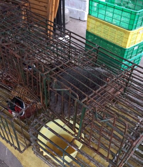
The Environmental Justice Foundation
(EJF) is a UK-based environmental and
human rights charity registered in England
and Wales (1088128).

1 Amwell Street
London, EC1R 1UL
United Kingdom
www.ejfoundation.org

Comments on the report, requests for
further copies or specific queries about
EJF should be directed to:
info@ejfoundation.org

This document should be cited as: EJF (2003)
Viral diseases from wildlife in China.
Could SARS happen again?

Cover pictures:
Upper left: Machs Einfach (CC BY-NC 2.0)
Upper right: Clurross (CC BY-NC-ND 2.0)
Lower left: Scott Edmunds (CC BY 2.0)
Lower right: Gavin Anderson (CC BY-SA 2.0)

This report was researched and written by Dr Lynn Dicks for EJF in 2003.

Dr Lynn Dicks, Lecturer in Animal Ecology, Department of Zoology, University of Cambridge

It was peer reviewed by:

Professor Robin A. Weiss, Emeritus Professor of Viral Oncology, University College London, UK
Professor Wolfgang Preiser, Professor of Medical Virology, University of Stellenbosch / National Health
Laboratory Service (NHLS) Tygerberg, Cape Town, South Africa
Professor Malcolm Bennett, School of Veterinary Medicine and Science, University of Nottingham, UK

                                                        2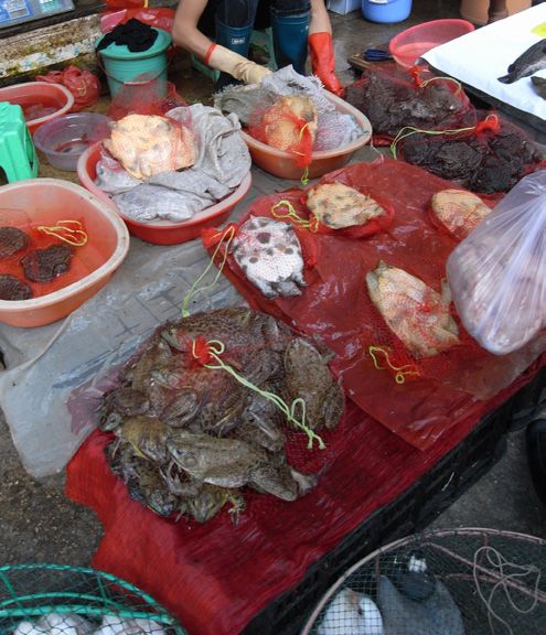
CONTENTS
EXECUTIVE SUMMARY 5
INTRODUCTION 6
CHAPTER 1 – BASIC VIRUS BIOLOGY 7
Structure and Replication 7
Types of Virus 7
A Word on Epidemiology 8
Virus Transfers Between Humans and Animal Species (Zoonoses) 9
CHAPTER 2 – CASE STUDIES OF EMERGING VIRAL ZOONOSES 11
CASE STUDY ONE: INFLUENZA A 11
CASE STUDY TWO: EBOLA 13
CASE STUDY THREE: HIV 14
CASE STUDY FOUR: HANTAVIRUS PULMONARY SYNDROME 15
CASE STUDY FIVE: HENDRA VIRUSES 17
CASE STUDY SIX: SARS 18
CHAPTER 3 – REVIEW OF DIRECT VIRAL ZOONOSES 20
What Types of Animal do they Come From? 20
What Factors Promote the Emergence of New Viruses? 21
Where Does SARS Fit? 24
CHAPTER 4 – IS CHINA A LIKELY AREA FOR FURTHER ZOONOTIC VIRUSES TO EMERGE? 25
1 - Widespread Consumption of Wildlife 25
2 - Ecological Change 28
3 - Immunosuppression Resulting from AIDS 30
4 - Selenium Deficiency 30
RECOMMENDATIONS 31
APPENDIX I: Virus families containing human and animal viruses 32
APPENDIX II: Review of viral zoonoses without arthropod vectors 34
APPENDIX III: Wildlife species reported to have been consumed or sold
in wildlife markets in China 41
APPENDIX IV: List of arboviruses 48
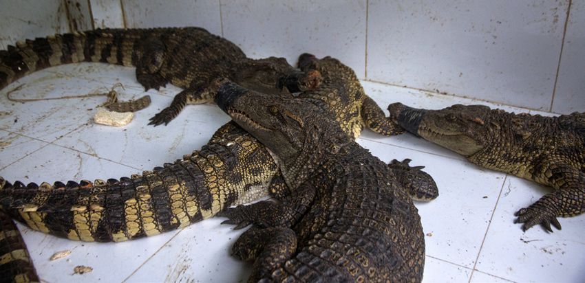
Wet market - Wuhan, China. © Animal Equality
3
PREFACE

It is important to note that this report was written 17 years ago in 2003. We are publishing it here as it was then.
There are, of course, sections that could be updated; for example, we could add information on ‘swine’ flu H1N1 in
2009, MERS in 2012, the 2014-2016 Ebola epidemic, and plenty of research which points to bats as the original source
of coronaviruses.

However, we publish it here exactly as it was in 2003 to show how much we did know at the time. Looking back from
2020, the six case studies in chapter 2 make sobering reading, especially those on Ebola and SARS. Consider the table
in Appendix II (pages 34-39), listing 76 viruses known then to be transmissible directly from animals, of which 52
were classed as public health problems and 49 were new or increasing. The six recommendations to reduce the risk
of another previously unknown viral disease emerging in China (page 31) could be repeated without much alteration.

We had all the knowledge we needed to curb dangerous practices and lower the risks of tragic pandemics such
as the one we are living through in 2020. Now we are scrambling to sequence the new coronavirus that causes
COVID-19, develop tests, drugs and conduct trials within a few weeks. It is amazing what science can achieve.
However, translation of knowledge into policies is seriously lacking. We are not even prepared when it comes to the
very basics of protective equipment for frontline medical staff. Nor did we heed the lessons of SARS in 2003. Instead
“business as usual” continued, including commerce and practices known to be risky. In 2003, we had a chance to
shut the stable door before the horse bolted. This time, we are not so lucky. It is imperative that we use the pandemic
to learn our lesson.

Steve Trent (EJF), Wolfgang Preiser (University of Stellenbosch) and Lynn Dicks (University of Cambridge), April 2020.

                                                          4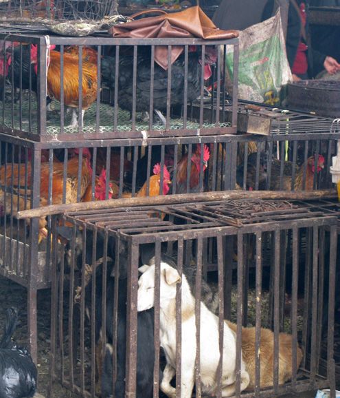
EXECUTIVE SUMMARY
As demonstrated by the recent outbreak of Severe Acute Respiratory Syndrome (SARS), viruses can emerge suddenly
in the human population and spread rapidly, with devastating effect. This report reviews knowledge of viruses that
transmit directly from animals to humans and asks whether anything can be done to reduce the risk of further virus
outbreaks from animals in China.

The report begins with an outline of virus biology and important concepts such as zoonosis (transfer of pathogens
between humans and animals). Six detailed case studies of emerging viral zoonoses are presented: influenza A; Ebola;
HIV; hantavirus pulmonary syndrome; hendra viruses and SARS. These illustrate the range of factors associated with
transfers of disease from animals to humans.

A detailed review of documented viral zoonoses not carried by arthropod vectors is given. 76 viruses are found to fit
this description. The review compiles scientific knowledge about the animal origins, severity and distribution of these
diseases and the factors responsible for the transfer to humans. 49 are new or increasing in the human population; 52
cause fatality or widespread infection; 36 are found in Asia. The majority come from primates, even-toed ungulates
and rodents. Factors associated with viral disease transfer from wildlife are reviewed from the literature. The most
important are ecological change, including climate change and changing agricultural practises; and increasing human
populations, both of which can entail increased contact with wildlife. RNA viruses, transmitted by arthropods, with a
broad host range are the most likely pathogens to emerge as new diseases from wildlife.

The SARS virus is assessed in the context of this review. It fits some of the categories of a virus likely to emerge, but
it could not have been predicted, because it comes from a group of viruses that do not have a broad host range, and
apparently from a group of animals with little history of zoonosis.

Three main factors are outlined that make China a likely place for further emergence of directly acquired zoonotic
viruses. The first is widespread consumption of wildlife species in China. The risk of zoonosis is during contact with
live animals, their blood and raw meat, or in eating poorly cooked meat. In a review of studies of species consumed in
China, 226 species were documented. 77% were birds, reptiles and amphibians; 23% were mammals. Since mammals
form the vast majority of sources of previous direct zoonoses, these are considered in more detail. Most are primates
and carnivores, with a significant proportion of rodents and ungulates. Of these groups, primates, ungulates and to a
lesser extent rodents have the greatest history of viral disease transfers. Primates, especially monkeys, are of particular
concern because they represent three of the seven most popularly eaten mammal species. This group are the source of
around a fifth of documented viral zoonoses, and Asian macaques are known to carry viruses with zoonotic potential
such as a strain of Ebola virus.

Secondly, China is found to be experiencing significant ecological changes that have been linked directly to increased
risk of zoonosis – specifically deforestation, climate change and increasing rodent populations. Finally, the high
and increasing number of Chinese carrying HIV (1.5 million) makes the population vulnerable to new viruses
establishing themselves.

Overall, the risk of further viral zoonoses emerging in China can be considered substantial. Six recommendations are
suggested to reduce the risk, including reducing the number and quantity of wild species eaten, particularly primates,
wild pigs and rodents; warning hunters, traders and consumers of the risk of viral zoonosis; encouraging well-inspected
farming rather than hunting of wildlife and further research to anticipate zoonoses.

                                                            5
INTRODUCTION
“If there is any conceivable way a germ can travel from one species to another, some microbe will find it”
(McNeill, 1976).
Viruses are probably the largest threat to the continued prosperity of mankind. Despite nominal success in combating
the smallpox virus (Variola), most viruses evade eradication, by continually changing or switching between hosts.
The majority of human viruses originated in animals. Some scientists claim there has been a dramatic increase in the
occurrence of new pathogenic viruses in the human population over the last 25 years (Crawford, 2000), and there has
been an increase in global mortality from infectious disease (Ludwig, Kraus et al., 2003). These increases can be expected
to continue as a result of increasing ecological change and environmental degradation, and increasingly mobile human
populations (Morse, 1995; Stöhr and Meslin, 1997; Osterhaus, 2001). As demonstrated by the recent outbreak of Severe
Acute Respiratory Syndrome (SARS), viruses can emerge suddenly in the human population and spread rapidly, with
devastating effect.
This report reviews our knowledge of viruses that can transmit directly from animals to humans and asks whether
anything can be done to reduce the risk of further virus outbreaks from animals. It does not include other human
parasites, such as bacteria, which also pose a threat of infection from animals and include well-known zoonoses such
as the plague bacteria Yersinia pestis, Weil’s disease from rats and Salmonella food poisoning. The report is divided into
four chapters. Chapter one outlines virus biology, describes some important principles and concepts in virology and
introduces the concept of zoonoses. Chapter two is a series of six case studies of emerging viral zoonoses. Chapter
three is a detailed review of documented viral disease transfers, their animal origins and the factors responsible for the
transfer to humans. Chapter four considers the current situation in China, and how it relates to risk factors identified in
chapter three. The report ends with a set of recommendations on how to reduce the likelihood of future viral zoonotic
diseases emerging in China.
6CHAPTER 1 – BASIC VIRUS BIOLOGY
Structure and Replication
Viruses are distinct from other living organisms, because they do not carry the cellular machinery to metabolise. They
consist simply of genetic material and a protein coat, and rely on the cells of living organisms to reproduce. When
an infectious virus particle, known as a virion, infects an organism, it attaches onto its cells and hijacks the cellular
machinery by injecting virus genes into the nucleus. The cell then produces new virus particles, which in some cases
rupture the cell as they emerge.
Most viruses are specific to certain types of cell within their host. Hepatitis viruses recognize liver cells, for example,
while respiratory viruses infect epithelial cells in the respiratory tract and lungs. Viruses recognise specific receptor
molecules on the surfaces of cells. A wide variety of different receptor molecules have been identified for different
viruses, including proteins, carbohydrates or lipids involved in a variety of functions from cellular signaling to adhesion.
Closely related viruses do not necessarily use the same type of receptor, and some viruses such as hepatitis C use more
than one (Baranowski, Ruiz-Jarabo et al., 2001).
Proteins that stick out from the outer coat of the virus bind to these receptors and initiate infection. These ‘spike’ proteins
are often called glycoproteins, because they have a sugar molecule attached. They may be a target of the host’s immune
response, which learns to recognize the virus by particular sequences in these surface proteins. In the case of influenza,
a change in a single amino acid can change the cell surface receptor that is used (Baranowski, Ruiz-Jarabo et al., 2001).
This is why the same person can repeatedly suffer the disease, because the target for the immune system changes.
Types of Virus
According to the universal system of virus taxonomy, devised by the International Committee on Taxonomy of Viruses
(ICTV), there are currently 71 families of viruses. 24 of these contain viruses that infect humans and animals (listed in
Appendix I). The universal system classifies viruses according to their biochemical structure and method of replication.
Specifically, they are divided according to what type of nucleic acid they have (DNA or RNA, single or double stranded);
whether their genome is a single piece of nucleic acid, or segmented into separate sections, a bit like our chromosomes;
which way the genome is read (positive or negative) and whether the virus particle is surrounded by a membranous
envelope or not. Most human pathogenic viruses use RNA although there are notable exceptions, such as pox- and
herpes viruses.
To some extent, this method of classification is reflected in the epidemiology of the viruses, in that viruses in the same
taxonomic group often have similar methods of transmission, clinical signs and affect similar types of organism.
For example, viruses in the family Orthomyxoviridae, the influenza type viruses, are respiratory infections, acquired
by inhalation or direct contact with mucous membranes. The family Flaviviridae contains viruses causing fever and
encephalitis, and transmitted by arthropod vector, such as yellow fever, dengue fever and rocio. Viruses in the family
Arenaviridae usually cause haemorrhagic fever in humans. All but one are carried by rodents mostly from the Muridae
(mice and rats) and the Cricetidae (voles, lemmings and gerbils) families. The exception, Tacaribe virus from Trinidad, is
carried by the fruit bat Artibeus sp (Howard, 1998). This pattern is not without exceptions. Almost all the viruses in the
family Bunyaviridae, for example, are arthropod-borne except for those in the genus Hantavirus, which are transmitted
by murid rodents (Clement, McKenna et al., 1998).
7Viruses can also be classified according to their mode of transmission. Enteric viruses are intestinal and acquired by
ingestion. Respiratory viruses are acquired by inhalation of ‘fomites’ – small droplets transmitted between hands, nose,
mouth and eyes. Arboviruses enter directly into the bloodstream via the bites of arthropod vectors, such as ticks or
mosquitoes. Many other viruses are acquired by close contact, such as sexual contact, involving host body fluids. Most
virus families contain examples of several of these categories.
Within a virus family, viruses are classified into genus, then species. Proper names for viral species are decided by the
ICTV and listed on Index Virum (ICTV, 2003). Identifying and separating different species is difficult, especially because
viruses cannot be cultured outside host cells and do not survive long alone. A variety of methods are used, falling into
three categories: serological methods, structural characterisation and genetic methods.
Serological techniques involve using antibodies created by the host immune system to identify and distinguish different
viruses. Antibodies are virus-specific molecules produced by an infested organism, which bind to virus particles and are
instrumental in disabling them. In most cases, they appear in large quantities in host serum, during primary infection
with a virus. Some serological techniques, such as neutralization, directly measure the capacity of the antibody to block
a specific viral function. Others focus on the chemical reaction between the antibody and virus. Serological techniques
are particularly useful in circumstances where isolation of the virus itself is difficult, or in demonstrating previous
infection rates of non-persistent viruses (see Epidemiology section below). Certain viruses show cross-reactivity with
antibodies created against other virus species. For example, cowpox antibodies provide protection against smallpox,
which is why dairy workers largely escaped the latter disease. In other cases, such as influenza, different strains of a
single virus species produce different antibodies – the strains are called different serotypes.
Virus groups have different shapes and can be viewed under an electron microscope. Coronaviruses, for example, are
round particles covered in surface projections, or spikes. Filoviruses such as Ebola are long filaments.
Genetic characterization of viruses plays an increasingly important part in diagnostics and taxonomy. Genome
sequences specific to many viruses are now available and small quantities of virus can be detected in clinical samples
either by hybridization with a nucleic acid probe, or by amplifying the viral genome using the polymerase chain reaction
(PCR). Since virus genomes can mutate and change rapidly, there is currently considerable effort to identify sequences
that remain stable within virus groups (e.g. Rota, Oberste et al., 2003).
A Word on Epidemiology
Epidemiology is the study of the dynamics of infectious diseases: how they survive and change as populations, and
interact with their host populations. An important dichotomy in epidemiology is between pathogens that are short-
lived in their host, usually leaving them immune but virus-free, and those that persist, creating a population of more or
less healthy hosts that carry the virus.
Short-lived pathogens need to infect at least one other host after each infection for the population to persist (Anderson
and May, 1992). Since they leave the host either dead or immune, they need a host population large enough for there
always to be naïve host individuals available. The number of new individuals infected after each case can be used to
predict whether novel viruses, such as the SARS coronavirus, are likely to create an epidemic in a new host population
(Bull and Dykhuizen, 2003). It is believed that in low density populations, with small social groups isolated from
one another, as in pre-agricultural humans and many wildlife species, microbes tend to co-evolve with their hosts,
establishing the latter type - persistent infections that are not highly pathogenic (Weiss, 2001). Examples in humans are
the herpes viruses and the chickenpox virus. Similarly, many viruses carried by wild animals do not make them sick.
8When humans developed agriculture, our population densities became high enough to support non-persistent,
pathogenic parasites of the former type (Anderson and May, 1992).
Virus Transfers Between Humans and Animal Species (Zoonoses)
Viruses that naturally transfer between humans and animal species are known as zoonotic viruses, or zoonoses (Palmer,
Soulsby et al., 1998). Most human infections originally came from an animal source (Weiss, 2001). 61% of all human
pathogens and 76% of all human viruses are zoonotic (Taylor, Latham et al., 2001). Some, such as the ‘flu virus, are well
known and have been present in human populations for a very long time. Others are brand new arrivals. The dynamics
of such diseases are complex. Often there is a ‘reservoir’ population of one or more species, which holds the disease
permanently in a stable equilibrium (Haydon, Cleaveland et al., 2002). From the reservoir, the disease can occasionally
transmit to a different host and establish itself in the new population. In other zoonoses, like rabies, each new case
represents a new infection from an animal.
Many of our most deadly zoonotic pathogens are arboviruses, carried by insects. They frequently change hosts and
their dynamics can be affected by changes in patterns of settlement, agriculture or irrigation that affect populations
of the vectors, and their contact with humans. A list of known arboviruses is included in this report (Appendix IV), but
the diseases are not dealt with in detail. Prevention and control of arbovirus infections is built almost exclusively on
diagnosis and treatment in humans, and control of invertebrate hosts (Stöhr and Meslin, 1997), indicating that direct
transmission from animals is not an important route.
Viruses transmitted directly1 between vertebrate animals and humans are of interest because the likelihood of this kind
of transfer is closely related to the degree of contact between humans and animals. A detailed review of these viruses is
given in chapter three.
Changes in virulence when a virus switches host, are frequent, and difficult to predict (Frank and Jeffrey, 2001). Many
zoonotic viruses, such as the arenaviruses, hendraviruses and hantaviruses, produce a tolerated persistent infection
in the reservoir host, with no ill-effects and no detectable immune response (Clement, McKenna et al., 1998; Howard,
1998). On transfer to humans, they are deadly killers. The opposite can also happen. The arbovirus Sindbis, for example,
causes disease in birds, but is only dangerous to humans that are immunosupressed (Weiss, 2001). Herpes B virus
(Cercopithecine herpes virus type 1) is highly prevalent in wild Asian macaques, in 80-100% of which it causes small
skin lesions of low severity (Meurens, Gallego et al., 2002). In a human bitten by an infected monkey, the virus often
leads to fatal encephalitis. The disease has killed 24 humans since its discovery in 1932 out of around 40 reported cases
(Meurens, Gallego et al., 2002). Considering the widespread use of macaques in biomedical science, this low rate of
transmission between human and animal host has led one author to suggest there may be an unknown immunological
mechanism preventing cross species transfer of this type of virus (Skinner, Ahmad et al., 2001).
Many authors writing on the subject of zoonotic viruses include a stark warning about the likelihood of future incidences
producing highly transmissible or pathogenic epidemics (Morse, 1993; Weiss, 2001; Ludwig, Kraus et al., 2003). Close
surveillance and careful preparation of medical and public health institutions is consistently suggested, rather than any
attempt to reduce the risk of new zoonoses occurring.
1
‘ Direct’ in this context, and later ‘direct zoonoses’, refers to the lack of intermediate arthropod vector, and should not be understood to exclude viruses transmitted
between humans and animals via body fluids or environmental reservoirs.
9There are virus types common in animals that have not transferred to humans but may have the potential to. Canine
Distemper virus (Paramyxoviridae), for example, seems to have expanded its host range this century, infecting many
species of carnivore, including marine mammals (Baumgartner, Alldinger et al., 2003). There has long been concern
about the zoonotic potential of the cancer causing retroviruses which occur in cows, birds and cats, because of our close
contact with these animals, and the existence of a similar virus (HTLV-1) already infecting humans (Platt, 1994). The
latter has since been suggested to be a primate zoonosis (Voevodin, Johnson et al., 1997). Mice have been experimentally
infected with cow retrovirus via their milk. Analysis of genetic sequences shows that these retroviruses have switched
species several times over the last 70 million years (Platt, 1994).
How are zoonoses detected?
Identifying a zoonosis event is not easy. Five lines of evidence are available: prevalence in the natural host; geographic
coincidence; plausible routes of transmission; similarities in viral genome organization and phylogenetic relatedness
to viruses in animals.
In some cases, the coincidence of events alone provides very strong evidence. For example, a recent outbreak of
monkeypox in the mid-West United States was traced to African rats imported as pets (Lutz, 2003). Similarly, Ebola virus
has been transferred in shipments of monkeys for biomedical research on a number of occasions, although primates are
probably not the main reservoir. In others cases, this sort of evidence is weak, such as the unproven link between bats
and Ebola (see case study two).
Molecular genetic ways of identifying the source of a virus can be applied retrospectively. A virus that has co-evolved
with the human species, rather than being a zoonotic infection, will have a relationship with similar viruses in other
animals that reflects our own evolutionary relationship with them. Our herpes viruses, for example, are more similar
to chimpanzee herpes than to a rabbit version (Weiss, 2001). But if a virus appears in humans that is very similar, or
indistinguishable from one present in an animal species not particularly closely related to ours, this is strong evidence
of a zoonotic transfer, especially if the virus is associated with an illness not previously observed. A zoonotic origin for
measles is postulated on the basis of the genetic structure of the virus, although it is an event that probably happened
7000 to 8000 years ago, when humans first domesticated ruminants (Diamond, 1997).
For some viruses, scientists are unsure. There is uncertainty, for example over the origin of the hepatitis B virus
(Bollyky, Rambaut et al., 1997; MacDonald, Holmes et al., 2000; Odemuyiwa, Mulders et al., 2001; Simmonds, 2001).
In 1997, Bollyky et al postulated it was a zoonosis from new world monkeys in the last 500 years. More recently, the
same group has shown that the related viruses are dispersed throughout the Old World Apes, suggesting we carried
the virus before we were humans (Macdonald 2000). But one group of human hepatitis B viruses in West Africa is very
similar to a local chimpanzee form (Odemuyiwa, Mulders et al., 2001).
To avoid catching zoonoses, the best strategy is to identify the natural host reservoir, and sever links with it (Haydon,
Cleaveland et al., 2002). Those links could be direct or via an intermediate host such as a domestic animal. As we shall
see, this is not always possible.
10CHAPTER 2 – CASE STUDIES OF EMERGING VIRAL ZOONOSES
An emerging virus is one that has appeared in a new host population, or whose incidence is increasing (Woolhouse,
2002). Six case studies of well-known emerging viral zoonoses are presented below. These have been chosen to illustrate
the range of factors that can influence the transfer of disease from wildlife to humans.
CASE STUDY ONE: INFLUENZA A
Influenza is a highly contagious acute respiratory illness. It is not usually fatal, although a flu epidemic
often causes death in the old and weak. Three types of the virus are known to infect humans: influenza A,
B, C. They have very high epidemic potential. Up to 25% of the world’s population could become ill during a
single epidemic, and the virus has the potential to kill 30% of its victims (Enserink, 2003). There are 50,000
influenza deaths per year in the United States (Murphy, 1994).
Until recently, influenza A was the only one known to move between species under natural conditions. It is
commonly found in humans, horses, pigs, chickens, aquatic birds, and more rarely occurs in seals, whales,
mink and cattle. Current understanding is that wild ducks and shorebirds are the primary and most stable
reservoir of the disease, but versions of it frequently transfer to other animals. Influenza A is known as a
re-emergent virus, because of this dynamic. The best evidence for transfer of influenza A between species
is the sporadic transmission of swine influenza to humans (Slemons and Brugh, 1994). Normally, such
infections occur in people in direct contact with pigs. They are no more serious than ordinary human ‘flu and
transmission to other humans is limited. There is also direct evidence of transfers of influenza A between
humans and chickens and humans and seals. In the latter case, the seal flu virus infected the eyes of men
investigating the outbreak, causing conjunctivitis (Webster, Geraci et al., 1981). In both instances, the infection
died out of its own accord.
The danger of a pandemic, or worldwide epidemic, is when a new and highly virulent strain arises. The labels
given to different strains of influenza A refer to the type of two surface glycoproteins on the outer envelope
– hemagglutinin (HA) and neuraminidase (NA). Currently there are 15 HA and 9 NA subtypes identified
(Horimoto and Kawaoka, 2001). Human and swine influenzas are limited to a few of these varieties, but birds
can be infected by every combination, forming a vast global reservoir of the virus.
New strains usually occur when two different versions of the virus meet in a single host and the genome
segments are re-sorted in a process called antigenic shift. For example, if a human already carrying the
flu virus becomes infected with an avian variant, a new combination of envelope glycoproteins can arise.
Sometimes the new strain is particularly virulent, as in the catastrophic ‘Spanish influenza’ epidemic in
1918. This was the most devastating viral disease of the twentieth century, and killed more than 20 million
people worldwide (Granoff and Webster, 1999).
Since the first human influenza virus was isolated in 1933, three more major pandemic strains have been
described: ‘Asian flu (1957) H2N2, Hong Kong flu (1968) H3N2 and Russian flu (1977). These strains caused
global outbreaks and high death tolls. The first two strains arose in China, when a human flu acquired new
genes from the wild duck flu. The same process brought us the Spanish flu. Russian flu was an identical strain
to the 1968 Hong Kong flu and may have been stored in a freezer in the intervening years. The low death rate
in this pandemic could be explained by immunity of people over 20 years old acquired during the Hong Kong
pandemic (Horimoto and Kawaoka, 2001).
11It has been suggested that the emergence of new flu strains often occurs in China because of the practice of
combined pig and duck agriculture there (Brown and Alexander, 1998). In this context, natural hosts of influenza
A are in direct and continuous contact, increasing the chances of a new strain arising by antigenic shift.
Avian influenza is enteric and can be spread in water contaminated with faeces. It is less host-specific than
versions of the virus in mammals and is found in a very wide variety of birds, including poultry, game and
wild birds. The current strategy for controlling flu involves watching for new strains in pigs or chickens,
particularly in China. The regularity of pandemics suggests that the likelihood of another increases over time.
Although significant measures are taken to control avian, swine and equine influenza, there is no feasible
method for control of influenza in wild free-flying birds.
Zoonosis of influenza A has generally been considered a rare event. A review of the literature reveals only
18 cases of pure swine fever directly crossing into people (Wuethrich, 2003). But this is probably a serious
underestimate of the actual incidence of such a transfer. Usually, only molecular biology can distinguish
between the different strains of flu, and in the absence of mortality or unusual infection pattern, most
cases are assumed to be ordinary human flu. A study last summer found that 23% of pig farm workers and
their families carried antibodies to swine flu strains, compared with less than 1% of urban dwellers (Olsen,
Brammer et al., 2002).
In the last seven years, outbreaks of two new strains of avian-derived influenza have been reported in humans, in
1997 (H5N1) (Osterhaus, 2001), 1999 (H9N2) (Lin, Shaw et al., 2000) and 2003 (H5N1) (WHO, 2003a). Fortunately
none of these developed into a pandemic, partly because human-human transmission was inefficient and
partly due to the widespread slaughter of poultry in the affected areas (Shortridge 2001). In each case, the flu
was passed directly from birds to humans, without any involvement of pigs as a ‘mixing vessel’. It is normally in
pigs that the switch happens from the enteric avian form to the respiratory mammalian form. In each case, the
transfer occurred in Hong Kong. In the case of H5N1 in 1997, the event was traced to the Hong Kong waterfowl
markets, where the virus had transferred from wild birds to chickens. Eighteen people caught the disease, six
of them died. There is increasing concern among scientists that a new pandemic strain of influenza could arise
at any time (Horimoto and Kawaoka, 2001; Li, Xu et al., 2003; Webster and Walker, 2003).
This year an old strain (H7N7) has reappeared in poultry farms in the Netherlands with apparently new
potential for human infection. This virus has caused conjuncitivitis in at least 83 people and killed one person.
Although human-human transmission seems weak, it is possible. Flu specialists are worried, because of the
potential for an avian flu to recombine with genes from a human strain of flu inside a person. In addition,
antibodies to the H7N7 strain have already been found in Dutch pigs (Enserink, 2003).
12CASE STUDY TWO: EBOLA
Ebola virus and its cousin Marburg virus are killers. Their source and natural ecology have vexed the scientific
community for 35 years and remain a mystery. Both cause haemorrhagic fever, with massive internal
bleeding often resulting in death. Marburg virus first appeared in 1967. 31 laboratory workers in Germany
and Yugoslavia were infected. They had been exposed to tissues from African green monkeys (Cercopithecus
aethiops) imported from Uganda. Seven of them died. When the virus was identified, it was placed in a new
viral family, the Filoviridae. There have been three subsequent outbreaks of Marburg in South Africa (1975),
Kenya (1980 and 1987) and in the Democratic Republic of Congo and Zaire in the 1990s.
In 1976 and 1979 there were several outbreaks of another Filovirus causing lethal hameorrhagic fever in Zaire
and Sudan. It was named Ebola virus. It was mostly confined to hospitals, and transmission between people
appeared to be by direct contact with infected tissue, or close personal contact. Mortality rates in the initial
outbreak were 88% in Zaire and 53% in Sudan. There have been seven subsequent outbreaks of the disease in
Africa, with mortality rates ranging from 50-81%. All have been confined to relatively small areas. Currently
an epidemic is occurring in Gabon and the Republic of Congo, which so far has affected 143 people, and killed
128 (90%) (WHO, 2003a). In 1994, a scientist became infected with a different strain of Ebola, the Ivory Coast
strain, after performing an autopsy on a wild chimpanzee.
Scientists suspect the disease is initially caught in each outbreak from an infected animal, perhaps a primate
hunted for ‘bushmeat’ (CDC, 2003). But monkeys and chimpanzees are unlikely to be the natural reservoir
of Filoviruses, because they become ill and die when they are infected (Ludwig, Kraus et al., 2003). Extensive
surveys of wildlife, including mammals, birds and insects in the outbreak areas have identified no natural
host. In 1987, a boy was infected with Marburg virus after spending considerable time in a cave on Mount
Elgon filled with bats. Bats were also present in large numbers in a cotton factory where there were outbreaks
of Ebola in 1976 and 1979. They are a strong candidate for the reservoir, because they can be artificially infected
without becoming ill (Ludwig, Kraus et al., 2003). But no one has managed to isolate the virus from wild bats.
Scientists remain convinced that the disease is zoonotic, and that outbreaks are triggered by initial infection
from wildlife (CDC, 2003). Temporal patterns of the outbreaks suggest that the natural host has contact with
monkeys and or humans for only a short period of the year. Most outbreaks begin in November or December
(Guenno, 1997). The lack of a natural host is mystifying, leading some scientists to speculate that the disease
may even be an arthropod or plant virus (Monath, 1999).
In 1989 a new Ebola-type virus appeared in America, in a shipment of macaques (Macaca fascicularis) from
the Phillipines. No African connection could be found for these monkeys, but they themselves were dying. To
the horror of scientists, antibodies to the virus were also found in some of the human laboratory workers, but
they showed no disease symptoms. Rapid euthanasia of monkeys and disease control tactics prevented the
spread of the outbreak. The disease, named the Reston strain of Ebola, after the quarantine site in America,
was subsequently found to be present in captive macaques in the Philippines. During a further outbreak of
the same virus in 1990, also at Reston, the disease was noted to have respiratory involvement. There was high
concentration of viral antigens in pulmonary secretions and post mortem showed the virus to be reproducing
in the lung tissue. 80% of infected monkeys died and several workers at the laboratory were infected without
having had direct contact with the monkeys (Peters, Johnson et al., 1993). The same virus was isolated from
monkeys from the Philippines in Italy in 1999 and again in the US in 1996 (Rollin, Williams et al., 1999) but
no humans were infected, due to employment of careful screening and barrier precautions. Again, captive
macaques and primate facilities in the Philippines were investigated, and the virus was concluded to be rare
(Miranda, Ksiazek et al., 1999). There have been no surveys for this virus in other Asian wildlife species.
13This case demonstrates the epidemic potential of the Filoviruses. It happens that the Reston strain does not
cause human disease. But in the words of Peters, Johnson et al (1993), “the seriousness of the efficient (airborne)
spread of a filovirus cannot be overestimated.” There seems to have been little further investigation into the
natural ecology of this strain of Ebola, perhaps due to the lack of immediate public health implications. Ebola
research is concentrated in Africa.
It is not clear whether there are increasing numbers of Ebola virus outbreaks in central Africa or whether more
are recorded due to better surveillance from the international health community. If outbreaks are increasing
in frequency, this has been linked to ecological perturbations caused by deforestation (Guenno, 1997).
CASE STUDY THREE: HIV
HIV-1 is currently responsible for an extremely serious global pandemic, particularly affecting the poor and
undeveloped nations of the world, with no end in sight (Weiss and Weiss, 2001). 42 million people are infected
and the numbers are still increasing. In some African countries more than 30% of the adult population have
the virus (Ludwig, Kraus et al., 2003).
The disease caused by HIV, Acquired Immune Deficiency Syndrome, AIDS, is invariably fatal. It was first
recognized in the USA in 1981 (Gottlieb, Schroff et al., 1981), and a few years later, scientists realized the disease
was already widespread in Africa (Bayley, Cheingsongpopov et al., 1985; Serwadda, Sewankambo et al., 1985).
It is a particularly formidable pandemic, because it presents opportunities for other infections, and possible
zoonoses, to establish themselves in populations with severely weakened immunity. The virus is transmitted
by direct contact of body fluids and evolves extremely rapidly, both within an individual host and around the
world (Piot, Bartos et al., 2001).
It is well established that HIV-1 came to humans from the simian immunodeficiency virus, chimpanzee SIV
(Gao, Bailes et al., 1999). Analysis of the genetic sequences of the different strains of HIV indicates there
have been seven separate zoonotic incidents, in the last 100 years. All happened in Africa, where there is
the greatest variation. HIV-1 appears to have crossed the species barrier 3 times, resulting in the M, N and O
groups. These groups are as distinct from one another as they are from the chimpanzee SIV (Gao, Bailes et al.,
1999). It is the group M that has spread around the world and caused the pandemic. Another form of HIV, HIV-
2, which is less transmissible but causes local epidemics, also appears to be of primate origin. It arrived from
the sootey mangabey, Cercocebus atys, and can be subdivided into six or more groups that appear to represent
separate zoonoses (Gao, Yue et al., 1992). HIV-2 is endemic in West Africa, but has spread to Europe (Portugal)
and India (Weiss, 2001).
The exact route of HIV zoonosis has been a matter of heated debate amongst scientists. In the case of HIV-
2, the contact between humans and host is through mangabeys hunted for food and orphans kept as pets
(Hahn, Shaw et al., 2000). Hunting and field-preparation of chimpanzees, a common practice in West Africa,
is also blamed for the transfer of HIV-1. However, some scientists argue that the ‘cut-hunter’ theory, as it is
known, has not been carefully studied. It is assumed to be possible, but not proven (Martin, 2001; Weiss and
Weiss, 2001). Another theory suggests that HIV originally infected humans through the preparation of live
polio vaccines using chimpanzee kidneys (Hahn, Shaw et al., 2000; Hooper, 2000; Martin, 2001; Weiss and
Weiss, 2001). The polio vaccine theory has been intensely scrutinized and now looks unlikely. Comparison
14between human and primate HIV/SIV viruses suggest that HIV-1 was present in the human population 10-50
years before the polio vaccines trials began. It is not clear why HIV remained geographically isolated in the
intervening years, before suddenly enlisting a pandemic in the 1970s.
The natural primate hosts of HIV/SIV harbour similar levels of the virus to infected humans, but they do not
become ill or die of AIDS-like illnesses. Twenty-four other species of African primate harbour types of SIV, but
there is no evidence that any of these have infected humans (Hahn, Shaw et al., 2000). Asian macaques do not
have SIVs (Granoff and Webster, 1999).
CASE STUDY FOUR: HANTAVIRUS PULMONARY SYNDROME
Until 1993, Hantaviruses were recognized zoonoses, mostly carried by Murine rodents (Old World rats and
mice) and causing haemorrhagic fever with renal syndrome, HFRS – that is acute renal failure in the context
of a flu-like febrile illness. Epidemics of this disease have been documented as far back as the American Civil
War, but the virus was first described in the 1950s, during the Korean War (Earle, 1954). A number of outbreaks
have since been documented in Asia, Europe and America, largely caused by three strains of hantavirus
named Hantaan virus, Seoul and Puumala virus (Chu, Rossi et al., 1994). There are tens of thousands of cases
of Hantaan virus annually in China. But the disease was not particularly feared. Rather, it was considered a
“somewhat arcane zoonosis, of interest to kidney and rodent specialists” (Jacobson, 2003).
Then, in the early 1990s, a group of new forms of the virus exploded on the scene in Europe and North America,
prompting global concern. These viruses attacked not the kidneys, but the lungs. The viruses were classified in the
same genus, Hantavirus, and a new syndrome was named, hantavirus pulmonary syndrome (HPS), characterized
by acute respiratory distress in adults, and often fatal pulmonary or cardiac failure (Clement, McKenna et al.,
1998) (Täger, Vial et al., 2003). From 1993 until now, new HPS viruses have been recognised almost every year and
there are approximately 200 HPS cases annually throughout the Americas (Khan and Khan, 2003).
The outbreaks of HPS were not caused by new viruses, but by viral agents having long existed in their
Sigmodontine (New World) rodent hosts (Nerurkar, Song et al., 1994). Hantaviruses are single-stranded RNA
viruses, with a genome segmented into three. Studies have shown that the newly emerged hantaviruses are
not a re-assortment of segments from other viruses. Each of the three segments are quite independent of
previously known viruses (Spiropoulou, Morzunov et al., 1994). The diversity and evolutionary relationships
of the viruses reflect the evolutionary relationships of the rodent species, indicating a close and ancient
relationship between rodents and viruses (Clement, McKenna et al., 1998; Monroe, Morzunov et al., 1999).
However, this also seemed to be true for HIV/SIV viruses, but a recent paper has suggested that frequent
transmissions between closely related species of host could produce the same pattern (Charleston and
Robertson, 2002). Overlapping distributions between different rodent species in South America provide
opportunities for host-switching events (Plyusnin and Morzunov, 2001) and there is increasing recognition
that host switching may have a role in generating hantavirus diversity (Bohlman, Morzunov et al., 2002).
Some authors suspect other types of hantavirus may occur in human populations, but are not noted because
they do not cause severe disease (Täger, Vial et al., 2003).
Hantaviruses are all transmitted from infected rodents to man via excretions in urine, faeces or aerosolized
15respiratory droplets (Clement, McKenna et al., 1998). Most sufferers of the disease report sightings of rodents,
but almost never mention a physical contact. The rodent carriers appear healthy. Person to person transmission
of hantaviruses has rarely been reported, despite extensive epidemiological studies. As a result, the diseases
remain localized. However, it seems to be possible in the case of Andes virus. During an outbreak in Argentina
in 1996 two people were infected who had been in contact with infected people but neither visited the locality
of the outbreak nor had high exposure to rodents (Wells, Estani et al., 1997). This also happened in Chile the
following year (Toro, Vega et al., 1998) and research is underway to establish the mechanism of transmission
(Khan and Young, 2001).
The New World hantaviruses are causing serious public health problems in South America in the early 21st
Century, having been first discovered in North America. Two main factors are thought responsible for the
repeated outbreaks. One is changing weather conditions. The original outbreak of Sin Nombre virus in
1993 was clearly related to unusual weather conditions. Heavy rains and snow in the previous spring, after
a long drought, led to an abundance of rodent food, such as piñon nuts and grasshoppers. The numbers of
deer mouse (Peromyscus maniculatus), the Sin Nombre virus host, were up tenfold on normal levels during
that year (Stone, 1993). During the nineties, several years of high precipitation caused by El–Niño events
increased rodent populations generally in the Americas and may have been associated with other outbreaks
of hantavirus disease (Childs, Ksiazek et al., 1994; Hjelle, Jenison et al., 1994; Spiropoulou, Morzunov et al.,
1994; Hjelle, Jenison et al., 1995; Engelthaler, Mosley et al., 1999; Hjelle and Glass, 2000; Täger, Vial et al., 2003)
The second factor is changes in land use, which have increased people’s contact with rodents. A recent study
(Täger, Vial et al., 2003) found infection with the Andes strain of Hantavirus to be correlated with those
working in the forestry industry. The natural host of this strain is the long-tailed pygmy rice rat (Olgoryzomys
longicauatus), a species that occurs primarily in temperate forest and is associated with the abundant bamboo-
like forest understorey plant Chusquea quila. The increasing development of the forestry industry has caused
humans to interact more closely with this rice rat species, and acquire its virus.
Hantaviruses are prevalent in wild rodent populations all around the world. In China for example, Old World
hantavirus isolates or antigen were found in 55 species of vertebrate, including 37 rodent species (Chen and
Qui, 1993). The field mouse (Apodemus agrarius) and the brown rat (Rattus norvegicus) were the most important
vectors with 5.3% and 4.9% testing positive, respectively. A more recent study have found Hantaan-like
viruses in 9.9% of wild rodents in Ningxia province, China (Kariwa, Zhong et al., 2001). Hantaan virus causes
an estimated 50-100,000 infections a year in China (Khan and Khan, 2003). Rates of 2.1% have been found in
rodents in Thailand (Nitatpattana, Henrich et al., 2002). In Uruguay, hantavirus Central Plata strain is carried
in 2.6% of its host, the yellow pygmy rice rat Oligoryomys flavescens (Delfraro, Clara et al., 2003). Seoul virus
persists at even higher levels, having been found at frequencies of 33.9% in Rattus norvegicus in Taiwan (Chin,
Chiueh et al., 2000) and up to 56% in Brazil (Leduc, Smith et al., 1985).
Although rodents are considered the primary reservoir, the viruses have been detected in other mammals, and
birds – a total of 164 different species from eight different orders, including bats and cats (Clement, McKenna
et al., 1998). Cat ownership has been described as a risk factor for hantavirus disease in China (Xu, Tang et al.,
1987). Although a recent study in the Netherlands searched for Puumala virus in over 2000 domestic animals,
including dogs and cats, and found none (Groen, Gerding et al., 1995).
No-one understands why the old and new world strains of Hantavirus cause different diseases. But the virus
genetics and host range suggest they have evolved independently for a very long time and make it relatively
unlikely that an HPS-type virus will naturally emerge in the Old World.
16CASE STUDY FIVE: HENDRA VIRUSES
In the 1990s, a group of three previously unknown viruses suddenly appeared in the human population in
Australasia, causing highly pathogenic encephalitis in one case. These viruses – Hendra (1994), Menangle
(1997) and Nipah (1998) virus – belong to the family Paramoxyviridae, and are unusual for that family in
being able to switch between hosts from different vertebrates classes. Nipah virus was the most serious
public health incident, killing 105 people, with separate outbreaks in Malaysia and Singapore (Chua, Bellini
et al., 2000; Field, Young et al., 2001). Menangle virus causes embryonic mortalities, stillbirths, mummified
foetuses and congenital malformation in pigs (Love, Philbey et al., 2001). Similar symptoms have not been
recorded in humans, but all the people found carrying antibodies to this virus were men, not women (Chant,
Chan et al., 1998).
The natural host reservoir for all three turned out to be flying foxes, or fruit bats (Pteropid bats). They had
transferred to humans via an intermediate. In the case of Nipah and Menangle viruses, outbreaks were traced
to pig farms and abbatoirs (Bowden, Westenberg et al., 2001; Chua, 2003). Hendra virus came from horses
(Barclay and Paton, 2000). Why did these viruses all suddenly emerge, within a few years of one another?
Molecular genetic studies of Hendra and Nipah viruses indicate they are not new or newly recombinant
viruses. Rather they are ‘old’ and have persisted unknown and non-pathogenic in their hosts, for perhaps
millennia. Field et al (Field, Young et al., 2001) argue that ecological change is the most plausible explanation
for their sudden emergence as people killers. Fruit bats are in decline in Australia and southeast Asia, due to
habitat loss, particularly deforestation, and hunting. They are struggling to find food, and increasingly living
in closer proximity to man. Nipah virus jumped the species barrier into pigs when pig farms were established
in the hosts’ natural range in the mid 1990s. It is a classic case of a zoonotic virus emerging in a newly
encountered host species and may be a direct result of the clearance of rainforest, the bats’ natural habitat
(Weiss, 2001). The virus then spread rapidly within and between farms due to the intensity of the husbandry
and frequent transfer of animals. Extensive sampling of wildlife has shown that these viruses reside in no
other peridomestic species, except for dogs (Field, Young et al., 2001).
Although the viruses themselves have only so far been isolated from species of Pteropus (flying fox), antibodies
to Nipah virus were found in five species of bat: four species of fruit bat, two of which were not flying foxes and
one insectivorous bat (Field, Young et al., 2001).
Scientists warn that this could happen again. The stresses on fruit bat populations have not gone away. During
the investigations on bats, two further new viruses were found. One, Tioman virus (Chua, Wang et al., 2001)
comes from the same virus family, but has not been recorded in other species. The other, Australian bat
lyssavirus, is in the Rhabdoviridae and closely related to the rabies virus. Either of these has the potential also
to emerge as a serious disease in humans.
17CASE STUDY SIX: SARS
Severe acute respiratory syndrome (SARS) appeared in the Chinese province of Guangdong in late 2002,
and was first reported by the World Health Organisation in February 2003. It is a respiratory disease akin to
pneumonia, easily transmitted between people and with an apparent fatality rate of nearly 10% (WHO, 2003e).
A previously unknown coronavirus was isolated from SARS patients in March 2003, and is now identified as
the principle cause of the disease (Kuiken, Fouchier et al., 2003; Drosten, Günther et al, 2003; Ksiazek, Erdman
et al., 2003; Peiris, Lai et al., 2003).
Where did the SARS virus come from? Although there is no direct evidence that SARS infected humans via
wildlife, most experts, including Zhong Nanshan (China’s leading respiratory disease expert), are convinced
there is a link (Anon, 2003a) and evidence of SARS as an emerging zoonosis is amassing.
The genome of the SARS coronavirus, which has been fully sequenced (Marra, Jones et al., 2003), shows that
it belongs to a completely new group (group 4) of coronaviruses. Analysis of the sequence suggests it is an
animal virus for which the natural reservoir host is still unknown and that has recently developed the ability
to infect humans (Tobler, Ackermann et al., 2003). It is not a mutant of a known coronavirus or a recombinant
between known viruses. However, a genetic mutation or recombination of an unknown animal virus may
have happened in a new animal host, as commonly happens with flu (Harnden and Mayon-White, 2003;
Shaila, 2003). The genome has been stable as the virus passed between humans, suggesting it is well adapted
to its new host (Kamps and Hoffman, 2003).
Research shows that SARS coronavirus isolated from humans can easily infect both ferrets (Mustela furo)
and domestic cats (Felis domesticus), two distantly related carnivores, and transmit from these animals to
uninfected co-habiting animals (Martine, Haagmans et al, 2003). The ferrets become ill, but the cats do not.
This research suggests the virus can pass easily between humans and animals and the reservoir may involve
a range of animal species.
In May 2003, Chinese researchers isolated viruses genetically very similar to the SARS coronavirus from masked
palm civets (Paguma larvata) and racoon dogs (Nyctereutes procyonoides) collected at Dongmen wildlife market
in Guangdong (WHO, 2003d; Guan, Zheng et al., 2003). The virus isolated from masked palm civets reacts
serologically with the human SARS virus and its genome is virtually identical, with the exception of a small
additional sequence of 29 nucleotides. Four of the six civets included in the study harboured the virus. The
team also found antibodies reactive against human SARS coronavirus in one Chinese ferret badger (Melogale
moschata. The researchers carried out the survey following reports that a disproportionate number of the first
SARS patients were working in southern China’s food industry, often as chefs (Normile and Enserink, 2003).
This evidence does not mean the civet, or other animals harbouring the virus are the reservoir of the SARS
coronavirus, nor that it arrived in the human population from this source. The survey was limited, involving
only 25 animals, in eight species, from one market. A subsequent survey by a team from the China Agriculture
University in Beijing apparently found no trace of SARS in civets or sixty-four other domestic and wild species
(Enserink, 2003; Normile and Enserink, 2003), despite testing 732 animals. But the specificity and reliability
of these tests have been questioned (Enserink and Normile, 2003). The WHO want to send four teams of
international animal-virus hunters into China to work with local researchers and test hundreds of species
in many markets, but the progress of negotiations is slow according to one report (Enserink and Normile,
2003). So far 100 species have been tested and ‘some’ are positive (Ansfield, 2003). In June 2003, Yi Guan, a
virologist from the University of Hong Kong, said related viruses had been found in about 6 species, apparently
18including pigs, snakes, monkeys and bats (News24, 2003). In October, PCR evidence of SARS coronavirus in
fruit bats (Cynopteryx sphinx), Rhesus macaques (Macaca mulatta), Chinese water snakes (Enhydris plumbea)
and Chinese pangolins (Manis pentadactyla) collected at markets in Guangdong, was presented to the WHO
SARS Scientific Advisory Committee (SARS Animal Reservoir Studies Working Group, 2003). This research
is not yet published with peer review (Cyranoski, 2003). Evidence of infection with the SARS virus has been
found in a pet dog and domestic cats in Hong Kong (Martina and Haagmans, 2003; Ng, 2003).
Masked palm civets are generally captured from the wild and raised in farms. They could have caught the
virus from another wild animal, as pigs catch avian flu viruses. But the predominance of the virus in civets
suggests that this species was its ‘springboard’ from wildlife to humans (Enserink, 2003). It is possible that
the loss of 29 nucleotide sequence made the virus able to prey on humans. Two isolates of the human SARS
virus, early cases from Guangdong, still contain these additional nucleotides (Enserink, 2003). More research
is required to establish the true dynamics and zoonotic nature of the SARS coronavirus (WHO, 2003e). If a
natural reservoir host is found, steps can be taken to minimize contact with that host, either direct or indirect,
as they have with the flying foxes that carry hendra viruses.
Like other coronaviruses, SARS is transmitted between people via respiratory droplets. It is also stable for
up to four days in faeces and urine from infected people at room temperature, and for one to two days on dry
surfaces, suggesting there may be a chance of catching the disease indirectly from environmental sources
(Kamps and Hoffman, 2003; WHO, 2003c). A large outbreak in a private housing estate, Amoy Gardens, in
Hong Kong in March was traced to an overnight visit by a single patient, leading scientists to suggest the
disease may have been carried in the ventilation system, and thus is airborne. WHO officials have since
announced this was unlikely (Anon., 2003b). Instead, Stephen Ng from the Mailman School of Public Health,
USA, has argued the disease was spread by rats (Ng, 2003). The involvement of black rats, Rattus rattus,
explains several unusual features of this outbreak, including the distribution of sufferers in floors above the
apartment visited by the infected man. However, there is no direct evidence – the rats of the apartment block
were exterminated when the people were evacuated. Rats caught subsequently showed no sign of the disease
and scientists have been unable to infect other rats with SARS. Ng believes the local Hong Kong rats may have
been susceptible, or carried a viral cofactor that made them susceptible to the disease. He has strong support
amongst his colleagues for this hypothesis (pers. comm. Ng, August 2003).
A combination of global panic and well-coordinated scientific and public-health effort seems to have
succeeded in containing SARS. The last reportable case was detected in Taiwan on June 15th (WHO, 2003f).
The disease infected 8437 people and killed 813 in over 30 countries (WHO, 2003b). Re-emergence cannot be
ruled out, either as part of a seasonal pattern (Normile and Enserink, 2003), or via re-infection from an animal
reservoir, and some scientists think it likely because there has been no reduction in contact between people
and potential reservoirs in southern China (Enserink and Normile, 2003; pers. comm. Ng 2003; MacKenzie,
2003). Continued surveillance is intense.
The SARS outbreak cost China an estimated $2.2 billion dollars, and the entire far-east economic region $10.6
- $15 billion dollars (WHO, 2003e).
19You can also read