Rehabilitation Protocols for Children with Dysfunctional Voiding
←
→
Page content transcription
If your browser does not render page correctly, please read the page content below
Chapter

Rehabilitation Protocols for
Children with Dysfunctional
Voiding
Vesna D. Zivkovic, Ivona Stankovic, Lidija Dimitrijevic,
Hristina Colovic, Dragan Zlatanovic and Natasa Savic

Abstract

    Dysfunctional voiding is a functional voiding disorder characterized by an
intermittent uroflow rate due to involuntary intermittent contractions of the
striated muscle of the external urethral sphincter or pelvic floor muscles (PFMs)
during voiding in neurologically normal children. Symptoms include voiding
difficulties as well as urgency, voiding frequency and, in some instances, urinary
incontinence and/or nocturnal enuresis. Recurrent urinary tract infections, chronic
constipation and/or fecal incontinence and vesicoureteral reflux (VUR) contribute
to this condition. Urotherapy is the mainstay of the treatment. It starts with educa-
tion and demystification and simple behavioral modifications. Specific measures
include PFM exercises with various forms of biofeedback concentrating at the
recognition of PFM function and their relaxation. However, the PFMs are part of
the abdominal capsule and they act in coordination with lower abdominal muscles.
These muscles need to be relaxed during voiding. Diaphragmatic breathing exer-
cises were introduced to teach children abdominal muscle relaxation. Easy to learn
exercises do not require any specific equipment and can be performed at all health
care levels. Children from five years of age could benefit from these exercises.
In children resistant to standard treatment, botulinum toxin type A application,
intermittent catheterization and surgery in children with VUR are recommended.

Keywords: dysfunctional voiding, urotherapy, pelvic floor exercises, diaphragmatic
breathing exercises, pelvic floor electromyography biofeedback

1. Introduction

    Disorders of bladder and bowel control are among the most common problems
in childhood. At the age of 7, 10% of children get wet during the night, 2 to 3% wet
their clothes during the day, while 1 to 3% of children have fecal incontinence [1].
These disorders often occur together. Despite a high rate of spontaneous remission,
1 to 2% of adolescents have nocturnal enuresis and less than 1% have daily urinary
incontinence or fecal incontinence [2]. Most of these disorders are functional, i.e.
they are not caused by neurological, structural, or medical factors.
    Functional voiding disorders are some of the main causes of daily wetting in
children, the development of recurrent urinary tract infections and vesicoureteral
reflux (VUR). In addition to the risk of developing structural changes in the bladder

1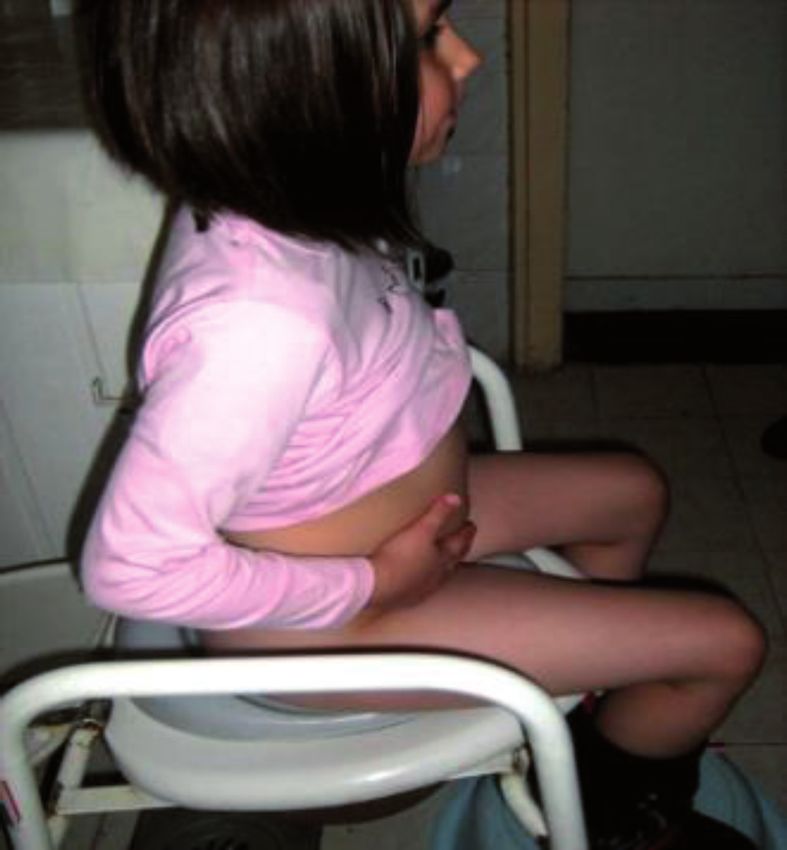
Pelvic Floor Disorders

wall and upper urinary tract, voiding disorders, accompanied by urinary inconti-
nence, can be a severe psychosocial problem. Children describe wetting at school as
the third most stressful event in life after the death of a parent and loss of sight [3].
It is evident that urinary incontinence causes significant psychological morbidity,
and treatment is crucial.
     Functional voiding disorders can be treated in a number of ways, includ-
ing pharmacological therapy, urotherapy, and surgical treatment in the most
severe cases.

2. Dysfunctional voiding

2.1 Definition

    Dysfunctional voiding (DV) refers only to the disorder of the bladder emptying
phase, and is characterized by intermittent contraction of the external urethral
sphincter and/or PFMs during the voiding phase of a micturition cycle [4]. A typi-
cal finding is interuppted or staccato uroflowmetry curve with increased electro-
myography (EMG) pelvic floor muscle activity during urination.
    The more severe form is referred to as Hinman syndrome by the author who first
described it [5]. Other terms previously used for DV are detrusor-sphincter dis-
coordination, non-neurogenic neurogenic bladder and occult neurogenic bladder.
In the United States, the term “dysfunctional voiding” has been used for all types
of voiding disorders, even bladder filling phase disorders. According to the ICCS
standardization of terminology from 2016, DV refers exclusively to the disorder of
the voiding phase [4]. It is thought to be the result of excessive PFM activity in an
attempt to prevent urination that occurs due to uninhibited detrusor contractions in
the early stage of bladder filling.

2.2 Epidemiology

     Epidemiological data on DV are lacking. Dysfunctional voiding was found
to occur in 4.2% of children referred for urinary incontinence [6]. In published
studies, the prevalence was estimated to be between 5 and 25% and 32% [7, 8].
Dysfunctional voiding was observed in 65% of children aged 5-9 years with urinary
tract infections, and in 23% of children who were urinary tract infections free [9].
It is evident that the criteria for including children in the studies were different, as
well as the accuracy of their evaluation, which indicates the need to conduct new
research to determine accurate data.

2.3 Etiology

    Dysfunctional voiding was first observed in 1973 by Hinman and Baumann
[5]. Hinman describes it as an acquired, reversible behavioral disorder that can be
ameliorated by suggestion and changes in behavior. He defined it as a bad habit for
special people in a bad family environment. Allen stated in 1977 that hyperactivity of
a child is a typical sign and that psychological factors play a key role in at least 50% of
the 21 children described [10]. He also points to the importance of stressful situations
in the family, such as parental alcoholism, parental divorce and father dominance.
    Contrary to these considerations, Van Gool points out that DV is not related
to emotional or psychosocial problems, but is caused by delayed CNS matura-
tion and external urethral sphincter dysfunction [11]. Hjalmas considers the
importance of hereditary factors, as DV was observed in several members of the

2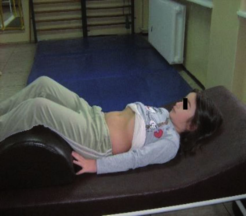
Rehabilitation Protocols for Children with Dysfunctional Voiding
DOI: http://dx.doi.org/10.5772/intechopen.98573

same family [12]. It is not known whether this is due to genetic factors or common
family habits.
   Most authors, however, believe that DV is a learned behavior [13]. It can develop
from overactive bladder (OAB) and voiding postponement as a result of PFM
contractions in an attempt to prevent urination, but it can also exist without these
precursors. In some children, urgency, voiding frequency and, in some instances,
urinary incontinence and signs of DV are present at the same time [14].
   From the review of the literature so far, it can be concluded that the etiology is
probably multifactorial. Possible risk factors are:

    1. Inadequate toilet training process

      Wiener et al. suggest that functional voiding disorders may be caused by inad-
      equate toilet habits and postures [15], which was also confirmed in the study
      by Bakker et al. [16]. This study indicated that the use of adult toilets in the
      process of children’s toilet training may increase the risk of developing func-
      tional voiding disorders. Thus, most of the programmes used in the treatment
      of DV involve taking an adequate position when urinating, i.e. the use of a
      toilet insert and footrests to ensure the stability of the trunk and the relaxation
      of the PFMs, and thus enable the physiological emptying of the bladder.

    2. Small structural anomalies in the anatomy or innervation of the lower
       urinary tract.

    3. Delay in maturation: detrusor overactivity as a component of DV may rep-
       resent the persistence of normal infantile mode of urination even after toilet
       training. It is possible that delays in CNS maturation reduce the ability of these
       children to take voluntary control of the micturition reflex.

    4. Impact of school: more than half of the time children spend in school, which
       suggests that teachers can positively or negatively influence the acquisition of
       toilet habits. In a study by Cooper et al., the influence of schools on the develop-
       ment of DV in children was examined and it was pointed out that most teachers
       allow going to the toilet only during rest [17]. Such a ban on going to the toilet
       for a child with an urgency who has not yet developed a complete inhibition of
       the micturition reflex may impair the normal function of the urinary bladder
       and sphincter. In addition, most children with daily urinary incontinence avoid-
       ed going to the school toilet due to lack of privacy or poor toilet hygiene [17].

    5. Unpleasant events during toilet training and/or personal stress: serious emo-
       tional stresses, such as sexual abuse, mostly of girls, should be considered in the
       event of sudden onset of DV, and in the absence of other etiological factors [18].

2.4 Clinical signs and symptoms

     Children are usually referred for wetting clothes, but not for DV. Children and
parents usually do not register specific symptoms of DV, so the physician must
insist on them. Typical signs are difficulty initiating micturition (hesitancy), as
well as straining to overcome the resistance of the contracted urinary sphincter. The
urinary stream is usually not strong because the PFMs do not relax completely, and
it is often intermittent.
     In an attempt to postpone voiding or suppress urgency and/or urinary inconti-
nence, children assume characteristic positions such as crossing their legs, standing

3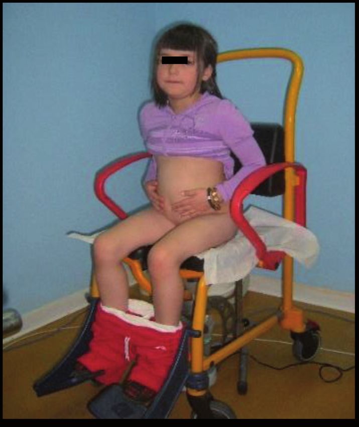
Pelvic Floor Disorders

on tiptoes, squatting, or manual compression of the genitals (pelvic holding maneu-
vers). It is typical for girls to squat by pressing their heel against the perineum [19].
   Stool retention, chronic constipation and fecal incontinence occur in more than
50% of children as a result of repeated and habitual contractions of the PFMs [9].
   Plenty of data from the literature indicate an association between DV and recur-
rent urinary tract infections [8, 20–22]. Treatment of DV reduces recurrent urinary
tract infections. About 50% of children with DV may have VUR [10].

2.5 Hinman-Allen syndrome

    The most severe form of DV is Hinman-Allen syndrome, according to the
authors who first described it in 1973 [5, 10]. The old term “non-neurogenic neuro-
genic bladder” can also be found in the literature.
    This syndrome occurs in children who voluntarily and habitually contract the
external urethral sphincter during uninhibited detrusor contractions, which leads
to the inability of emptying the bladder. The condition is characterized by detrusor
overactivity and possibly its decompensation. It is clinically manifested by daytime
and nighttime wetting, urgency and overflow incontinence, chronic constipation,
as well as recurrent urinary tract infections. Voiding cystourethrography reveals a
trabeculated bladder, high-grade bilateral VUR and large post-void residual urine.
If not treated in time, it leads to reflux nephropathy followed by kidney scarring,
hypertension and progressive renal failure.

3. Evaluation of a child with dysfunctional voiding

    Evaluation of children with DV includes taking anamnestic data, physical
examination, filling in the bladder and bowel diary, urinalyses and urine culture,
ultrasound examination of the bladder and kidneys and uroflowmetry with deter-
mination of post-void residual urine. Cystometry, voiding cystourethrography and
MRI of the spine are indicated only in certain cases.

3.1 History

    The evaluation process begins with anamnesis, data on perinatal factors,
developmental course, current mental state, school success and events during toilet
training. Every child should be asked how he urinates, whether he has difficulty
starting to urinate, whether urinating is difficult and whether he strains when uri-
nating. Also, one should insist on the characteristics of the urinary stream, such as
strength and continuity. A weak and intermittent stream indicates the existence of
DV. The child should be asked if he has a feeling of incomplete urination or urinary
retention (inability to urinate). Questions regarding urgency, voiding frequency
and severity of daytime and nighttime wetting, pelvic holding maneuvers, and
bowel emptying should follow.

3.2 Physical examination

3.2.1 Abdominal examination

   Palpation of the left iliac fossa is necessary in order to determine fecal impaction.
Suprapubic tenderness may indicate the presence of cystitis. If urine leaks when
the bladder is pressed, the existence of a neurogenic bladder with sphincter damage
should be suspected.

4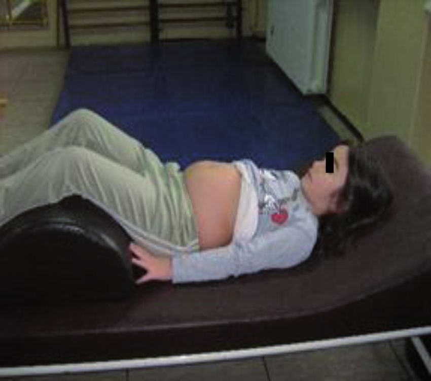
Rehabilitation Protocols for Children with Dysfunctional Voiding
DOI: http://dx.doi.org/10.5772/intechopen.98573

3.2.2 Neurological examination

    It is necessary to perform a careful inspection of the lower part of the spine in
order to determine the cutaneous manifestations of occult spinal dysraphism and/or
sacral agenesis (lipomas, nevi, increased hairiness, low intergluteal cleft, flattened
buttocks). After that, it is necessary to test the tendon reflexes on the lower extremi-
ties as well as the existence of the Babinski sign. Hyperreflexia, asymmetry in reflexes
or positive Babinski indicate spinal cord damage. It is also important to examine the
strength of the muscles of the lower extremities, ataxia and gait.

3.2.3 Inspection of genitals and perianal region

    Genital inspection is necessary in every patient with DV. Inspection of the
perineum reveals the position of the anus, fissure, fistula or perianal inflammation
of the skin in children with constipation.

3.2.4 Rectal examination

    Digital rectal examination can determine the tone and function of the anal
sphincter, the width and content of the rectal ampulla, the amount and consistency
of feces in the ampulla, and the presence of pain. It is recommended that anorectal
examination be performed only in those children who meet 1 of 6 Rome III criteria
for the diagnosis of constipation [9].

3.3 Bladder and bowel diary

    The bladder diary is a crucial diagnostic and therapeutic instrument for children
with voiding disorders. After the anamnesis and physical examination, this is the
third most important part of the evaluation. It is used to understand and quantify the
function of the bladder at home, because the memory of the elements of voiding is
unreliable. A complete bladder diary is kept for 7 nights and urinary incontinence is reg-
istered, as well as nocturnal urine volume, in order to evaluate nocturnal enuresis [4].
    The 48 hr. daytime frequency and volume chart is kept for 48 hours, preferably
on weekends for practical reasons [4]. The data obtained are frequency of void-
ing, volume of individual urination, maximum voided volume, urine production,
nocturia, nocturnal urine production and fluid intake. The frequency of voiding in
children with DV is variable.
    A bowel diary is kept for 7 days to rule out the presence of constipation [4].
The frequency of defecation, the appearance of the stool according to the Bristol
scale, pain and tension during defecation, whether the child has previously delayed
defecation and whether fecal incontinence is present are entered.

3.4 Urinalysis and urine culture

    Urinalyses and urine culture are initial tests to rule out urinary tract infection.

3.5 Urodynamics

3.5.1 Uroflowmetry

    Uroflowmetry is the simplest form of urodynamics. As a non-invasive method, it
is ideal for pediatric needs (Figure 1). It enables the examination of the function of
the detrusor and the sphincter in the voiding phase of the child’s micturition.

5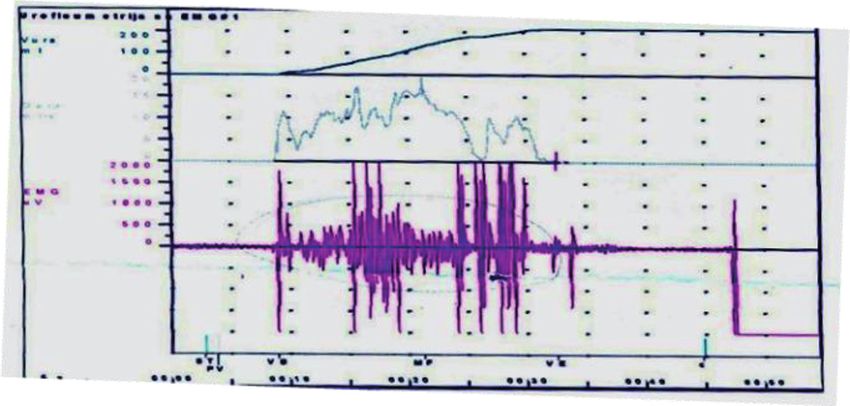
Pelvic Floor Disorders

Figure 1.
Uroflowmetry.

   The uroflowmeter was first described in the 1950s [23]. The method can be
applied from the fourth year of life of a child with established micturition control. In
order to get reliable results, it is necessary for the child to urinate at a capacity that is
not less than 50% nor more than 115% of the bladder capacity expected for age [4]. In
addition, it is necessary to repeat examination to make the result accurate and reliable.
   The method consists in urinating the patient in a uroflowmeter, continuously
measuring the urine flow rate (ml/s) and at the same time graphically showing the
curve. The placement of two superficial EMG electrodes on the perineum enables
the recording of the activity of the PFMs in the micturition phase. In this way,
significant data are obtained, especially in children with DV. The uroflowmetry
curve is of the staccato or interrupted type with an increase in the EMG activity of
the PFMs during urination (Figure 2).
   Upon completion of this study, an ultrasound examination of the bladder is per-
formed to determine post-void residual urine. In a child aged 7 to 12 years, residual
urine greater than 20 ml or 15% of the bladder capacity expected for age is consid-
ered elevated [4]. Residual urine that is larger than 10 ml or 6% of bladder capacity
expected for age during several measurements is also considered elevated [4].

3.5.2 Cystometry

   Cystometry is the only method by which bladder function can be examined
directly and in detail. The method is invasive. In order to obtain the data necessary

6
Rehabilitation Protocols for Children with Dysfunctional Voiding
DOI: http://dx.doi.org/10.5772/intechopen.98573

Figure 2.
Interrupted uroflowmetry curve with increased pelvic floor EMG activity during voiding.

for urodynamic analysis of bladder function, simultaneous measurement of pres-
sure in the bladder, urethra and abdomen is required. This continuous measurement
of detrusor pressure and sphincter activity during bladder filling and emptying
allows an accurate diagnosis to be made for most lower urinary tract disorders.
    In children with DV, cystometry is indicated only in certain cases, primarily
because the diagnosis is made using non-invasive methods. It should be performed
only in severe cases of DV, which should be differentially and diagnostically distin-
guished from neurogenic bladder with detrusor sphincter dyssynergia, suspicion of
Hinman-Allen syndrome and DV resistant to the applied therapy.

3.6 Voiding cystourethrography (VCUG)

    Voiding cystourethrography is not routinely performed in children with DV. It
is used in children with recurrent urinary tract infections to rule out the presence
of VUR. A typical finding in DV is a ballooned proximal urethra. If ultrasound
examination reveals bladder wall thickening and hydronephrosis and/or a plateau
uroflowmetry curve, it is necessary to perform VCUG to exclude the presence of a
posterior urethral valve in boys.

3.7 Magnetic resonance imaging

   Magnetic resonance imaging (MRI) of the spine should be performed in every
child with a suspected neurogenic voiding disorder or in whom a clinical examina-
tion has established the existence of spinal dysraphism. Suspicion of tethered cord
syndrome, as well as spinal tumors, are also indications for referring a child for an
MRI of the spine.

4. Urotherapy

   Functional voiding disorders should be actively treated due to the possibility of
developing structural changes in the bladder and upper urinary tract. In the Anglo-
Saxon literature, the term “urotherapy” has been used since 1980 in children with void-
ing disorders, which is synonymous with rehabilitation of the lower urinary tract [24].

4.1 Definition

   Urotherapy is a conservative, non-surgical treatment of the lower urinary tract
and can be defined as re-education of the bladder or rehabilitation programme
7
Pelvic Floor Disorders
aimed at correcting the phase of filling and emptying the bladder [4]. Urotherapy,
first of all, includes changing the habits that the child has acquired in the process of
toilet training, as well as establishing motor control over the micturition reflex. It
can also include drug therapy, because most children have some kind of pharmaco-
therapy during urotherapy.
4.2 Goals
The goals of urotherapy are clearly defined and include normalization of the
phase of filling and emptying the bladder, facilitation of normal bowel function
and normalization of the dynamics of defecation. The goals of treatment in clini-
cal practice are aimed at reducing urgency and urinary incontinence, nocturnal
enuresis, post-void residual urine, high intravesical pressure, PFM activity in the
micturition phase, normalization of uroflowmetry curve, cure urinary tract infec-
tions and constipation, and reduce the degree of VUR.
4.3 Indications
Urotherapy is used in functional voiding disorders that are accompanied by an
altered phase of bladder filling and emptying, in children in whom satisfactory
results have not been achieved with the use of pharmacological therapy, and in
children who are just starting therapy.
4.4 Components of urotherapy
Urotherapy consists of standard urotherapy and specific measures that include
neuromodulation, PFM relaxation exercises, biofeedback and intermittent
catheterization [4].
4.4.1 Standard urotherapy
In the literature, we often come across the term “standard urotherapy”, which
includes education, behavioral modifications, keeping a bladder and bowel diary
and regular check-ups [4]. Before starting urotherapy, it is necessary to dedicate
a lot of time to educating the child, examining his motivation to carry out the
treatment, and eliminating the shame and other effects that urinary incontinence
can cause.
4.4.1.1 Education and demystification
The child should be acquainted with the structure and function of the bladder,
external urinary sphincter, colon and anorectum in a way that is appropriate for
his age. He also needs to be explained the etiology of his voiding and defecation
disorders. It is important to examine the motivation for treatment. If the child is not
motivated, the results of the treatment are much worse.
4.4.1.2 Behavioral modifications
The goal is to improve the control of urination and defecation through changes
in the child’s behavior. This includes regular voiding and hydration, taking the
correct position during urination and defecation, as well as changing the diet. With
the application of this initial treatment, a cure can be achieved in about 20% of
children [21].
8Rehabilitation Protocols for Children with Dysfunctional Voiding
DOI: http://dx.doi.org/10.5772/intechopen.98573
4.4.1.3 Regular hydration
Children with voiding disorders are often voluntarily dehydrated. Concentrated
urine can cause irritation and burning when urinating. The introduction of ade-
quate hydration (200 ml 5-6 times a day) increases urine production and bladder
capacity, reduces urine concentration and improves bowel function. It is forbidden
to drink beverages such as Coca-Cola, coffee, tea, cold juices, because they can
induce detrusor overactivity [25].
4.4.1.4 Regular voiding
It is the basis of bladder rehabilitation and includes the adoption of a voiding
schedule every 2-3 hours. The goal is to prevent bladder distension, to restore
the feeling of fullness of the bladder, and to reduce the hyperactivity of the
bladder. The child and parents should know that the ability to start micturition,
even when the child does not want to urinate, is an important step in controlling
the continence and emptying of the bladder. The child should try to urinate
only at a predetermined time, and avoid delaying urination. It is necessary
to analyze the child’s daily activities and determine the time of micturition
accordingly.
4.4.1.5 Optimal bladder emptying
The position when urinating has a significant effect on the ability to empty
the bladder. Toilet bowls are intended for adults and as such are not suitable for
children. Relaxation of the PFMs is difficult or impossible with various irregular
positions that the child assumes during urination.
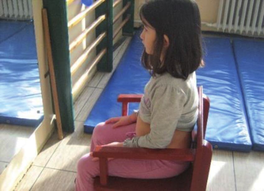
Proper voiding position means urinating in a sitting position on the toilet bowl
for girls and boys. For smaller children, it is important to require the use of an
adequate toilet seat insert and footrests to ensure trunk stability [26]. When urinat-
ing, the child should be slightly bent forward, with the spine in extension, the hips
in abduction and the relaxed abdominal muscles (Figure 3). It is necessary for
children to listen to the sound of the stream when urinating and the goal is for the
stream to be strong, long and sonorous.
4.4.1.6 Treatment of chronic constipation/fecal incontinence
It is important to recognize the defecation disorder, because it has been proven
that the treatment of constipation alone significantly reduces the symptoms of the
lower urinary tract. In the group of children with increased post-void residual urine
and constipation, 66% had an improvement in bladder emptying after constipation
treatment. Urinary incontinence, nocturnal enuresis, and recurrent urinary tract
infections were cured in most children treated only for constipation [20]. Therefore,
treatment begins with chronic constipation management.
In the treatment of chronic constipation, four steps are applied: education,
disimpaction of fecal mass, prevention of its re-accumulation and follow-up [27].
Treatment is usually applied for 3-6 months, but the relapses are frequent [28]. The
cure rate of chronic functional constipation after application of standard treatment
that includes laxatives and behavioral approaches is only 50-60% [29].
More than half of children with chronic constipation have an abnormal
defecation pattern because they contract the external anal sphincter and the M.
puborectalis during defecation [30]. This form of abnormal defecation is consid-
ered to be learned resulting from the habit of delaying defecations. Physiotherapy
9Pelvic Floor Disorders
Figure 3.
Correct position when urinating.
interventions such are diaphragmatic breathing exercises and pelvic floor exercises
with or without biofeedback were introduced in order to educate a child to relax
the external anal sphincter and the PFMs during defecation [31–35]. In refractory
cases, even botulinum toxin injections are administrated into the external anal
sphincter [36].
Interferential current stimulation (IFS) has been used in the treatment of
chronic constipation resistant to standard therapy in children. Significant improve-
ments in clinical symptoms (increased frequency of defecation, reduction of fecal
incontinence and abdominal pain) were seen in 67% of children and lasted for more
than two years in one third of the treated patients [37]. In addition, the time of
colonic transit on colonic scintigraphy was shorter after the application of IFS [38].
Although the mechanism of action of IFS is still insufficiently known, the proposed
theories are the activation of local sensory nerves in the skin, spinal nerves (sensory
and motor T9-L2), sympathetic and parasympathetic nerves in the intestine, enteric
nerves, pacemaker cells (Cajal’s interstitial cells) or smooth muscle cells in the
intestinal wall [39].
5. Rehabilitation protocols for dysfunctional voiding
Although there is no general approach to treatment and treatment varies from
one patient to another, there are several ways to treat children with DV, including
urotherapy, pharmacotherapy, botulinum toxin application, and surgical treatment
in children with VUR.
10Rehabilitation Protocols for Children with Dysfunctional Voiding
DOI: http://dx.doi.org/10.5772/intechopen.98573
In addition to standard urotherapy, special measures are applied, such as
exercises for relaxation of PFMs with or without biofeedback and exercises for
relaxation of abdominal muscles.
5.1 Pelvic floor muscle relaxation exercises and biofeedback
Pelvic floor muscle exercises were first used in pediatric urology by Wennergren
and Oberg, with the aim of developing the child’s awareness of their function [40].
During the exercises, the child learns to contract and relax these muscles without
activating auxiliary muscles (gluteal and hip adductors). In order to improve vol-
untary control, the exercises can be combined with different types of biofeedback,
such as visual (observation of the abdomen in front of a mirror), tactile (palpation
of the PFMs or M. transversus abdominis), uroflowmetry or electromyography.
Biofeedback was first used by Maizels et al. in 1979, who implemented the use of
urodynamics devices in children with detrusor sphincter dyssynergia [41]. During
urination, children observed EMG activity of the sphincter. Improvement was
achieved in two of the three treated children.
Uroflowmetry biofeedback consists of the child observing the uroflowmetry
curve while urinating. During voiding, the child is advised to make sure that the
curve is bell-shaped. Kjolseth examined the efficacy of uroflowmetry biofeedback
in 32 children with DV [42]. The number of applied sessions was 1-9, while 47% of
children required 4-5 sessions. Cure was achieved in 50% of children, improvement
in 8 children, and 7 children were unchanged. The uroflowmetry curve was com-
pletely normalized in 55% of children. It has been shown that this type of biofeed-
back requires a smaller number of sessions compared to EMG biofeedback and leads
to faster normalization of the act of urination [43].
5.1.1 Animated pelvic floor EMG biofeedback
Mc Kenna et al. in 1999 applied biofeedback in the form of interactive computer
games that enabled the active participation of patients [44]. Computer play main-
tained the child’s interest and motivation for the exercise programme. The method
consists of placing superficial EMG electrodes on the child’s perineum, and then
the child is taught to properly contract and relax the PFMs by watching a game
on a computer monitor. In this way, children become aware of the activity of the
PFMs and learn to control them by controlling the activities of their favorite heroes
(dolphin, monkey, fish, bee).
In a study by Herndon et al. interactive computer games were used in 160
children with DV [45]. In 87% of patients, subjective improvement of symptoms
was achieved. In a study by McKenna et al. improvement of nocturnal enuresis
was achieved in 90%, daily wetting in 89%, constipation and fecal incontinence in
100% [44].
Kaye and Palmer did not find significant differences in efficacy after applica-
tion of non-animated (biofeedback without animation using only EMG tracing)
and animated biofeedback [46]. However, a group of children who had animated
biofeedback required a smaller number of sessions to normalize the uroflowmetry
curve and reduce residual urine. In a study by Desantis et al. there was an improve-
ment in urinary tract infections in 83%, diurnal incontinence in 80%, constipation
from 18–100%, urinary frequency from 67–100%, urgency from 71–88% and VUR
from 21–100% of children [47].
In a study by Palmer et al. in children with DV and VUR, the use of biofeedback
accelerated the resolution of VUR or reduced the degree of VUR in 71% of children
[48]. Similar results were presented by Khen-Dunlop et al. and Kibar et al. [49, 50].
11Pelvic Floor Disorders
Adequate patient selection seems to be the most important for biofeedback success.
Parents and children should be motivated and compliant to continue practicing
exercises at home [51].
Although numerous studies highlight the positive effects of PFM relaxation
exercises with or without biofeedback, there is no clear recommendation of an
exercise protocol to use in the rehabilitation of children with DV. The number of
sessions, the number of repetitions, the duration of the contraction and relaxation
phase, as well as the period of performing the exercises differ significantly between
the studies.
De Paepe et al. applied PFM relaxation exercises with EMG biofeedback [52].
The protocol consisted of 30 submaximal contractions lasting 3 seconds, followed
by a relaxation phase of 30 seconds. One session per week was applied for 6 months
(maximum 20-24 sessions). In a study by Vasconcelos et al., 24 home exercise
sessions lasting 20 minutes were applied, three times a week during a three-month
period [53]. The contractions lasted for 5 seconds, followed by a 10-second relax-
ation period. Shei Dei increased the duration of contractions to 10 seconds and
extended the relaxation period to 30 seconds [54]. Yagci et al. applied submaximal
contractions of 3 seconds, followed by a relaxation period of 30 seconds [55].
The children repeated the exercises at home three times a day for 6 months. In a
retrospective study, Drzeviecki et al. analyzed a programme in which the contrac-
tions lasted 10 seconds, followed by a relaxation of 10 seconds [56]. After that, fast
contractions lasting 5 seconds and 5 seconds of relaxation followed. On average, 3
sessions (1-8) were applied.
5.2 Abdominal capsule
Sapsford et al. showed that the PFMs are not an isolated unit, but a part of the
abdominal capsule that surrounds the abdominal and pelvic organs [57]. The struc-
tures that make this capsule are the lumbar vertebrae, M. multifidus, diaphragm,
M. transversus abdominis and PFMs. These muscles contribute to maintaining the
posture of the body in an upright position and act synergistically.
Coactivation of the abdominal and PFMs is necessary for the development of
intra-abdominal pressure and contributes to the stability of the spine. It is shown
that M. transversus abdominis contributes the most to the development of intra-
abdominal pressure in relation to other abdominal muscles [58]. This muscle is first
activated during functions related to the increase in intra-abdominal pressure, such
as spinal stabilization and expiratory tasks [58]. Coactivation of the abdominal
capsule muscles has been demonstrated during weight lifting, coughing and forced
expiratory tasks [58, 59].
Pelvic floor muscle dysfunction can present as hyperactivity, leading to the
development of voiding and defecation disorders, such as DV, chronic constipation,
perianal and perineal pain. Many of these children have hyperactivity of the lower
abdominal muscles, which do not relax during urination and defecation and thus
prevent the relaxation of the PFMs [57].
5.3 Diaphragmatic breathing exercises
As lower abdominal muscles (M. transversus abdominis and M. obliquus internus
abdominis) and PFMs act synergistically, it is necessary for them to relax together
during urination and defecation.
The simplest way for children to learn how to relax their abdominal muscles
is through diaphragmatic breathing exercises. During diaphragmatic breathing,
in inspiration, the diaphragm moves caudally and pushes the abdominal organs
12Rehabilitation Protocols for Children with Dysfunctional Voiding
DOI: http://dx.doi.org/10.5772/intechopen.98573
forward. The anterior abdominal wall relaxes, as do the PFMs. This forward bulg-
ing of the anterior abdominal wall has been shown to reduce urethral pressure in
healthy women and thus facilitate urination and defecation [58] .
Our institution was the first to incorporate this novel approach to treating DV.
In a prospective clinical study of 43 children, in addition to standard urotherapy
that included education on the importance of regular urination and hydration,
proper voiding position and pattern, diaphragmatic breathing exercises and PFM
relaxation exercises were performed in hospital settings for two weeks and then
continued at home [60].
Diaphragmatic breathing exercises were performed in a supine position with
the lower extremities supported by a pillow and hands placed on the abdominal
muscles. The patient is required to inhale air through the nose, expel the anterior
abdominal wall, hold the breath for a few seconds, and then exhale the air through
the mouth (Figures 4 and 5). The exercises were repeated in both lateral posi-
tions, in the prone position, and then in the sitting position in front of the mirror
(Figure 6). Children are required to observe the anterior abdominal wall during
inspiration and then apply this exercise before urinating and defecating.
In addition, exercises for relaxation of the PFMs were performed. The child
was placed in a lateral position with the upper leg flexed at the hip and knee and
the lower leg extended. To enhance the proprioception of the PFMs, the examiner
placed two fingers on the child’s perineum and demanded that the child contract
the PFMs without activating adjacent muscles such as the gluteus and hip adductor
muscles. In this way, the child learned to localize and control the PFMs. The child
was then required to perform submaximal contractions for 3 seconds followed by
prolonged relaxation for about 30 seconds, for a total of 20 contractions. Children
are required to perform these exercises daily at home for 6 months.
Control examinations were performed monthly for 12 months. Clinical manifes-
tations (daytime urinary incontinence, nocturnal enuresis, urinary tract infections,
Figure 4.
Diaphragmatic breathing exercises in supine position (expiration).
13Pelvic Floor Disorders
Figure 5.
Diaphragmatic breathing exercises in supine position (inspiration).
Figure 6.
Diaphagmatic breathing exercises in front of the mirror.
constipation) were analyzed on a monthly basis and uroflowmetry was performed.
The performance of diaphragmatic breathing exercises was controlled and the
importance of daily exercise at home was emphasized. The children are encouraged
to continue with the treatment.
After one year of monitoring and treatment, reevaluation of clinical manifesta-
tions and uroflowmetry parameters was performed. Urinary incontinence was
cured in 83% of children, nocturnal enuresis in 63%, and urinary tract infections
14Rehabilitation Protocols for Children with Dysfunctional Voiding
DOI: http://dx.doi.org/10.5772/intechopen.98573
in 68%. Chronic constipation was cured in all 15 patients. In addition, an objective
improvement in uroflowmetry parameters was achieved. A normal uroflowmetry
curve was registered in 90% of children.
The authors suggested that examination of lower abdominal muscles, recogni-
tion of their function during voiding and their relaxation should be incorporated
in the treatment program of these children. Easy to learn diaphragmatic breathing
exercises did not require any specific equipment and could be performed in chil-
dren from five years of age. For centres that do not have access to pelvic floor EMG
biofeedback, this programme could provide a treatment alternative as success rates
are comparable to previous studies that used pelvic floor EMG biofeedback during
urotherapy [54–56]. In order to achieve subjective and objective progress, children
needed an average of 6.5 sessions, which is also equivalent to the average number of
sessions in programmes that included non-animated biofeedback [46].
In the following study, the effects obtained in this group were compared with the
effects in the group of children treated only with standard urotherapy (32 children)
[31]. The children had 10 sessions of urotherapy in a hospital setting, and then were
required to continue with it at home. After one year of follow-up, cure of urinary
incontinence was achieved in only two children, nocturnal enuresis in 5, and uri-
nary tract infections in 6 children. Constipation was cured in 6 out of 10 children.
Uroflowmetry parameters did not show significant improvements. The authors
concluded that diaphragmatic breathing exercises and PFM relaxation exercises,
in combination with standard urotherapy, are important for the treatment of daily
urinary incontinence, nocturnal enuresis and urinary tract infections, as well as for
normalizing bladder function in children with DV.
5.4 Pharmacological therapy
Pharmacological therapy is considered an adjunct to improve bladder emptying
in children with DV [43].
5.4.1 Α-1 adrenergic receptor blockers
The role of α-1 adrenergic receptor blockers in the treatment of children with
DV is controversial, as the mechanism of action at the level of the external urethral
sphincter is still insufficiently known [61]. The possible mechanism of action is
traditionally assumed to be relaxation of the periurethral, prostatic and bladder
neck smooth muscles. In the study of Yucel et al., it has been shown that the effects
of α-1 adrenergic blockers in reducing post-void residual urine can be compared
with the effect of biofeedback [62].
5.4.2 Muscle relaxants
As DV is characterized by the inability of relaxation of the external urinary
sphincter during urination, it was considered that muscle relaxant could be used in
treatment.
Baclofen has been shown to be effective in reducing skeletal muscle spasticity,
as well as in patients with striated sphincter dyssynergia [63]. However, the thera-
peutic effect is achieved only after the application of high doses. Serious adverse
effects, especially after abrupt withdrawal, reduce its efficacy and safety in children
with DV [64]. Therefore, tizanidine, a muscle relaxant used in many studies as a
short-acting muscle relaxant due to spasmolytic action, was used. In a prospec-
tive, randomized study, 40 children with DV were divided into two groups [65].
The first group was treated with tizanidine (an imidazole derivative), while the
15Pelvic Floor Disorders
second group of children was treated with α-blocker (doxazosin). After 6 months
of follow-up, both groups had similar improvement in symptoms and uroflowmetry
parameters. In the doxazosin-treated group, urgency was the only symptom that
showed a significant reduction after therapy. However, nocturnal enuresis, urgency,
and daytime incontinence were significantly reduced in the tizanidine-treated
group. Side effects were reported in 6 patients (15%). Epigastric pain was reported
in two children (10%) receiving doxazosin. In the tizanidin group, loss of appetite
was noted in two children (10%), epigastric pain in one (5%) and headache in one
child (5%).
5.5 Botulinum toxin type A (BT-A)
BT-A is one of the strongest known toxins. When injected directly into a
muscle, it causes flaccid paralysis by blocking the presynaptic release of acetylcho-
line [66].
The use of BT-A in patients with detrusor sphincter dyssynergia (DSD) was first
described by Dykstra et al. [67]. In this study, BT-A was injected into the external
urinary sphincter of adult patients with spinal cord injury and DSD. Positive
results, reduced urethral pressure, and volume of residual urine remained for an
average of 50 days.
Indications for the injection of BT-A in the external urinary sphincter have been
extended over time to adult patients with DV and detrusor hypocontractility. In the
study by Kuo et al. clinical and urodynamic improvement was registered in 83%
of patients with urethral sphincter non-relaxation and detrusor hypocontractility
[68]. Petit and co-workers reported a significant reduction in detrusor and urethral
pressure, as well as the volume of post-void residual urine after a single injection
of 150 units Dysport (BT-A) in patients with spinal cord injury and DSD [69]. The
beneficial effects of the therapy lasted for about 2-3 months.
In children treated with BT-A (amp. Dysport) due to spasticity, the most com-
monly reported adverse effects were local muscle weakness, urinary incontinence,
fatigue, somnolence, flu-like symptoms, fever, and rash [70].
BT-A is used in the treatment of DV in children who are resistant to standard
urotherapy. In the study of Radojicic et al., BT-A was applied in to the external
urinary sphincter in children with DV [71]. The residual urine decreased signifi-
cantly in 17 of 20 patients after 6 months of follow-up. The authors emphasized
that temporary inhibition of the external urinary sphincter and/or PFMs may
interrupt the DV cycle. The use of urotherapy during this period could help
the child to re-adopt a normal urination pattern and thus reduce the need for
re-injections.
In our institution, a prospective clinical study included 9 neurologically
healthy girls with DV, aged 3-11 years, who had previously been treated with
standard urotherapy without improvement [72]. Application of BT-A (amp.
Dysport) in a dose of 500 units was performed transperineally into the external
urethral sphincter. After two weeks of application, rehabilitation treatment
consisting of standard urotherapy and PFM relaxation exercises was included.
Six months after Dysport administration, there was a statistically significant
improvement in clinical manifestations (urinary incontinence, voiding difficul-
ties, urinary tract infections), and a significant reduction in post-void residual
urine. No significant improvement in uroflowmetry parameters was registered.
No children had systemic side effects with Dysport. The authors concluded that
the act of urination in children with DV resistant to standard therapy can be
significantly improved and maintained for at least 6 months after the use of amp.
Dysport and urotherapy.
16Rehabilitation Protocols for Children with Dysfunctional Voiding
DOI: http://dx.doi.org/10.5772/intechopen.98573
5.6 Manual physical therapy
Manual physical therapy with an osteopathic approach (MPT-OA) entails
palpation and receptive manipulation of body tissues to relieve restraints that limit
mobility and health. Biomechanical, myofascial, and articular constraints can
contribute to DV by altering alignment, distorting the pelvis, restricting mobility,
and thus affecting pressures within the abdominal and pelvic cavities [73]. Altered
pressure relationships can affect neurological, vascular, lymphatic, and hormonal
functions. In the randomized controlled trial that involved 21 children with DV, it
has been shown that children with additional 4 sessions of MPT-OA demonstrated
better short-term results compared to children that had only standard treatment
[73]. The authors speculated that MPT-OA treatment helped restoring more natural
alignment and mobility which helped the abdominal and PFMs to function more
efficiently. However this single-centre promising results should be confirmed by
multi-centre randomized controlled trials in order to draw definitive conclusions of
MPT-OA in children with DV.
6. Conclusion
Urotherapy is the cornerstone of DV care for children. The treatment begins
with standard urotherapy, after which specific measures are added. Rehabilitation
programmes with diaphragmatic breathing exercises and pelvic floor relaxation
exercises are superior in curing lower urinary tract symptoms and normalizing
urinary function than programmes that only include standard urotherapy. The
success rates of programmes that included pelvic floor relaxation exercises and
diaphragmatic breathing exercises without the use of pelvic floor EMG biofeedback
were equivalent to those that included pelvic floor EMG biofeedback, proposing a
treatment choice for centres that do not have access to EMG biofeedback. However,
no standardized pelvic floor exercise protocol egists. Therefore, new prospective
multicentric randomized trials with a larger number of children are needed to
determine the most appropriate programme that will have the best therapeutic
outcome.
Acknowledgements
This work has been supported by the Ministry of Science and Technological
Development of the Republic of Serbia, under the project 43011.
Conflict of interest
None declared.
17Pelvic Floor Disorders Author details Vesna D. Zivkovic1,2*, Ivona Stankovic1,2, Lidija Dimitrijevic1,2, Hristina Colovic1,2, Dragan Zlatanovic1,2 and Natasa Savic3 1 Faculty of Medicine, University of Nis, Nis, Serbia 2 Clinic of Physical Medicine and Rehabilitation, University Clinical Centre, Nis, Serbia 3 College of Health Studies Cuprija, Cuprija, Serbia *Address all correspondence to: petvesna67@gmail.com © 2021 The Author(s). Licensee IntechOpen. This chapter is distributed under the terms of the Creative Commons Attribution License (http://creativecommons.org/licenses/ by/3.0), which permits unrestricted use, distribution, and reproduction in any medium, provided the original work is properly cited. 18
Rehabilitation Protocols for Children with Dysfunctional Voiding
DOI: http://dx.doi.org/10.5772/intechopen.98573

References

[1] Gontard A, Neveus T. Introduction.           [8] Hoebeke P, Van Laecke AR, Van
In: Hart M (ed). Management of                   Camp C, Raes A, Van De Walle J. One
disorders of bladder and bowel control           thousand video-urodynamic studies in
in childhood. London: Mac Keith Press;           children with non-neurogenic bladder
2006. p. 1-2.                                    sphincter dysfunction. BJU Int 2001;
                                                 87(6): 575-80. doi: 10.1046/j.1464-410x.
[2] Sillen U. Epidemiology. In:                  2001.00083.
Proceedings of the ICCS course on „
Multidisciplinary management of                  [9] Burgers RE, Mugie SM, Chase J,
nocturnal and diurnal wetting problems           Cooper CS, von Gontard A, Rittig CS et
in children“ & ESPU course on „Surgery           al. Management of functional
for the incontinent child and bowel              constipation in children with lower
problems“; 14-17 September Antalya,              urinary tract symptoms: Report from
Turkey, 2006. p. 136-40.                         the standardization committee of the
                                                 International Children’s Continence
[3] Hjalmas K, Arnold T, Bower W,                Society. J Urol. 2013; 190: 29-36. doi:
Caione P,Chiozza LM, von Gontard A,              10.1016/j.juro.2013.01.001
et al. Nocturnal enuresis: an
international evidence based                     [10] Allen TD. The nonneurogenic
management strategy. J Urol                      neurogenic bladder. J Urol 1977;117:23.
2004;171:2545-61. doi: 10.1097/01.               doi: 10.1016/s0022-5347(17)58412-8
ju.0000111504.85822.b2
                                                 [11] Van Gool JD, Kuitjen RH,
[4] Austin PF, Bauer SB, Bower W,                Donckerwolcke RA, Messer AP,
Chase J, Franco I, Hoebeke P et al. The          Vijverberg M. Bladder-sphincter
standardization of terminology of lower          dysfunction, urinary infection and
urinary tract function in children and           vesico-uretheral reflux with special
adolescents: Update Report from the              reference to cognitive bladder training.
standardization committee of the                 Contrib Nephrol 1984;39: 190-210. doi:
International Children's Continence              10.1159/000409249.
Society. Neurourol Urodyn. 2016;
35(4):471-81. doi: 10.1002/nau.22751             [12] Hjalmas K. Is dyscoordinated
                                                 voiding in children an hereditary
[5] Hinman F, Baumann FW. Vesical and            disorder. Scand J Urol Nephrol Suppl
ureteral damage from voiding                     1995;173:31-5.
dysfunction in boys without neurologic
or obstructive disease. J Urol                   [13] Koff S, Jayanthi V. Non-neurogenic
1973;109:727-32. doi: 10.1016/                   lower urinary tract dysfunction. In:
s0022-5347(17)60526-3.                           Walsh PC, Retik AB, Vaughn ED, Wein
                                                 AJ (eds). Cambell’s Urology. 8th Ed.
[6] Von Gontard A. Enuresis im                   Philadelphia: WB Saunders Co; 2002. p.
Kindesalter – psychiatrische, somatische         2262-83.
und molekulargenetische
Zusammenhange [thesis]. Keln:                    [14] Vijverberg MAW, Elzinga-Plomp A,
Universitat zu Koln; 1995.                       Messer AP, van Gool JD, de Jong TP.
                                                 Bladder rehabilitation, the effect of
[7] Hoang-Bohm J, Lusch A, Sha W,                cognitive training programme on urge
Alken P. Biofeedback for urinary bladder         incontinence. Eur Urol 1997 ;31:68-72.
dysfunctions in childhood. Indications,          doi: 10.1159/000474421.
practice and the results of therapy.
Urologe A 2004;43:813-9. doi: 10.1007/           [15] Wiener JS, Scales MT, Hampton J,
s00120-004-0617-3.                               King LR, Surwit R, Edwards CL.

19
Pelvic Floor Disorders
Long-term efficacy of simple behavioral [23] Kaufman JJ. A new recording
therapy for daytime wetting in children. uroflowmeter: a simple automatic device
J Urol 2000; 164 (3 pt 1): 786-90. doi: for measuring voiding velocity. J Urol
10.1097/00005392-200009010-00048. 1957;78:97-9. doi: 10.1016/
s0022-5347(17)66405
[16] Bakker E, van Gool JD, van
Sprudel M, King LR, Surwit R, [24] Hellstrom AL, Hjalmas K, Jodal U.
Edwards CL. Results of a questionnaire Rehabilitation of the dysfunctional
evaluating the effects of different bladder in children: method and 3-year
method of toilet training on achieving follow-up. J Urol 1987; 138:847-9. doi:
bladder control. BJU int 2002;90:456-61. 10.1016/s0022-5347(17)43395-7.
doi: 10.1097/00005392-200009010-
00048. [25] De Paepe H, Renson C, Hoebeke P,
Raes A, Van Laecke E, Vande Walle J.
[17] Cooper C, Abousally C, Austin C, The role of pelvic-floor therapy in the
Boyt M, Hawtrey C. Do public schools treatment of lower urinary tract
teach voiding dysfunction? Results of an dysfunctions in children. Scand J Urol
school teacher survey. J Urol Nephrol 2002; 36(4): 260-7. doi:
2003;170:956-8. doi: 10.1097/01. 10.1080/003655902320248218.
ju.0000075916.55446.ee.
[26] Wennergren HM, Oberg BE,
[18] Ellsworth P, Merguerian P, Sandstedt P. Importance of leg support
Copening ME. Sexual Abuse: Another for relaxation of the pelvic floor
causative factor in dysfunctional muscles. A surface electromyographic
voiding. J Urol 1995;153:773-6. study in healthy girls. Scand J Urol
Nephrol 1991;25:205-13. doi:
10.3109/00365599109107948
[19] Vincent SA. Postural control of
urinary incontinence. The curtsey sign.
[27] Mugie SM, Di Lorenco C,
Lancet 1966; 2 (7464) : 631-2. doi:
Benninga MA. Constipation in
10.1016/s0140-6736(66)91942-8.
childhood. Nat Rev Gastroenterol
Hepatol 2011;8:502-11. doi: 10.1038/
[20] Koff SA, Wagner TT, Jayanthi VR. nrgastro.2011.130.
The relationship among dysfunctional
elimination syndromes, primary [28] Bongers ME, van Wijk MP,
vesicoureteral reflux and urinary tract Reitsma JB, Benninga MA. Long-term
infections in children. J Urol prognosis for childhood constipation:
1998;160:1019-22. doi: 10.1097/ clinical outcomes in adulthood.
00005392-199809020-00014. Pediatrics. 2010;126(1):e156-62. doi:
10.1542/peds.2009-1009.
[21] McKenna LS, McKenna PH. Modern
management of nonneurologic pediatric [29] Van Dijk M, Bongers ME, de
incontinence. J WOCN 2004; 31(6): Vries GJ, Grootenhuis MA, Last BF,
351-6. doi: 10.1097/00152192- Benninga MA. Behavioral therapy for
200411000-00006. childhood constipation: a randomized,
controlled trial. Pediatrics. 2008;
[22] Hoebeke P. Voiding dysfunction, 121(5):e1334-41. doi: 10.1542/
recurrent UTI, constipation, and peds.2007-2402.
vesicoureteric reflux: a common disease
complex. In: Fourth course on paediatric [30] Van Dijk M, Benninga MA,
urodynamics, Utrecht. Edited by Grootenhuis MA, Nieuwenhuizen AM,
European Society of Paediatric Urology, Last BF. Chronic childhood
2005. p. 61-5. constipation: a review of the literature
20Rehabilitation Protocols for Children with Dysfunctional Voiding
DOI: http://dx.doi.org/10.5772/intechopen.98573
and the introduction of a protocolized J Pediatr Surg. 2009; 44: 1791-98. doi:
behavioral intervention program. 10.1016/j.jpedsurg.2009.02.056.
Patient Educ Couns. 2007; 67: 63-77. doi:
10.1016/j.pec.2007.02.002. [37] Leong LC, Yik YI, Catto-Smith AG,
Robertson VJ, Hutson JM, Southwell BR.
[31] Zivkovic V, Lazovic M, Stankovic I, Long-term effects of transabdominal
Vlajkovic M, Slavkovic A. The electrical stimulation in treating
evaluation of combined standard children with slow-transit constipation.
urotherapy, abdominal and pelvic floor J Pediatr Surg. 2011; 46: 2309-12. doi:
retraining in children with 10.1016/j.jpedsurg.2011.09.022.
dysfunctional voiding. J Pediatr Urol.
2011; 7: 336-41. doi: 10.1016/j. [38] Clarke MC, Chase JW, Gibb S,
jpurol.2011.02.028. Robertson VJ, Catto-Smith A,
Hutson JM, Southwell BR. Decreased
[32] Kajbafzadeh AM, LidaSharifi- colonic transit time after transcutaneous
Rad L, Ghahestani SM, Ahmadi H, interferential electrical stimulation in
Kajbafzadeh M, Mahboubiet AH. children with slow transit constipation.
Animated biofeedback: an ideal J Pediatr Surg. 2009; 44: 408-12. doi:
treatment for children with 10.1016/j.jpedsurg.2008.10.100.
dysfunctional elimination syndrome. J
Urol. 2011; 186: 2379-84. doi: 10.1016/j. [39] Chase J, Robertson VJ, Southwell B,
juro.2011.07.118. Hutson J, Gibb S. Pilot study using
transcutaneous electrical stimulation
[33] Loening-Baucke V. Modulation of (interferential current) to treat chronic
abnormal defaecation dynamics by treatment-resistant constipation and
biofeedback treatment in chronically soiling in children. J Gastroenterol
constipated children with encopresis. J Hepatol. 2005; 20: 1054-61. doi:
Pediatr. 1990; 116:214-22. doi: 10.1016/ 10.1111/j.1440-1746.2005.03863.x.
s0022-3476(05)82877-x.
[40] Wennerrgren H, Oberg B. Pelvic
[34] Zivkovic VD, Stankovic I, floor exercises for children: A method of
Dimitrijevic L, Kocic M, Colovic H, treating dysfunctional voiding. BJU
Vlajkovic M, et al. Are interferential 1995;76:9-15. doi: 10.1111/j.1464-
electrical stimulation and 410x.1995.tb07823.x.
diaphragmatic breathing exercises
beneficial in children with bladder and [41] Maizels M, King LR, Firlit CF.
bowel dysfunction? Urology 2017; 102: Urodynamic biofeedback: a new
207-12. doi: 10.1016/j. approach to treat vesical sphincter
urology.2016.12.038. dyssynergia. J Urol. 1979;122:205-209.
[35] Savic N, Zivkovic V, Stankovic I, [42] Kjolseth D, Knudsen LM, Madsen B,
Stojkovic T, Milenkovic M, Stojanovic Z, Nørgaard JP, Djurhuus JC. Urodynamic
Balov B. The role of physical therapy in biofeedback training for children with
the treatment of children with chronic bladder-sphincter-discoordination
functional constipation. Military- during voiding. Neurourol Urodyn
Medical and Pharmaceutical Review 1993;12:211-21. doi: 10.1002/
2020;77:87-91. doi: 10.2298/ nau.1930120303.
VSP170503039S.
[43] Chase J, Austin P, Hoebeke P,
[36] Keshtgar AS, Ward HC, Clayden GS. McKenna P. The management of
Transcutaneous needle-free injection of dysfunctional voiding in children: A
botulinum toxin: a novel treatment of report from the standardisation
childhood constipation and anal fissure. committee of the International
21You can also read


