Renal angiomyoadenomatous tumor (RAT): a rare distinct entity with diagnostic challenges-a case report - Journal of the ...
←
→
Page content transcription
If your browser does not render page correctly, please read the page content below
Majumder et al. Journal of the Egyptian National Cancer Institute
https://doi.org/10.1186/s43046-020-00056-y
                                                                                   (2021) 33:1
                                                                                                                           Journal of the Egyptian
                                                                                                                           National Cancer Institute

 CASE REPORTS                                                                                                                                        Open Access

Renal angiomyoadenomatous tumor (RAT):
a rare distinct entity with diagnostic
challenges—a case report
Ankur Majumder1, Ravi Hari Phulware2* , Arvind Ahuja1, Anurag Singla3 and Pawan Kumar4

  Abstract
  Background: Renal angiomyoadenomatous tumor (RAT) is a recently described rare renal neoplasm with variations
  in the presentation, gross, and microscopic findings, and having a benign course and good prognosis. It is characterized
  microscopically by the admixture of three components—epithelial cells arranged in tubules and nests, angiomyomatous
  stroma, and capillary-sized interconnecting vascular channels in close association with the epithelial cell clusters.
  Microscopically, these tumors can be confused with clear cell carcinoma, papillary carcinoma, mixed epithelial and stromal
  tumors, and angiomyolipoma. RAT differs from conventional clear cell carcinomas, which can rarely be associated with an
  identical leiomyomatosis stroma occasionally forming abortive vascular structures. RAT is a distinct morphologic entity,
  being different morphologically, immunohistochemically, and genetically from all renal tumors including conventional
  clear cell carcinoma and mixed epithelial and stromal tumor of the kidney.
  Case presentation: Here, we report a case of a 21-year-old man with renal angiomyoadenomatous tumor, a rare
  neoplasm with only a few previous cases reported in the literature. Unlike our case, most tumors have been identified in
  middle-aged males; they present as well-circumscribed, encapsulated tan-brown masses with variably prominent cystic
  areas.
  Conclusion: Diagnosis of RAT is challenging because of the rarity of the disease and common presenting symptoms
  to other renal pathology and is supplemented with histopathology and immunohistochemistry. A multidisciplinary
  team approach for diagnosis and management along with long-term follow-up are warranted.
  Keywords: Rare renal tumor, Renal angiomyoadenomatous tumor, Renal cell carcinoma, Mixed epithelial and stromal
  tumor, Case report

Background                                                                              ducts, a stroma that is leiomyomatosis in nature and in-
Renal angiomyoadenomatous tumor (RAT) is a very rare                                    terspersed by abortive vascular channels [1, 2].
neoplasm with fewer than 15 cases reported in literature                                  Here, we present a case of RAT in a 21-year-old male
till now. Newer entities are being added continuously to                                with a distinctive presentation and gross features in an
the already existing database of tumors and RAT is one                                  attempt to include diversity to the pathological profile of
such entity that has not yet found its place in the World                               this, particularly rare neoplasm.
Health Organization (WHO) classification of kidney tu-
mors. It has been microscopically described as a neo-
                                                                                        Case presentation
plasm containing an epithelial component in the form of
                                                                                        A 21-year-old male patient presented to the Ram Manohar
                                                                                        Lohia (RML) Hospital, Post Graduate Institute of Medical
* Correspondence: ravipaarti@gmail.com                                                  Sciences (PGIMER), New Delhi, India, with pain in left
2
 Department of Pathology, All India Institute of Medical Sciences (AIIMS),
Room no. C-2, Level 3, Rishikesh, India                                                 flank pain intermittent hematuria. Non-contrast computed
Full list of author information is available at the end of the article                  tomography (NCCT) scan of the abdomen showed gross
                                        © The Author(s). 2021 Open Access This article is licensed under a Creative Commons Attribution 4.0 International License,
                                        which permits use, sharing, adaptation, distribution and reproduction in any medium or format, as long as you give
                                        appropriate credit to the original author(s) and the source, provide a link to the Creative Commons licence, and indicate if
                                        changes were made. The images or other third party material in this article are included in the article's Creative Commons
                                        licence, unless indicated otherwise in a credit line to the material. If material is not included in the article's Creative Commons
                                        licence and your intended use is not permitted by statutory regulation or exceeds the permitted use, you will need to obtain
                                        permission directly from the copyright holder. To view a copy of this licence, visit http://creativecommons.org/licenses/by/4.0/.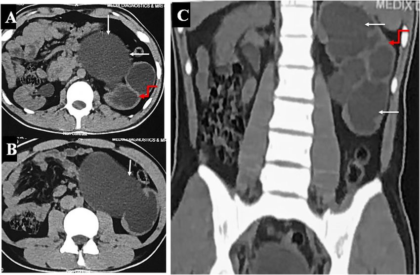
Majumder et al. Journal of the Egyptian National Cancer Institute     (2021) 33:1                                                     Page 2 of 5

hydronephrosis of the left kidney (white arrows) with severe              CD10 (Fig. 2f) and HMB-45 (Fig. 2g), the stromal com-
parenchymal thinning with pelvic ureteric junction obstruc-               ponent was positive for smooth muscle actin (SMA)
tion (PUJO) (Fig. 1). A left radical nephrectomy was done                 (Fig. 2h) and was immunonegative for CD34, estrogen
with hilar lymph node dissection with a clinical diagnosis of             receptor (ER); thereby confirming the smooth muscle
non-functioning kidney secondary to left PUJO.                            differentiation. Therefore, based on the histomorpholo-
   Gross examination showed a specimen of the kidney                      gical and immunohistochemical staining pattern diagno-
measuring 14 × 8 × 6 cm. The external surface was bos-                    sis of renal angiomyoadenomatous tumor was given.
selated. The capsule was easily stripped off. No scars
were noted externally. Serial slicing revealed multiple                   Discussion
interconnecting cysts with few thickened areas replacing                  RAT is a rare and distinct neoplasm. The average age
the entire parenchyma of the kidney. The cysts ranged                     group in reported cases was 46 years. No sex predilec-
in size from 1.5 to 4 cm. Cortico-medullary differenti-                   tion has been noted in the tumor. Rare studies have
ation could not be made out (Fig. 2a). A small part of                    shown a tumor with cystic changes. Common differen-
the ureter was seen measuring 1 cm. Hilar lymph nodes                     tials for mixed renal carcinomas to be kept in mind are
measuring 2.5 × 1 was received separately.                                mixed epithelial and stromal tumor of the kidney
   Microscopic examinations from multiple sections                        (MESTK), angiomyolipomas, clear cell renal cell carcin-
showed a tumor composed of epithelial, smooth muscle                      omas with angioleiomyomatous stroma, and clear cell
and vascular components. The epithelial component was                     papillary renal cell carcinomas (ccpRCC) [3–5].
arranged in the form of tubules. The tumor cells had a                       According to Michal et al. [2], RAT epithelial compo-
moderate amount of clear to eosinophilic cytoplasm with                   nent consists of adenomatous structures composed of
a basally placed round to oval vesicular nucleus and ap-                  cells that are secretory having basophilic nuclei alienated
ical snouting. Intervening stromal areas showed smooth                    along the basal membrane and prominent apical snouts,
muscle differentiation arranged in fascicles and bundles                  resulting in a characteristic appearance of “Shark’s
(Fig. 2c, d). For the vascular component, both thick and                  smile.” This epithelial component is usually shown
thin blood vessels were seen which were lined by plump                    immunopositivity for all cytokeratins like CK-7 more
endothelial cells. The resected end of the ureter and the                 than CK-20, CAM 5.2, and cytokeratins AE1-AE3. In
hilar structures were free of the tumor. The specimen la-                 addition to cytokeratins positivity, epithelial membrane
beled as hilar lymph node showed features of reactive                     antigen (EMA) and vimentin are also positive. While
lymphoid hyperplasia. Immunohistochemistry (IHC) was                      IHC for CD10, Melan-A, and HMB-45 is immunonega-
done for the tumor. The epithelial component was posi-                    tive. The present case showed immunopositivity for pan-
tive for pan-cytokeratins (Fig. 2e) and was negative for                  cytokeratin, while IHC foe CD10 and HMB-45 were

 Fig. 1 Axial (a, b) and coronal (c) non-contrast-enhanced CT (NCCT) scan images showing gross hydronephrosis of the left kidney (white arrows)
 with severe parenchymal thinning (curved red arrows)
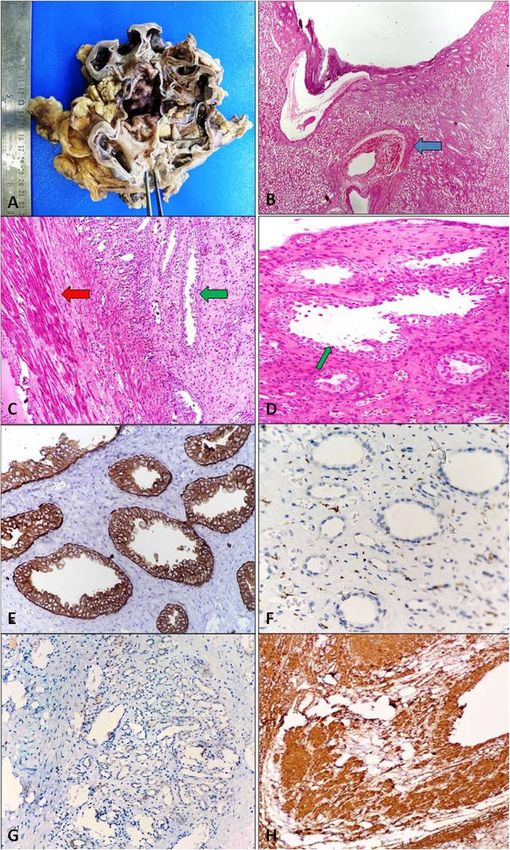
Majumder et al. Journal of the Egyptian National Cancer Institute        (2021) 33:1                                                        Page 3 of 5

 Fig. 2 Histopathological examination of renal angiomyoadenomatous tumor (RAT): gross image demonstrating multiple interconnecting cystic
 spaces with intervening thickened areas with patchy hemorrhagic areas (a). Hematoxylin and eosin (H&E) stained lower magnification image
 showing cystic spaces (epithelial component) along with thick-walled blood vessels (blue arrow) in renal parenchyma (b, h&e, × 40). Epithelial
 component arranged in adenomatous/glandular/tubular pattern with basally located bland small beaded nuclei and intimately surrounded by
 thin vascular channels. The cytoplasm of the cells is optically clear with blisters like apical snouts giving the appearance of “Shark’s Smile/moth-
 eaten” (green arrow). Stromal smooth muscle component (red arrow) (H&E; × 100, c; and × 400, d). The epithelial component is immunopositive
 for pan-cytokeratin (e, × 400), immunonegative for CD10 (f, × 400), and immunonegative for HMB-45 (g, × 40). The stromal smooth muscle was
 immunopositive for smooth muscle actin (SMA) (h, × 100)

negative. Focal solid and clear cell areas may be seen. It                   resistant, and mucicarmine negative [2–4]. Our case
may resemble conventional clear cell carcinoma Fuhr-                         shows similar morphology.
man grade 1. These secretory cells usually contain glyco-                      RAT shows a unique relation between the capillary net-
gen which is periodic acid-Schiff (PAS) positive, diastase                   work and the epithelial component. The capillaries tightly
Majumder et al. Journal of the Egyptian National Cancer Institute   (2021) 33:1                                                         Page 4 of 5

surround the basal membrane of the adenomatous struc-                  of ccpRCC will be evident with a minor component of
tures. The identification of these endothelial cells of the            the RAT-like area [10]. Immunohistochemical feature of
capillary network is possible mostly by the immunohisto-               RAT may overlap with ccpRCC, but morphologically
chemistry for CD34. This intimate capillary network is not             ccpRCC will be having protuberant papillary architecture
seen elsewhere [2, 6]. The stromal muscular component is               with thick cellular core and the large, generous clear
made up of strands that grow among the epithelial com-                 cells lining the papillary structures so that the cells of
ponent, resembling abortive vessels without the elastic                one papilla may touch the cells of the adjacent papilla.
layer. Occasionally myxoid, hyaline, or metaplastic change             In RAT, papillary structures are absent and the clear cell
(ossification) is seen. This leiomyomyomatous stroma can               component is less prominent [6, 7, 10].
also be seen in conventional clear cell renal cell carcin-               RAT usually a solid tumor with some microcystic
omas [2].                                                              areas. The presence of macro-cystic areas in RAT is a
  Leiomyomatous components in the tumor stroma of                      very rare occurrence. Michal et al. [2] studied five cases
RAT are usually immunopositive for SMA, vimentin,                      of RAT in his initial study out of which only one showed
and h-caldesmon while negative for HMB-45 and                          marked cystic areas. The present case tumor replaced
Melan-A. In the index case, the leiomyomatous compo-                   the whole kidney and showed marked cystic changes
nent in the tumor stroma was positive for SMA. Accord-                 which is an uncommon finding in RAT and not reported
ing to literature, few studies have reported renal cell                before. However, some studies like Deml et al. [11] have
tumors with glandular elements and leiomyomatosis                      postulated that RAT and clear cell papillary renal cell
stroma as a metachronous renal cell carcinoma with “an                 carcinoma (ccpRCC) are two entities of the same
abnormally large quantity of smooth muscle, not related                spectrum of disease and it is difficult to distinguish on
to the pelvis or calyces, nor to blood vessels” or de-                 the grounds of morphology, immunohistochemistry
scribes them as “hamartomas” or “fibroleiomuscular”                    markers, and molecular changes. They have described
component. Kuhn et al. reported five cases of renal cell               that RAT is a tumor with “varying amounts of tubular,
carcinomas with angioleiomyomas-like components and                    papillary, and cystic architecture.” Other differentials in-
a desmoplastic reaction in the stroma, unlike what we                  clude Xp11 and transcription factor E3 (TFE3) trans-
see in RAT [1–3, 5].                                                   location cancer [8, 9, 11].
  MESTK is usually seen in middle-aged, peri-                            Precise diagnosis is crucial since this neoplasm has an
menopausal women and is related to estrogen. It was                    excellent prognosis. Fluorescence in situ hybridization
earlier grouped under the broader term of “Cystic                      studies in four cases by Kuroda et al. have revealed that
nephroma.” The stroma in MESTK is identical to ovar-                   monosomy of chromosomes 1, 11, and 16 can be consid-
ian stroma with few leiomyomatosis areas. There can be                 ered to be diagnostic in RAT. The preferred treatment is
various Müllerian epithelial type differentiations, e.g.,              surgical resection and there have no reported cases of
tubal, endometrial, squamous. Intestinal mucinous glan-                recurrence or death due to the neoplasm [10].
dular epithelium and Paneth cells may be seen. These
features are not seen in RATs [6, 7]. Angiomyolipomas
                                                                       Conclusion
usually occur in association with tuberous sclerosis.
                                                                       To summarize, RAT is a rare renal neoplasm with varia-
They contain adipose tissue with thick blood vessels de-
                                                                       tions in the presentation, gross, and microscopic find-
void of the elastic layer, which can sometimes be seen in
                                                                       ings, and having a benign course and good prognosis.
RATs. However, they have a typical arrangement of
                                                                       However, owing to its distinct morphological, immuno-
myoid stromal cells which are perpendicular to vascular
                                                                       histochemical, and genetic profile, a correct diagnosis
lumens. Also, angiomyolipomas, tumors stain positive
                                                                       needs to be made.
for melanocytic markers like HMB45 and Melan-A,
which is not seen in RATs. None of the melanocytic                     Abbreviations
markers tested positive in the angioleiomyomatous                      RAT: Renal angiomyoadenomatous tumor; WHO: World Health Organization;
stroma of RATs [7–9].                                                  NCCT: Non-contrast computed tomography; PUJO: Pelvic ureteric junction
                                                                       obstruction; IHC: Immunohistochemistry; SMA: Smooth muscle actin;
  Conventional clear cell carcinomas may rarely show                   MESTK: Mixed epithelial and stromal tumor of the kidney; CcpRCC: Clear cell
leiomyomatosis stroma. However, the characteristic                     papillary renal cell carcinomas; PAS: Periodic acid-Schiff
Shark’s Smile is not seen. The VHL gene mutation and
CD10 marker positivity are seen consistently in clear cell             Acknowledgements
carcinomas and not found in RATs. Finally, we need to                  N/A
differentiate RATs from ccpRCC. Grossly both the
tumor may show either cystic or papillary architecture.                Authors’ contributions
                                                                       AM: manuscript writing and data collection. RP: manuscript revision and
Rare cases of clear cell ccpRCC with RAT-like areas                    editing. PK: radiological supervision. AA: pathology revision. AS: manuscript
have been reported in the literature. In these cases, areas            revision and supervision. All authors had read and approved the manuscript.
Majumder et al. Journal of the Egyptian National Cancer Institute (2021) 33:1 Page 5 of 5
Funding
No funds were received.
Availability of data and materials
The datasets used and/or analyzed during the current study are available
from the corresponding author on reasonable request.
Ethics approval and consent to participate
Informed written consent was taken from the patient. Internal review board
approval is not needed as it is a case report.
Consent for publication
Written informed consent was obtained from the patient included in the
study.
Competing interests
The authors declare that they have no conflicts of interest.
Author details
1
Department of Pathology, ABVIMS, PGIMER, RML Hospital, New Delhi, India.
2
Department of Pathology, All India Institute of Medical Sciences (AIIMS),
Room no. C-2, Level 3, Rishikesh, India. 3Department of Urology and Renal
Transplant, PGIMER, ABVIMS, RML Hospital, New Delhi, India. 4Department of
Radiology, Goa Medical College, Bambolim, Goa, India.
Received: 26 September 2020 Accepted: 15 December 2020
References
1. Michal M, Hes O, Havlicek F. Benign renal angiomyoadenomatous tumor: a
previously unreported renal tumor. Ann DiagnPathol. 2000;4:311–5.
2. Michal M, Hes O, Nemcova J, Sima R, Kuroda N, Bulimbasic S, et al. Renal
angiomyoadenomatous tumor: morphologic, immunohistochemical, and
molecular genetic study of a distinct entity. Virchows Arch. 2009;454:89–99.
3. Verine J. Renal angiomyoadenomatous tumor: morphologic,
immunohistochemical, and molecular genetic study of a distinct entity.
Virchows Arch. 2009;454:479–80.
4. Singh C, Kendi AT, Manivel JC, et al. Renal angiomyoadenomatous tumor.
Ann DiagnPathol. 2012;16:470–6.
5. Kuhn E, De Anda J, Manoni S, Netto G, Rosai J. Renal cell carcinoma
associated with prominent angioleiomyoma-like proliferation. Report of 5
cases and review of the literature. Am JSurgPathol. 2006;30:1372–81.
6. Petersson F, Grossmann P, Hora M, Superga M, Perez D, Martinek P. Renal
cell carcinoma with areas mimicking renal angiomyoadenomatous tumor/
clear cell papillary renal cell carcinoma. Hum Pathol. 2013;44:1412–20.
7. Mohanty SK, Parwani AV. Mixed epithelial and stromal tumors of the kidney:
an overview. Arch Pathol Lab Med. 2009;133:1483–6.
8. Tan G, Liu L, Qiu M, Chen L, Cao J, Liu J. Clinicopathologic features of renal
epithelioid angiomyolipoma: report of one case and review of the literature.
Int JClinExpPathol. 2015;8:1077–80.
9. Williamson SR, Eble JN, Cheng L, et al. Clear cell papillary renal cell
carcinoma: differential diagnosis and extended immunohistochemical
profile. Mod Pathol. 2013;26:697–708.
10. Kuroda N, Michal M, Hes O, et al. Renal angiomyoadenomatous tumor:
fluorescence in situ hybridization. PatholInt. 2009;59:689–91.
11. Deml KF, Schildhaus HU, Comperat E, et al. Clear cell papillary renal cell
carcinoma and renal angiomyoadenomatous tumor: two variants of a
morphologic, immunohistochemical, and genetic distinct entity of renal cell
carcinoma. Am J Surg Pathol. 2015;39:889–901.
Publisher’s Note
Springer Nature remains neutral with regard to jurisdictional claims in
published maps and institutional affiliations.You can also read