Review Article Update on the epidemiology, genetics, and therapeutic options of hyperuricemia
←
→
Page content transcription
If your browser does not render page correctly, please read the page content below
Am J Transl Res 2020;12(7):3167-3181
www.ajtr.org /ISSN:1943-8141/AJTR0109399

Review Article
Update on the epidemiology, genetics,
and therapeutic options of hyperuricemia
Lijun Li1, Yipeng Zhang2, Changchun Zeng3

Departments of 1Quality Control, 2Clinical Laboratory, 3Medical Laboratory, Shenzhen Longhua District Central
Hospital, Guangdong Medical University, Shenzhen 518110, Guangdong, P. R. China
Received February 17, 2020; Accepted June 17, 2020; Epub July 15, 2020; Published July 30, 2020

Abstract: Hyperuricemia may occur when there is an excess of uric acid in the blood. Hyperuricemia may result
from increased production or decreased excretion of uric acid. Elevated uric acid levels are a risk factor for gout,
and various risk factors, including some medications, alcohol consumption, kidney disease, high blood pressure,
hypothyroidism, and pesticide exposure, as well as obesity, are associated with an elevated risk of hyperuricemia.
Although the mechanisms underlying the pathogenesis of hyperuricemia are complex, previously reported studies
have revealed that hyperuricemia is involved in a variety of biological processes and signaling pathways. In this
review, we summarize common comorbidities related to hyperuricemia and describe an update of epidemiology,
pathogenesis, and therapeutic options of hyperuricemia. This systematic review highlights the epidemiology and
risk factors of hyperuricemia. Moreover, we discuss genetic studies on hyperuricemia to uncover current status
and advances in the pathogenesis of hyperuricemia. Additionally, we conclude with a reflection on the underlying
mechanisms of hyperuricemia and present the alternative drug strategies for the treatment of hyperuricemia to of-
fer more effective clinical interventions.

Keywords: Hyperuricemia, epidemiology, genetics, mechanisms, treatment

Introduction                                                  development of obesity, chronic kidney disease,
                                                              diabetes mellitus, and hypertension [11]. Hy-
Uric acid, a nitrogenous component of urine, is               peruricemia may occur when there are high lev-
a poorly soluble final product of protein and                 els of uric acid in the blood due to excess pro-
purine metabolism in humans. The level of                     duction, or more commonly, inefficient excre-
serum uric acid maintained the balance be-                    tion of uric acid. Defects in the key enzymes of
tween its production and excretion [1-3]. Uric                purine metabolism lead to impaired purine utili-
acid is a by-product of amino acid metabolism,                zation or enhanced purine oxidase activity. The
and the breakdown of amino acids produces                     increased reabsorption or decreased secretion
uric acid in the liver. Moreover, the breakdown               of uric acid in proximal renal tubules results in a
of purines releases uric acid in small quanti-                decrease in the rate of uric acid excretion. The
ties. Uric acid in human urine and the blood                  regulation of the enzymes is responsible for uric
may form sharp crystals and bring about an                    acid breakdown and production. Multiple trans-
increased risk of gout [3-6]. Hyperuricemia                   port proteins related to uric acid transport are
mainly caused by metabolic disorders of purine                crucial in the treatment of hyperuricemia [12].
and closely associated with the increases in                  An absolute lack of comprehension of the me-
the risk of cardiovascular disease, kidney dis-               chanism is a limiting factor in the management
ease, diabetes, obesity can be congenital or                  of hyperuricemia. However, the cellular and mo-
acquired [7-9]. Hyperuricemia is the main fac-                lecular processes and its implications are still
tor that leads to long-term systemic inflamma-                not completely elucidated. New insights to-
tion in patients with gout [10]. Inflammatory                 wards understanding the mechanisms may pro-
reactions in patients with asymptomatic hyper-                vide more precise treatment options for hy-
uricemia induced by urate can contribute to the               peruricemia.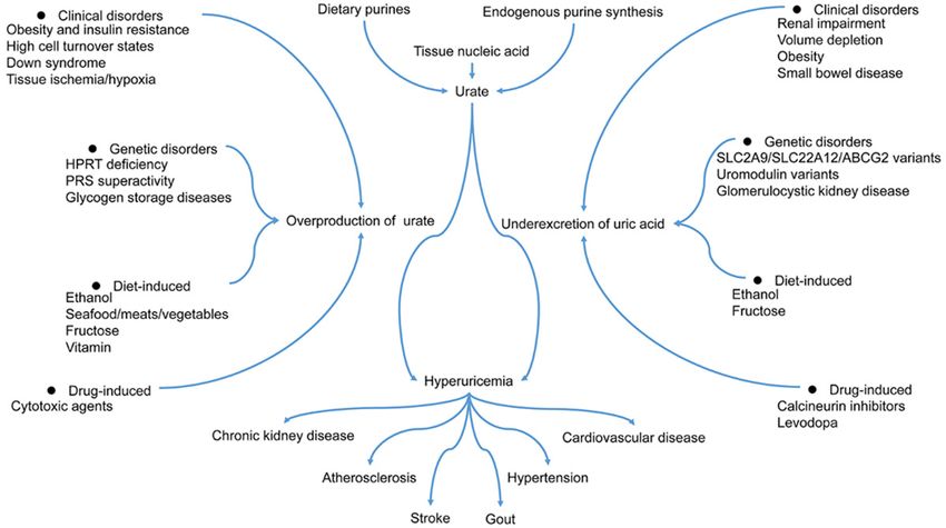
Update on hyperuricemia

Epidemiology                                        aged population in Tibet Autonomous Region is
                                                    1.83%, relatively lower than other places in
Worldwide prevalence of hyperuricemia               China. The prevalence in men is 2.86%, where-
                                                    as it is only 0.75% in women [22]. Additionally,
In recent years, the disease burden of hyperuri-    the age-standardized prevalence rate of hyper-
cemia is increasing, especially in high-income      uricemia in Henan rural population is 12.60%
countries and economically developing world         from July 2015 to September 2017, consistent
with a Western lifestyle [13, 14]. The preva-       with the primary meta-analysis (11.7%). The
lence and incidence of hyperuricemia sub-           prevalence and risk of hyperuricemia increa-
stantially differ across geographical areas [15].   se significantly with age. Interestingly, the de-
According to the data from the National Health      creased prevalence of hyperuricemia is ob-
and Nutrition Examination Survey (NHANES)           served in older men, while the contradictory
2007-2016, a nationally representative survey       trend is found in women [23].
showed that the prevalence rates of hyperuri-
cemia were 20.2% among men and 20.0%                Risk factors
among women between 2015 to 2016 in the
United States and the incidence of hyperurice-      The levels of urate hinge on the dynamic bal-
mia remained stable in 2007-2016 [16].              ance between purine-rich foods intake, synthe-
                                                    sis of urate within the body, the excretion of
The prevalence of hyperuricemia increases in        urate via urine or the gastrointestinal tract
both men (19.7% to 25.0%) and women (20.5%          (Figure 1) [24]. Prospective epidemiologic stud-
to 24.1%) from 2006 to 2014 in Ireland [17]. In     ies have pointed to obesity, hypertension, met-
the United States, an epidemiological survey        abolic syndrome, diuretic use, dietary factors,
has shown that the prevalence of hyperurice-        and chronic kidney disease as risk factors for
mia substantially increased from 19.1% (1988-       gout and hyperuricemia [9, 25-29]. Recently, it
1994 years) to 21.5% (2007-2008 years). The         turns out that iron overload can enhance serum
National Health and Nutrition Examination           uric acid levels, indicating a causal connection
Survey (NHANES) 2007-2008 found a similar           between hyperferritinaemia and hyperuricemia
prevalence of hyperuricemia between women           [30]. It is well known that chronic noncommuni-
(21.6%) and men (21.2%) [15]. Most epidemio-
                                                    cable diseases, such as cardiovascular and
logical studies show that the prevalence of
                                                    rheumatic diseases, are associated with the
hyperuricemia is generally higher in high-in-
                                                    development of hyperuricemia [31-33]. An on-
come countries than economically developing
                                                    set sequence study revealed that hyperurice-
world [15, 17-20]. The reduction of hormone
                                                    mia is an earlier-onset metabolic disorder than
estrogen production in postmenopausal wo-
                                                    hypertriglyceridemia, diabetes mellitus, and
men can decrease the removal of urate from
                                                    hypertension [34]. The study of epidemiological
the body result in an increase in urate levels
                                                    aspects of hyperuricemia in Poland shows that
and an elevated risk of developing hyperurice-
                                                    doctors often underestimate the problem of
mia [21].
                                                    hyperuricemia in patients with a high risk of
Prevalence of hyperuricemia in China                cardiovascular disease [32]. Due to increased
                                                    comorbidity, moderate hyperuricemia has as-
China, as the largest developing country, is        sociations with increased cardiovascular mor-
characterized by marked regional disparities        tality [35]. A 5-year Japanese cohort study indi-
and diverse populations [14, 18]. A national        cated that hyperuricemia is an independent
cross-sectional survey has shown that the prev-     risk factor for developing hypertension, espe-
alence of hyperuricemia was 8.4% (9.9% in           cially in children and adolescents [36]. Uric ac-
men and 7.0% in women) among Chinese adults         id associated with pro-inflammatory immune
from 2009 to 2010 [14]. A systematic review         effects can not only contribute to microvascu-
and meta-analysis systematically have shown         lar injury within the intracellular environment
that the pooled prevalence of hyperuricemia         but also conduce to increased blood pressure
was 13.3% in Mainland China from 2000 to            [37, 38]. Hyperuricemia or gout has relation-
2014 [18]. According to a large-scale popula-       ships with a substantial comorbidity burden in
tion-based survey, the overall crude prevalence     patients with rheumatoid arthritis [35]. Previous
of hyperuricemia of the elderly and middle-         scientific investigations make general refer-

3168                                                       Am J Transl Res 2020;12(7):3167-3181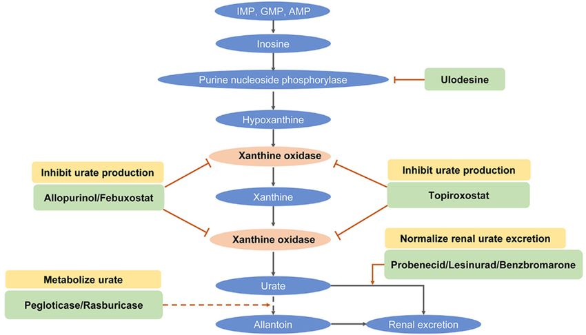
Update on hyperuricemia

Figure 1. Mechanisms of hyperuricemia. HPRT, Hypoxanthine-guanine phosphoribosyltransferase; PRS, Phosphori-
bosylpyrophosphate synthetase.

ence to the relationship between excess serum             sumption of high-protein animal foods can help
uric acid and gout [39, 40]. Gout, a common               lower the levels of plasma uric acid [46]. The
rheumatic disease, characterized by the depo-             association between hyperuricemia and the
sition of sodium monourate crystals in the peri-          intake of vitamin B12, vitamin B6, and folate
articular joints, can mainly result from hyperuri-        among American adults was assessed accord-
cemia [41]. Notably, some medicines used to               ing to the data from the National Health and
treat chronic diseases have the side effects of           Nutrition Examination Survey (NHANES) 2001-
elevated uric acid levels in the blood [24, 39,           2014. The result revealed that the intakes of
42].                                                      vitamin B12 and folate, but not vitamin B6,
                                                          were associated with a reduced risk of hyper-
Some high-purine foods, such as beer, seafood,            uricemia in males. The only intakes of food
sugar-sweetened beverages, dried beans, and               folate, folate, and total folate had associations
meat, can help raise serum uric acid levels.              with a lower risk of hyperuricemia in females
When purines break down to produce uric acid,             [47].
it can cause hyperuricemia in some individuals
[43-45]. However, there is limited information            According to a nationally representative survey,
on the exact content of purines contained in              body mass index, alcohol use, Dietary App-
foods mainly because several factors, such as             roaches to Stop Hypertension (DASH) diet, and
food processing procedures, can affect its con-           diuretic use are modifiable risk factors, and
tent. Some research suggests that purine-rich             they can be applied to illustrate the prevalence
foods intake is closely associated with the prev-         of hyperuricemia [48, 49]. Both endogenous
alence of hyperuricemia. There was a direct               and exogenous purine metabolism generates
and significant correlation between seafood               uric acid [42]. Previous studies capture much
intake and the prevalence of hyperuricemia,               attention to fructose consumption because,
and an antagonistic relationship between soy              during fructose metabolism, adenosine triphos-
food consumption and hyperuricemia among                  phate (ATP) hydrolysis can generate adenosine
middle-aged Chinese men. Moreover, protein                diphosphate (ADP) and adenosine monophos-
intake from animal or plant sources has the               phate (AMP), and the latter may stimulate the
opposite contribution to the prevalence of                increase of serum uric acid concentrations and
hyperuricemia. Additionally, reducing the con-            lead to hyperuricemia. Furthermore, fructose

3169                                                              Am J Transl Res 2020;12(7):3167-3181
Update on hyperuricemia
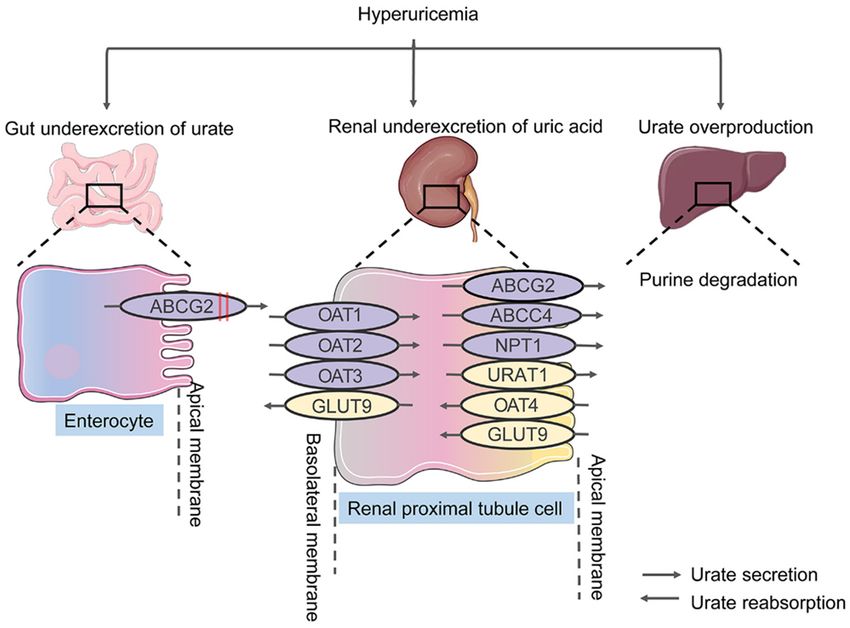
Figure 2. Urate transporters in humans. Overproduction and underexcretion of urate result in hyperuricemia. Under-
excretion of urate occurs mainly in the proximal renal tubules. Urate overproduction in the liver and underexcretion
of urate in the gut may lead to the occurrence of hyperuricemia. In the gut, Genetic variants in ABCG2 inhibit the ex-
cretion of urate and promote underexcretion. Numerous transmembrane transporters, such as ABCG2, URAT1, and
GLUT9, have crucial roles in urate reuptake and secretion. OAT, organic anion transporter; NPT1, sodium-dependent
phosphate cotransporter type 1; ABCG2, ABC transporter G family member 2; URAT1, urate anion exchanger 1;
GLUT9, glucose transporter type 9; ABCC4, ATP-binding cassette sub-family C member 4.
intake can contribute to the biosynthesis of uric [53]. As shown in Figure 2, most uric acid dis-
acid from amino acid precursors [50, 51]. Uric posal naturally occurs in the kidney. Insufficient
acid levels increase with the increase of body excretion of urate in the kidney accounts for
mass index (BMI), and obesity has an associa- about 90% of individuals with hyperuricemia.
tion with hyperuricemia. Additionally, hyperuri- Underexcretion associated with reduced glo-
cemia and obesity may ratchet up the likeli- merular filtration, impaired tubular secretion,
hood of type 2 diabetes [52]. and improved tubular reabsorption contribu-
tes to hyperuricemia. In the proximal tubular,
From the above, we can conclude that hyperuri- URAT1 (uric acid transporter 1) manage uric
cemia is closely related to multiple risk factors.
acid reabsorption. Besides, multiple medica-
However, its pathophysiology has not yet been
tions, extracellular fluid volume depletion, and
thoroughly investigated. We should pour
organic acids facilitating transport lead to
enough attention to the adverse effects caused
hyperuricemia. Furthermore, the kidney is held
by hyperuricemia.
accountable for 60-65% of urate elimination
Genetics associated with various transporters in the
intestinal mucosa, salivary glands, and the
Hyperuricemia, considered to result in a combi- proximal renal tubule. The filtered urate in the
nation of environmental and genetic factors, is kidney via the glomerulus is mainly reabsorbed
characterized by an excess of serum uric acid and secreted in the proximal tubule. Ultimately,
3170 Am J Transl Res 2020;12(7):3167-3181Update on hyperuricemia
only 3-10% of the filtered urate is eliminated in studies, offering novel insights into the genetic
the urine [54]. effects on hyperuricemia. However, current
research cannot fully account for the occur-
Uric acid, the end oxidation product of purine rence and progression of it. It is, therefore, cru-
breakdown, is primarily excreted in the urine. It cial to conduct more extensive and intensive
is clear that uric acid, a weak organic acid, cir- research on genetic studies.
culates in the ionized shape of urate under nor-
mal physiologic conditions. The metabolism of Genome-wide association studies provide new
purine chiefly happens in the liver, but it can perspectives on the genetic basis of hyperuri-
occur in the tissues with a wide distribution of cemia, which is dominated by loci containing
xanthine oxidase [3]. The excretion of uric acid urate transporters associated with urate excre-
from the kidney accounts for about two-thirds, tion. Genome-wide association studies in hy-
and the remaining proportion of uric acid is peruricemia identify more genetic variants and
mainly excreted into the intestine. Most uric delineate the pathogenesis of hyperuricemia,
acid is filtered in a free manner, with roughly providing novel opportunities for underlying
90% of the filtrate reabsorbed [55]. Enhanced clinical translation [60]. A meta-analysis show-
cellular breakdown, endogenous purine pro- ed gout and asymptomatic hyperuricemia loci
duction, and high-purine diets promote the pro- and reveal gout development by identifying the
duction of urate, responsible for a small portion gout risk loci associated with crystal-induced
of hyperuricemia. High Purine Foods chiefly inflammation [61, 62].
include some meats, some fish, seafood and
shellfish, and alcoholic beverages, strengthen- Previous research has revealed that various
ing the levels of uric acid by lowering renal transporter genes have associations with ser-
excretion. The higher phosphoribosylpyrophos- um uric acid levels, such as urate transporter 1
phate (PRPP) synthetase activity, as well as (URAT1), glucose transporter 9 (GLUT9), organ-
hypoxanthine phosphoribosyltransferase (HP- ic anion transporter 4 (OAT4), ATP-binding cas-
RT) deficiency, can not only improve endoge- sette transporter, subfamily G, member 2
nous purine production but also lead to uric (BCRP), and sodium-dependent phosphate co-
acid overproduction and accumulation. Cell transporter type 1 (NPT1). Among them, GLUT9
turnover or breakdown, such as tumor lysis, (SLC2A9) and BCRP (ABCG2) appear to have
rhabdomyolysis, and hemolysis, can improve the most significant impact on urate levels [63].
urate production. Ultimately, both environmen-
tal and physiological changes can influence the GLUT9
production of urate [56].
GLUT9 (SLC2A9), encoding a member of the
Patients who suffered from gout or hyperurice- SLC2A facilitative glucose transporter family to
mia have elevated urate reabsorption in the maintain glucose homeostasis, has an essen-
proximal tubule. According to renal urate excre- tial role in urate transporter and reabsorption.
tion, hyperuricemia is usually divided into urate The protein encoded by GLUT9 helps excrete
overproduction type and underexcretion type. urate into the urine or reabsorb urate into the
The urate reabsorption levels are driven by bloodstream. GLUT9 variants can reduce the
genetic variation in urate transporters or up- excretion of urate into the urine and enhance
stream regulators. Previous studies have con- the reabsorption of uric acid into the blood-
firmed that various genes are associated with stream, leading to hyperuricemia [64]. The
hyperuricemia [57-59]. The great mass of the rs7442295 single nucleotide polymorphism in
genes is involved in transporting urate, which is the SLC2A9 gene robustly associated with
a byproduct of natural biochemical processes. hyperuricemia, increased plasma uric acid lev-
Various hyperuricemia-associated genes regu- els, gout, and urate excretion has been pro-
late excretion or reabsorption of uric acid posed as a proxy measurement for uric acid. It
according to the body’s needs. Some hyperuri- has been used to explore its causal associa-
cemia-associated genes have relationships tions with ischaemic heart disease and blood
with the transport or breakdown of small mole- pressure [51, 65]. Loss-of-function mutations
cules [57, 59, 60]. Researchers in mounting in SLC2A9, interacting with the pore of the
numbers tend to pay much attention to genetic Glut9 urate transporter and the urate binding
3171 Am J Transl Res 2020;12(7):3167-3181Update on hyperuricemia
pocket, decreased the expression of Glut9 and observed [76]. The increased expression of
reduced the activity of Glut9 transport [66]. ALPK1 reduced the expression of URAT1. The
rs11726117 polymorphism in ALPK1 sup-
BCRP pressed urate reuptake and reduced the risk of
gout via SLC22A12 [77].
BCRP (ABCG2), a member of the superfamily of
ATP-binding cassette (ABC) transporters, is OAT4
involved in the transport of several molecules
across extra- and intra-cellular membranes. OAT4 (SLC22A11), encoding an integral mem-
BCRP, an ATP-driven efflux pump in the apical brane protein, is associated with the sodium-
membrane of the proximal tubule epithelial independent transport and excretion of organic
cells, excretes endogenous and exogenous anions [51]. NPT1 (SLC17A1), a urate exporter,
substrates. BCRP dysfunction reduces the associated with the transport of phosphate into
excretion of extra-renal urate, which is a signifi- cells via Na (+) cotransport has crucial roles in
cant contributor to hyperuricemia. Besides, the the resorption of phosphate by the proximal
protein encoded by BCRP helps excrete urate tubule of the kidney [78].
into the gut, and it, therefore, can be removed
from the body. Various functional variants of OAT10
BCRP were identified and enhanced the risk of
hyperuricemia and gout [67]. BCRP variants Organic anion transporter 10 (OAT10), also
can reduce the excretion of urate into the gut, known as SLC22A13, encodes a urate reab-
leading to hyperuricemia. Moreover, BCRP con- sorption transporter protein on the apical side
tributes to the clearance of urate in the gut [68, of the renal proximal tubular cells. OAT10 acts
69]. The BCRP 141K (rs2231142) variant con- as a critical part of urate transport from urine to
tributes to hyperuricemia and has a role in the the blood and the dysfunctional variants of
development of hyperuricemia to gout in OAT10 reduce serum uric acid levels [79].
Polynesian [70, 71]. Mutations in ABCG2 medi-
ating the intestinal excretion of uric acid lead to LDHD
hyperuricemia. In hereditary hemochromatosis
patients, iron/heme overload enhances the Lactate Dehydrogenase D (LDHD) as a member
activity of xanthine oxidase and accelerates the of the D-isomer specific 2-hydroxyacid dehydro-
degradation of p53, causing the reduction of genase family is involved in autosomal reces-
ABCG2 expression. In consequence, intestinal sive gout with hyperuricemia and decreased
excretion of uric acid through ABCG2 is reduced excretion of uric acid. The mutations of LDHD
and the production of uric acid is enhanced, result in the excessive production of blood
leading to the accumulation of uric acid in tis- D-lactate in exchange for uric acid reabsorp-
sue and serum and promoting the progression tion, eventually contributing to gout and hyper-
of hereditary hemochromatosis-associated uricemia [80].
arthritis [72].
UMOD
URAT1
Uromodulin encoded by UMOD is characterized
URAT1 (SLC22A12) as urate transporter gene as one of the most abundant proteins secreted
to adjust and control blood urate levels is a in mammalian urine. Lower concentrations of
disease-causing gene for renal hypouricemia urinary uromodulin associated with distal tubu-
type 1 [73, 74]. Glucocorticoids are crucial to lar cell damage are observed in individuals with
maintaining uric acid homeostasis through the UMOD-related diseases, such as familial juve-
glucocorticoid receptor signaling pathway. nile hyperuricemic nephropathy (FJHN), renal
Moreover, Glucocorticoids promote renal urate disorders medullary cystic kidney disease-2
excretion via downregulating URAT1 in mouse (MCKD2) and glomerulocystic kidney disease
kidney [75]. In a meta-analysis, the rs475688 with hyperuricemia and isosthenuria (GCKDHI).
polymorphism in SLC22A12 is associated with Besides, uromodulin excretion in urine pre-
gout susceptibility and a significant association vents urinary tract infections from uropatho-
between hyperuricemia susceptibility and the genic bacteria. UMOD mutations impair uro-
rs3825016 polymorphism in SLC22A12 was modulin protein folding and accumulate mis-
3172 Am J Transl Res 2020;12(7):3167-3181Update on hyperuricemia
folded proteins in the endoplasmic reticulum XDH
(ER) of renal tubular cells. The disruption of ER
function contributes to hyperuricemia and tu- Xanthine Dehydrogenase (XDH), a member of
bulointerstitial nephritis [81, 82]. the family of oxidoreductases, participates in
the oxidative metabolism of purines, catalyzing
HPRT1 the oxidation of hypoxanthine to xanthine and
the oxidation of xanthine to uric acid. Sulfhydryl
Hypoxanthine-guanine phosphoribosyltransfer- oxidation or proteolytic modification can con-
ase (HGPRT) encoded by hypoxanthine phos-
vert xanthine dehydrogenase to xanthine oxi-
phoribosyltransferase 1 (HPRT1) is a transfer-
dase. Xanthine dehydrogenase deficiency re-
ase, which catalyzes the conversion of hypo-
sults in xanthinuria, leading to adult respiratory
xanthine to inosine monophosphate and gua-
stress syndrome [87]. Downregulated XDH
nine to guanosine monophosphate. The cata-
expression can reduce the levels of xanthine
lytic reaction transfers the 5-phosphoribosyl
oxidoreductase, and it may be an underlying
group from 5-phosphoribosyl 1-pyrophosphate
(PRPP) to the purine. HPRT1 exerts a vital part treatment for hyperuricemia [88, 89].
in purine nucleotide biosynthesis via the purine
INS
salvage pathway. HGPRT deficiency caused by
HPRT1 mutation leads to elevated uric acid lev-
Insulin (INS) encoded by INS is considered as
els in the blood, which is associated with Kelley-
an essential anabolic hormone in the metabolic
Seegmiller syndrome, Lesch-Nyhan syndrome,
process, participating in the control of carbohy-
and hyperuricemia [83, 84].
drate and lipid metabolism. High levels of insu-
SARS2 lin in the blood hinder the production and
secretion of glucose. As an anabolic hormone,
The mitochondrial seryl-tRNA synthetase pre- it consolidates the conversion of small molecu-
cursor, a member of the class II tRNA synthe- lar substances in the blood into abundant
tase family, encoded by Seryl-TRNA Synthetase molecular substances inside the cells. Low
2, Mitochondrial (SARS2), which catalyzes the concentrations of insulin in the blood strength-
ligation of Serine to tRNA (Ser) and is involved en extensive catabolism. Insulin not only induc-
in selenocysteinyl-tRNA (sec) biosynthesis in es the change of cell permeability to fatty acids,
mitochondria. Mutations in SARS2 have been amino acids, and monosaccharides, but also
identified in Hyperuricemia, pulmonary hyper- facilitates the synthesis of glycogen, the pen-
tension, renal failure, and alkalosis syndrome tose phosphate cycle, and glycolysis in the liver.
(HUPRAS), which is a multi-system involvement Insulin resistance may be a potential metabolic
disease including pulmonary hypertension, disturbance explaining the relationship among
hyperuricemia, renal failure in infancy and alka- diverse elements of the metabolic syndrome
losis [85]. associated with glucose intolerance, hyperlipid-
G6PC emia, hypertension, and obesity. Besides,
hyperuricemia may be an underlying marker of
Glucose-6-Phosphatase Catalytic Subunit (G6- insulin resistance [90, 91].
PC), a multi-subunit integral membrane protein
of the endoplasmic reticulum, catalyzes the REN
hydrolysis of D-glucose 6-phosphate to D-glu-
cose and orthophosphate in the endoplasmic REN encoding renin causes autosomal domi-
reticulum and is one of the critical enzymes in nant tubulointerstitial kidney disease, REN-
blood glucose homeostasis, exerting a critical related (ADTKD-REN) characterized by low plas-
part in the regulation of gluconeogenesis and ma renin activity, bland urinary sediment, de-
glycogenolysis. Mutations in G6PC have been creased fractional excretion of urinary uric acid,
identified in Glycogen storage disease type I hyperuricemia, and hypoproliferative anemia.
(GSD1), a metabolic disorder, is marked by Hyperuricemia is depicted in 80% of patients
hypoglycemia, hyperlipidemia, hyperuricemia, with ADTKD-REN beginning in childhood due to
and lactic acidemia [86]. reduced renal excretion of uric acid [92, 93].
3173 Am J Transl Res 2020;12(7):3167-3181Update on hyperuricemia
Figure 3. Mechanism of urate-lowering therapy for hyperuricemia. Numerous agents can be applied to inhibit urate
production, promote renal urate excretion, and increase purine metabolism to allantoin.
GPATCH8 uricemia patients to decrease the levels of uric
acid [95]. The clinical symptoms of symptom-
G-Patch Domain Containing 8 (GPATCH8), a atic hyperuricemia can be gout, nephrolithiasis,
member of the G-patch domain family, is and uric acid renal disease [39].
encoded by GPATCH8. A potentially pathogenic
mutation in GPATCH8 is a conceivable candi- To effectively manage hyperuricemia, inhibiting
date for hyperuricemia [94]. uric acid synthesis and reabsorption, as well as
facilitating the excretion of uric acid, can be
Drugs for hyperuricemia alternative strategies. As shown in Figure 3,
urate-lowering medications can be roughly
According to the clinical scenarios, hyperurice- divided into three main categories: reducing
mia can be divided into asymptomatic or symp- the synthesis of uric acid (xanthine oxidase
tomatic hyperuricemia. Decreasing unneces- inhibitors), enhancing the excretion of uric acid
sary costs and avoiding underlying side effects (URAT1 inhibitors), and regulating the metabolic
take precedence over taking medication in hydrolysis of uric acid (uricase inhibitors).
careful consideration of the interests of the Xanthine oxidase inhibitors classified as purine
asymptomatic hyperuricemia patients. There analogs (including allopurinol) and non-purine
seems no need to carry medical therapy for a analog agents (including febuxostat and
multitude of asymptomatic hyperuricemia topiroxostat) can lower endogenous uric acid
patients. They accompany with an elevated production and further decrease the levels of
serum urate level but never experience signs or uric acid [96, 97].
symptoms of monosodium urate crystal depo-
sition disease, such as nephrolithiasis, gout, Allopurinol
and uric acid renal disease. Urate-lowering
drugs are likely to appeal to the asymptomatic Allopurinol, a purine-based competitive xan-
hyperuricemia patients subjected to cytolytic thine oxidase inhibitor, can be metabolized to
therapy for a malignant tumor to restrict tumor alloxanthine, an inhibitor of xanthine oxidase
lysis syndrome. It is sufficient to change in life- enzyme. Both allopurinol and alloxanthine can
styles, such as exercise, dietary changes, and restrain xanthine oxidase, converting hypoxan-
alcohol consumption, for asymptomatic hyper- thine to xanthine and xanthine to uric acid.
3174 Am J Transl Res 2020;12(7):3167-3181Update on hyperuricemia
Allopurinol promotes the secondary utilization amino acid residues in the solvent channel and
of hypoxanthine and xanthine for the synthesis bonds covalently to molybdenum (IV) ion, gen-
of nucleic acid and nucleotide by a metabolic erating a hydroxylated 2-pyridine metabolite to
reaction associated with hypoxanthine-guanine inhibit xanthine oxidase. Additionally, Topiro-
phosphoribosyltransferase (HGPRTase). This xostat can restrain ATP-binding cassette trans-
metabolic reaction accounts for an elevated porter G2 (ABCG2), which is involved in the res-
level of nucleotide, which leads to feedback toration of renal uric acid and uric acid secre-
suppression of de novo synthesis of purine. tion from the intestines [104].
Eventually, the reduced levels of urine and
serum uric acid are responsible for a reduction Probenecid
in the incidence of hyperuricemia [98, 99].
Based on the data from the Taiwan National Probenecid, a prototypical uricosuric agent,
Health Insurance Research Database, a retro- hinders the renal elimination of organic anions
spective nationwide population-based study and impairs tubular urate reabsorption. Addi-
showed that allopurinol could cause hypersen- tionally, it can decrease the renal elimination of
sitivity reactions in patients with asymptomatic other drugs, and therefore it is a promising uric
hyperuricemia accompanied by cardiovascular acid transporter 1 (URAT1) inhibitor for the
or renal diseases [100]. treatment of renal impairment. Probenecid
restrains the tubular urate reabsorption, facili-
Febuxostat tating urinary uric acid excretion, as well as
reducing serum urate concentrations. Besides,
Febuxostat, a non-purine xanthine oxidase Probenecid possibly decreases the binding of
inhibitor, can reduce the levels of serum uric urate by plasma proteins and reduces uric acid
acid, but not restrain different enzymes associ- secretion in the renal tubule [97, 105].
ated with the metabolism and synthesis of
pyrimidine and purine. The metabolism of Lesinurad
Febuxostat is dependent on uridine diphos-
phate glucuronosyltransferase (UGT) enzymes Lesinurad, a common URAT1 inhibitor, restrains
(including UGT1A1, UGT1A3, UGT1A9, and the levels of serum uric acid via the suppres-
UGT2B7), cytochrome P450 (CYP) enzymes sion of URAT1 and OAT4. URAT1, uric acid trans-
(CYP1A2, CYP2C8, and CYP2C9) and non-P450 porter, is associated with uric acid reabsorption
enzymes [99, 101]. Based on US Medicare from the renal tubule. Organic anion transport-
claims data (2008-2013), a cohort study on the er 4 (OAT4), uric acid transporter, is linked with
assessment of cardiovascular risk in 99744 the sodium-independent transport and excre-
older medicare patients with gout showed that tion of organic anions, involved in diuretic-
there was almost no difference in the risk of all- induced hyperuricemia [98]. The administra-
cause mortality, new-onset heart failure, myo- tion of Lesinurad in combination with allopuri-
cardial infarction, coronary revascularization, nol causes a considerable reduction in serum
or stroke between patients who used febuxo- concentrations of inflammatory cytokines, glu-
stat compared with allopurinol initiators. tathione peroxidase, catalase, urea nitrogen,
Nevertheless, the risk of heart failure exacer- and uric acid in a hyperuricemic mouse model,
bation was slightly lower in patients initiating ameliorating renal function of the hyperurice-
febuxostat compared with allopurinol [102]. A mic mice [106]. In vitro, lesinurad suppressing
randomized and controlled trial showed that the activity of OAT4 and URAT1 but not GLUT9,
treatment with febuxostat did not delay carotid OAT1, OAT3, or ABCG2 can reduce the levels of
atherosclerosis progression in Japanese pa- serum uric acid via restraint of urate transport-
tients with asymptomatic hyperuricemia and it ers in the human kidney [107].
did not support the use of febuxostat to delay
the progression of carotid atherosclerosis in Rasburicase
patients with asymptomatic hyperuricemia
[103]. Rasburicase, a recombinant uricase, catalyzes
the conversion of uric acid to allantoin, which is
Topiroxostat a metabolite existing in an inactive and soluble
form [108]. Rasburicase seems to have an
Topiroxostat, a non-purine xanthine oxidase advantage in the speedy correction of hyperuri-
inhibitor, makes interaction with numerous cemia compared with allopurinol. However, its
3175 Am J Transl Res 2020;12(7):3167-3181Update on hyperuricemia
clinical benefit in cancer patients with tumor Address correspondence to: Changchun Zeng,
lysis syndrome (TLS) is still confusing, especial- Department of Medical Laboratory, Shenzhen
ly for patients with concurrent renal failure and Longhua District Central Hospital, Guangdong
hyperuricemia [109]. Medical University, Shenzhen 518110, Guangdong,
P. R. China. Tel: +86-0755-28024426; E-mail:
Conclusions zengchch@glmc.edu.cn
Hyperuricemia seems to be rising steadily in References
prevalence over the last decades. High uric
[1] Borghi C and Piani F. Uric acid and estimate of
acid concentrations are involved in the elevat-
renal function. Let’s stick together. Int J Cardiol
ed risk of developing hyperuricemia. Hype- 2020; 310: 157-158.
ruricemia is a metabolic disease connected [2] Pan J, Shi M, Ma L and Fu P. Mechanistic in-
with Lesch-Nyhan syndrome and glycogen stor- sights of soluble uric acid-related kidney dis-
age disease-ia. Substantial evidence suggests ease. Curr Med Chem 2018; [Epub ahead of
that hyperuricemia is an underlying risk factor print].
for gout, and it can forecast the evolution of [3] Maiuolo J, Oppedisano F, Gratteri S, Muscoli C
chronic kidney disease, obesity, diabetes, and and Mollace V. Regulation of uric acid metabo-
hypertension. Although numerous studies have lism and excretion. Int J Cardiol 2016; 213:
demonstrated close correlations between hy- 8-14.
[4] Yu TY, Jin SM, Jee JH, Bae JC, Lee MK and Kim
peruricemia and multiple comorbidities such
JH. The protective effects of increasing serum
as acute and chronic kidney disease, diabetes, uric acid level on development of metabolic
metabolic syndrome, cardiovascular disease, syndrome. Diabetes Metab J 2019; 43: 504-
hypertension, and dyslipidemia, it is currently 520.
unclear that there is a causal relationship [5] Zhao T, Lv X, Cao L, Guo M, Zheng S, Xue Y, Zou
between hyperuricemia and multiple comorbid- H, Wan W and Zhu X. Renal excretion is a
ities. Genetic characteristics provide novel per- cause of decreased serum uric acid during
spectives on the physiology and pathophysiol- acute gout. Int J Rheum Dis 2018; 21: 1723-
ogy of hyperuricemia. More importantly, genetic 1727.
studies may provide more precision medicine [6] Andrade Sierra J and Flores Fonseca MM. Re-
nal handling of uric acid. Contrib Nephrol
for individuals. These are various approaches
2018; 192: 1-7.
that help manage hyperuricemia. Nevertheless,
[7] Lima WG, Martins-Santos ME and Chaves VE.
patient and health-care provider education is Uric acid as a modulator of glucose and lipid
the foundation of the successful treatment of metabolism. Biochimie 2015; 116: 17-23.
hyperuricemia. As a general rule, it is not indis- [8] Cai Z, Xu X, Wu X, Zhou C and Li D. Hyperurice-
pensable to treat most patients with asymp- mia and the metabolic syndrome in Hangzhou.
tomatic hyperuricemia in the absence of kidney Asia Pac J Clin Nutr 2009; 18: 81-87.
stones or gout. To effectively treat hyperurice- [9] Wang H, Zhang H, Sun L and Guo W. Roles of
mia, reducing the levels of uric acid is crucial, hyperuricemia in metabolic syndrome and car-
achieved by inhibiting uric acid synthesis and diac-kidney-vascular system diseases. Am J
reabsorption, as well as facilitating the excre- Transl Res 2018; 10: 2749-2763.
[10] Cabau G, Crisan TO, Kluck V, Popp RA and
tion of uric acid.
Joosten LAB. Urate-induced immune program-
ming: consequences for gouty arthritis and hy-
Acknowledgements peruricemia. Immunol Rev 2020; 294: 92-
105.
The study was supported by the National [11] Joosten LAB, Crisan TO, Bjornstad P and John-
Natural Science Foundation of China [No. son RJ. Asymptomatic hyperuricaemia: a silent
81660755], and the Science and Technology activator of the innate immune system. Nat
Project of Shenzhen of China [No. JCYJ2017- Rev Rheumatol 2020; 16: 75-86.
0307160524377]. [12] Mandal AK and Mount DB. The molecular
physiology of uric acid homeostasis. Annu Rev
Disclosure of conflict of interest Physiol 2015; 77: 323-345.
[13] Singh G, Lingala B and Mithal A. Gout and hy-
None. peruricaemia in the USA: prevalence and
3176 Am J Transl Res 2020;12(7):3167-3181Update on hyperuricemia
trends. Rheumatology (Oxford) 2019; 58: STROBE-compliant cross-sectional and longitu-
2177-2180. dinal study. Medicine (Baltimore) 2019; 98:
[14] Liu H, Zhang XM, Wang YL and Liu BC. Preva- e17597.
lence of hyperuricemia among Chinese adults: [27] Chaudhary NS, Bridges SL Jr, Saag KG, Rahn
a national cross-sectional survey using multi- EJ, Curtis JR, Gaffo A, Limdi NA, Levitan EB,
stage, stratified sampling. J Nephrol 2014; 27: Singh JA, Colantonio LD, Howard G, Cushman
653-658. M, Flaherty ML, Judd S, Irvin MR and Reynolds
[15] Roman YM. The Daniel K. Inouye college of RJ. Severity of hypertension mediates the as-
pharmacy scripts: perspectives on the epide- sociation of hyperuricemia with stroke in the
miology of gout and hyperuricemia. Hawaii J REGARDS case cohort study. Hypertension
Med Public Health 2019; 78: 71-76. 2020; 75: 246-256.
[16] Chen-Xu M, Yokose C, Rai SK, Pillinger MH and [28] Steiger S, Ma Q and Anders HJ. The case for
Choi HK. Contemporary prevalence of gout evidence-based medicine for the association
and hyperuricemia in the united states and between hyperuricaemia and CKD. Nat Rev
decadal trends: the national health and nutri- Nephrol 2020; 16: 422.
tion examination survey, 2007-2016. Arthritis [29] Sato Y, Feig DI, Stack AG, Kang DH, Lanaspa
Rheumatol 2019; 71: 991-999. MA, Ejaz AA, Sanchez-Lozada LG, Kuwabara M,
[17] Kumar AUA, Browne LD, Li X, Adeeb F, Perez- Borghi C and Johnson RJ. The case for uric ac-
Ruiz F, Fraser AD and Stack AG. Temporal id-lowering treatment in patients with hyperuri-
trends in hyperuricaemia in the Irish health caemia and CKD. Nat Rev Nephrol 2019; 15:
system from 2006-2014: a cohort study. PLoS 767-775.
One 2018; 13: e0198197. [30] Richette P and Latourte A. Hyperferritinaemia
[18] Liu R, Han C, Wu D, Xia X, Gu J, Guan H, Shan and hyperuricaemia-a causal connection? Nat
Z and Teng W. Prevalence of Hyperuricemia Rev Rheumatol 2018; 14: 628-629.
and Gout in Mainland China from 2000 to [31] Poletto J, Harima HA, Ferreira SR and Gimeno
2014: a systematic review and meta-analysis. SG. Hyperuricemia and associated factors: a
Biomed Res Int 2015; 2015: 762820. cross-sectional study of Japanese-Brazilians.
[19] Thompson MD. Insights in public health: hyper- Cad Saude Publica 2011; 27: 369-378.
uricemia and gout in Hawai’I. Hawaii J Med [32] Kostka-Jeziorny K, Widecka K and Tykarski A.
Public Health 2018; 77: 121-124. Study of epidemiological aspects of hyperuri-
[20] Zhu Y, Pandya BJ and Choi HK. Prevalence of cemia in Poland. Cardiol J 2019; 26: 241-252.
gout and hyperuricemia in the US general pop- [33] McHugh J. Hyperuricaemia or gout in patients
ulation: the national health and nutrition ex- with RA. Nat Rev Rheumatol 2019; 15: 384.
amination survey 2007-2008. Arthritis Rheum [34] Chiang KM, Tsay YC, Vincent Ng TC, Yang HC,
2011; 63: 3136-3141. Huang YT, Chen CH and Pan WH. Is hyperurice-
[21] Cho SK, Winkler CA, Lee SJ, Chang Y and Ryu mia, an early-onset metabolic disorder, caus-
S. The prevalence of hyperuricemia sharply in- ally associated with cardiovascular disease
creases from the late menopausal transition events in Han Chinese? J Clin Med 2019; 8:
stage in middle-aged women. J Clin Med 2019; 1202.
8: 296. [35] Chiou A, England BR, Sayles H, Thiele GM,
[22] Zhang Q, Gong H, Lin C, Liu Q, Baima Y, Wang Duryee MJ, Baker JF, Singh N, Cannon GW,
Y and Lin J. The prevalence of gout and hyper- Kerr GS, Reimold A, Gaffo A and Mikuls TR. Co-
uricemia in middle-aged and elderly people in existent hyperuricemia and gout in rheumatoid
Tibet autonomous region, China: a prelimin- arthritis: associations with comorbidities, dis-
ary study. Medicine (Baltimore) 2020; 99: ease activity and mortality. Arthritis Care Res
e18542. (Hoboken) 2019; 72: 950-958.
[23] Dong X, Zhang H, Wang F, Liu X, Yang K, Tu R, [36] Kuwabara M, Hisatome I, Niwa K, Hara S, Ron-
Wei M, Wang L, Mao Z, Zhang G and Wang C. cal-Jimenez CA, Bjornstad P, Nakagawa T, An-
Epidemiology and prevalence of hyperuricemia dres-Hernando A, Sato Y, Jensen T, Garcia G,
among men and women in Chinese rural popu- Rodriguez-Iturbe B, Ohno M, Lanaspa MA and
lation: the henan rural cohort study. Mod Johnson RJ. Uric acid is a strong risk marker for
Rheumatol 2019; 5: 1-24. developing hypertension from prehyperten-
[24] Robinson PC. Gout-an update of aetiology, ge- sion: a 5-year japanese cohort study. Hyper-
netics, co-morbidities and management. Ma- tension 2018; 71: 78-86.
turitas 2018; 118: 67-73. [37] Tatsumi Y, Asayama K, Morimoto A, Satoh M,
[25] Roddy E and Choi HK. Epidemiology of gout. Sonoda N, Miyamatsu N, Ohno Y, Miyamoto Y,
Rheum Dis Clin North Am 2014; 40: 155-175. Izawa S and Ohkubo T. Hyperuricemia predicts
[26] Ni Q, Lu X, Chen C, Du H and Zhang R. Risk fac- the risk for developing hypertension indepen-
tors for the development of hyperuricemia: a dent of alcohol drinking status in men and
3177 Am J Transl Res 2020;12(7):3167-3181Update on hyperuricemia
women: the Saku study. Hypertens Res 2020; of type 2 diabetes. Int J Obes (Lond) 2018; 42:
43: 442-449. 1336-1344.
[38] Stewart DJ, Langlois V and Noone D. Hyperuri- [53] Rivera-Paredez B, Macias-Kauffer L, Fernan-
cemia and hypertension: links and risks. Integr dez-Lopez JC, Villalobos-Comparan M, Marti-
Blood Press Control 2019; 12: 43-62. nez-Aguilar MM, de la Cruz-Montoya A,
[39] Dalbeth N, Choi HK, Joosten LAB, Khanna PP, Ramirez-Salazar EG, Villamil-Ramirez H, Quite-
Matsuo H, Perez-Ruiz F and Stamp LK. Gout. rio M, Ramirez-Palacios P, Romero-Hidalgo S,
Nat Rev Dis Primers 2019; 5: 69. Villarreal-Molina MT, Denova-Gutierrez E,
[40] Chen X and Ding X. Reference level of serum Flores YN, Canizales-Quinteros S, Salmeron J
urate for clinically evident incident gout. Ann and Velazquez-Cruz R. Influence of genetic and
Rheum Dis 2019; 78: e41. non-genetic risk factors for serum uric acid lev-
[41] Dalbeth N, Merriman TR and Stamp LK. Gout. els and hyperuricemia in Mexicans. Nutrients
Lancet 2016; 388: 2039-2052. 2019; 11: 1336.
[42] Benn CL, Dua P, Gurrell R, Loudon P, Pike A, [54] Taniguchi A and Kamatani N. Control of renal
Storer RI and Vangjeli C. Physiology of hyperuri- uric acid excretion and gout. Curr Opin Rheu-
cemia and urate-lowering treatments. Front matol 2008; 20: 192-197.
Med (Lausanne) 2018; 5: 160. [55] Bobulescu IA and Moe OW. Renal transport of
[43] Kan Y, Zhang Z, Yang K, Ti M, Ke Y, Wu L, Yang uric acid: evolving concepts and uncertainties.
J and He Y. Influence of d-Amino acids in beer Adv Chronic Kidney Dis 2012; 19: 358-371.
on formation of uric acid. Food Technol Bio- [56] de Oliveira EP and Burini RC. High plasma uric
technol 2019; 57: 418-425. acid concentration: causes and consequenc-
[44] Jakse B, Jakse B, Pajek M and Pajek J. Uric es. Diabetol Metab Syndr 2012; 4: 12.
acid and plant-based nutrition. Nutrients [57] Merriman TR. An update on the genetic archi-
2019; 11: 1736. tecture of hyperuricemia and gout. Arthritis
[45] Choi HK, Liu S and Curhan G. Intake of purine- Res Ther 2015; 17: 98.
rich foods, protein, and dairy products and re- [58] Cameron JS and Simmonds HA. Hereditary hy-
lationship to serum levels of uric acid: the third peruricemia and renal disease. Semin Nephrol
national health and nutrition examination sur- 2005; 25: 9-18.
vey. Arthritis Rheum 2005; 52: 283-289. [59] George RL and Keenan RT. Genetics of hyper-
[46] Villegas R, Xiang YB, Elasy T, Xu WH, Cai H, Cai uricemia and gout: implications for the present
Q, Linton MF, Fazio S, Zheng W and Shu XO. and future. Curr Rheumatol Rep 2013; 15:
Purine-rich foods, protein intake, and the prev- 309.
alence of hyperuricemia: the shanghai men’s [60] Major TJ, Dalbeth N, Stahl EA and Merriman
health study. Nutr Metab Cardiovasc Dis 2012; TR. An update on the genetics of hyperuricae-
22: 409-416. mia and gout. Nat Rev Rheumatol 2018; 14:
[47] Zhang Y and Qiu H. Folate, vitamin b6 and vita- 341-353.
min b12 intake in relation to hyperuricemia. J [61] Kawamura Y, Nakaoka H, Nakayama A, Okada
Clin Med 2018; 7: 210. Y, Yamamoto K, Higashino T, Sakiyama M, Shi-
[48] Choi HK, McCormick N, Lu N, Rai SK, Yokose C mizu T, Ooyama H, Ooyama K, Nagase M, Hi-
and Zhang Y. Population impact attributable to daka Y, Shirahama Y, Hosomichi K, Nishida Y,
modifiable risk factors for hyperuricemia. Ar- Shimoshikiryo I, Hishida A, Katsuura-Kamano
thritis Rheumatol 2020; 72: 157-165. S, Shimizu S, Kawaguchi M, Uemura H, Ibusuki
[49] Rai SK, Fung TT, Lu N, Keller SF, Curhan GC R, Hara M, Naito M, Takao M, Nakajima M, Iwa-
and Choi HK. The dietary approaches to stop sawa S, Nakashima H, Ohnaka K, Nakamura T,
hypertension (DASH) diet, western diet, and Stiburkova B, Merriman TR, Nakatochi M, Ichi-
risk of gout in men: prospective cohort study. hara S, Yokota M, Takada T, Saitoh T, Kamatani
BMJ 2017; 357: 1794. Y, Takahashi A, Arisawa K, Takezaki T, Tanaka
[50] Hannou SA, Haslam DE, McKeown NM and K, Wakai K, Kubo M, Hosoya T, Ichida K, Inoue
Herman MA. Fructose metabolism and meta- I, Shinomiya N and Matsuo H. Genome-wide
bolic disease. J Clin Invest 2018; 128: 545- association study revealed novel loci which ag-
555. gravate asymptomatic hyperuricaemia into
[51] Wang Z, Cui T, Ci X, Zhao F, Sun Y, Li Y, Liu R, gout. Ann Rheum Dis 2019; 78: 1430-1437.
Wu W, Yi X and Liu C. The effect of polymor- [62] Nakayama A, Nakaoka H, Yamamoto K, Saki-
phism of uric acid transporters on uric acid yama M, Shaukat A, Toyoda Y, Okada Y, Ka-
transport. J Nephrol 2019; 32: 177-187. matani Y, Nakamura T, Takada T, Inoue K, Ya-
[52] Han T, Meng X, Shan R, Zi T, Li Y, Ma H, Zhao Y, sujima T, Yuasa H, Shirahama Y, Nakashima H,
Shi D, Qu R, Guo X, Liu L, Na L, Li Y and Sun C. Shimizu S, Higashino T, Kawamura Y, Ogata H,
Temporal relationship between hyperuricemia Kawaguchi M, Ohkawa Y, Danjoh I, Tokumasu
and obesity, and its association with future risk A, Ooyama K, Ito T, Kondo T, Wakai K, Sti-
3178 Am J Transl Res 2020;12(7):3167-3181Update on hyperuricemia
burkova B, Pavelka K, Stamp LK, Dalbeth N, marsh J, Stamp LK, Dalbeth N and Merriman
Eurogout C, Sakurai Y, Suzuki H, Hosoyamada TR. Pleiotropic effect of the ABCG2 gene in
M, Fujimori S, Yokoo T, Hosoya T, Inoue I, Taka- gout: involvement in serum urate levels and
hashi A, Kubo M, Ooyama H, Shimizu T, Ichida progression from hyperuricemia to gout. Arthri-
K, Shinomiya N, Merriman TR, Matsuo H and tis Res Ther 2020; 22: 45.
Eurogout C. GWAS of clinically defined gout [71] Horvathova V, Bohata J, Pavlikova M, Pavelco-
and subtypes identifies multiple susceptibility va K, Pavelka K, Senolt L and Stiburkova B. In-
loci that include urate transporter genes. Ann teraction of the p.Q141K variant of the ABCG2
Rheum Dis 2017; 76: 869-877. gene with clinical data and cytokine levels in
[63] Xu L, Shi Y, Zhuang S and Liu N. Recent ad- primary hyperuricemia and gout. J Clin Med
vances on uric acid transporters. Oncotarget 2019; 8: 1965.
2017; 8: 100852-100862. [72] Ristic B, Sivaprakasam S, Narayanan M and
[64] Vitart V, Rudan I, Hayward C, Gray NK, Floyd J, Ganapathy V. Hereditary hemochromatosis
Palmer CN, Knott SA, Kolcic I, Polasek O, disrupts uric acid homeostasis and causes hy-
Graessler J, Wilson JF, Marinaki A, Riches PL, peruricemia via altered expression/activity of
Shu X, Janicijevic B, Smolej-Narancic N, Gor- xanthine oxidase and ABCG2. Biochem J 2020;
goni B, Morgan J, Campbell S, Biloglav Z, 477: 1499-1513.
Barac-Lauc L, Pericic M, Klaric IM, Zgaga L, [73] Enomoto A, Kimura H, Chairoungdua A, Shige-
Skaric-Juric T, Wild SH, Richardson WA, Hohen- ta Y, Jutabha P, Cha SH, Hosoyamada M, Take-
stein P, Kimber CH, Tenesa A, Donnelly LA, Fair- da M, Sekine T, Igarashi T, Matsuo H, Kikuchi Y,
banks LD, Aringer M, McKeigue PM, Ralston Oda T, Ichida K, Hosoya T, Shimokata K, Niwa
SH, Morris AD, Rudan P, Hastie ND, Campbell T, Kanai Y and Endou H. Molecular identifica-
H and Wright AF. SLC2A9 is a newly identified tion of a renal urate anion exchanger that regu-
urate transporter influencing serum urate con- lates blood urate levels. Nature 2002; 417:
centration, urate excretion and gout. Nat Gen- 447-452.
et 2008; 40: 437-442. [74] Sakiyama M, Matsuo H, Shimizu S, Nakashima
[65] Kobylecki CJ, Vedel-Krogh S, Afzal S, Nielsen H, Nakamura T, Nakayama A, Higashino T, Nai-
SF and Nordestgaard BG. Plasma urate, lung to M, Suma S, Hishida A, Satoh T, Sakurai Y,
function and chronic obstructive pulmonary Takada T, Ichida K, Ooyama H, Shimizu T and
disease: a Mendelian randomisation study in Shinomiya N. The effects of URAT1/SLC22A12
114,979 individuals from the general popula- nonfunctional variants, R90H and W258X, on
tion. Thorax 2018; 73: 748-757. serum uric acid levels and gout/hyperuricemia
[66] Ruiz A, Gautschi I, Schild L and Bonny O. Hu- progression. Sci Rep 2016; 6: 20148.
man mutations in SLC2A9 (Glut9) affect trans- [75] Li G, Han L, Ma R, Saeed K, Xiong H, Klaassen
port capacity for urate. Front Physiol 2018; 9: CD, Lu Y and Zhang Y. Glucocorticoids increase
476. renal excretion of urate in mice by downregu-
[67] Stiburkova B, Pavelcova K, Zavada J, Petru L, lating urate transporter 1. Drug Metab Dispos
Simek P, Cepek P, Pavlikova M, Matsuo H, Mer- 2019; 47: 1343-1351.
riman TR and Pavelka K. Functional non-synon- [76] Zou Y, Du J, Zhu Y, Xie X, Chen J and Ling G.
ymous variants of ABCG2 and gout risk. Rheu- Associations between the SLC22A12 gene and
matology (Oxford) 2017; 56: 1982-1992. gout susceptibility: a meta-analysis. Clin Exp
[68] Kannangara DR, Phipps-Green AJ, Dalbeth N, Rheumatol 2018; 36: 442-447.
Stamp LK, Williams KM, Graham GG, Day RO [77] Kuo TM, Huang CM, Tu HP, Min-Shan Ko A,
and Merriman TR. Hyperuricaemia: contribu- Wang SJ, Lee CP and Ko YC. URAT1 inhibition
tions of urate transporter ABCG2 and the frac- by ALPK1 is associated with uric acid homeo-
tional renal clearance of urate. Ann Rheum Dis stasis. Rheumatology (Oxford) 2017; 56: 654-
2016; 75: 1363-1366. 659.
[69] Ichida K, Matsuo H, Takada T, Nakayama A, [78] Chiba T, Matsuo H, Kawamura Y, Nagamori S,
Murakami K, Shimizu T, Yamanashi Y, Kasuga Nishiyama T, Wei L, Nakayama A, Nakamura T,
H, Nakashima H, Nakamura T, Takada Y, Sakiyama M, Takada T, Taketani Y, Suma S,
Kawamura Y, Inoue H, Okada C, Utsumi Y, Ike- Naito M, Oda T, Kumagai H, Moriyama Y, Ichida
buchi Y, Ito K, Nakamura M, Shinohara Y, Ho- K, Shimizu T, Kanai Y and Shinomiya N. NPT1/
soyamada M, Sakurai Y, Shinomiya N, Hosoya SLC17A1 is a renal urate exporter in humans
T and Suzuki H. Decreased extra-renal urate and its common gain-of-function variant de-
excretion is a common cause of hyperurice- creases the risk of renal underexcretion gout.
mia. Nat Commun 2012; 3: 764. Arthritis Rheumatol 2015; 67: 281-287.
[70] Wrigley R, Phipps-Green AJ, Topless RK, Major [79] Higashino T, Morimoto K, Nakaoka H, Toyoda Y,
TJ, Cadzow M, Riches P, Tausche AK, Janssen Kawamura Y, Shimizu S, Nakamura T, Hoso-
M, Joosten LAB, Jansen TL, So A, Harre Hind- michi K, Nakayama A, Ooyama K, Ooyama H,
3179 Am J Transl Res 2020;12(7):3167-3181Update on hyperuricemia
Shimizu T, Ueno M, Ito T, Tamura T, Naito M, fat food (Lipid Emulsion). Med Sci Monit 2017;
Nakashima H, Kawaguchi M, Takao M, Kawai 23: 1129-1140.
Y, Osada N, Ichida K, Yamamoto K, Suzuki H, [89] Schumacher HR Jr. Febuxostat: a non-purine,
Shinomiya N, Inoue I, Takada T and Matsuo H. selective inhibitor of xanthine oxidase for the
Dysfunctional missense variant of OAT10/SL- management of hyperuricaemia in patients
C22A13 decreases gout risk and serum uric with gout. Expert Opin Investig Drugs 2005;
acid levels. Ann Rheum Dis 2020; 79: 164- 14: 893-903.
166. [90] Vuorinen-Markkola H and Yki-Jarvinen H. Hy-
[80] Drabkin M, Yogev Y, Zeller L, Zarivach R, Zalk peruricemia and insulin resistance. J Clin En-
R, Halperin D, Wormser O, Gurevich E, Landau docrinol Metab 1994; 78: 25-29.
D, Kadir R, Perez Y and Birk OS. Hyperuricemia [91] Han T, Lan L, Qu R, Xu Q, Jiang R, Na L and Sun
and gout caused by missense mutation in d- C. Temporal relationship between hyperuri-
lactate dehydrogenase. J Clin Invest 2019; cemia and insulin resistance and its impact
129: 5163-5168. on future risk of hypertension. Hypertension
[81] Kottgen A, Hwang SJ, Larson MG, Van Eyk JE, 2017; 70: 703-711.
Fu Q, Benjamin EJ, Dehghan A, Glazer NL, Kao [92] Zivna M, Hulkova H, Matignon M, Hodanova K,
WH, Harris TB, Gudnason V, Shlipak MG, Yang Vylet’al P, Kalbacova M, Baresova V, Sikora J,
Q, Coresh J, Levy D and Fox CS. Uromodulin Blazkova H, Zivny J, Ivanek R, Stranecky V, So-
levels associate with a common UMOD variant vova J, Claes K, Lerut E, Fryns JP, Hart PS, Hart
and risk for incident CKD. J Am Soc Nephrol TC, Adams JN, Pawtowski A, Clemessy M, Gasc
2010; 21: 337-344. JM, Gubler MC, Antignac C, Elleder M, Kapp K,
[82] Rao RV and Bredesen DE. Misfolded proteins, Grimbert P, Bleyer AJ and Kmoch S. Dominant
endoplasmic reticulum stress and neurode- renin gene mutations associated with early-
generation. Curr Opin Cell Biol 2004; 16: 653- onset hyperuricemia, anemia, and chronic kid-
662. ney failure. Am J Hum Genet 2009; 85: 204-
[83] Chavarriaga J, Ocampo M, Fakih N and Silva 213.
Herrera J. Kelley-seegmiller syndrome: uroli- [93] Devuyst O, Olinger E, Weber S, Eckardt KU,
thiasis, renal uric acid deposits, and gout: Kmoch S, Rampoldi L and Bleyer AJ. Autoso-
what is the role of the urologist? Urol Int 2019; mal dominant tubulointerstitial kidney dis-
102: 233-237. ease. Nat Rev Dis Primers 2019; 5: 60.
[84] Agrahari AK, Krishna Priya M, Praveen Kumar [94] Kaneko H, Kitoh H, Matsuura T, Masuda A, Ito
M, Tayubi IA, Siva R, Prabhu Christopher B, M, Mottes M, Rauch F, Ishiguro N and Ohno K.
George Priya Doss C and Zayed H. Understand- Hyperuricemia cosegregating with osteogene-
ing the structure-function relationship of sis imperfecta is associated with a mutation in
HPRT1 missense mutations in association GPATCH8. Hum Genet 2011; 130: 671-683.
with Lesch-Nyhan disease and HPRT1-related [95] Brucato A, Cianci F and Carnovale C. Manage-
gout by in silico mutational analysis. Comput ment of hyperuricemia in asymptomatic pa-
Biol Med 2019; 107: 161-171. tients: a critical appraisal. Eur J Intern Med
[85] Linnankivi T, Neupane N, Richter U, Isohanni P 2020; 74: 8-17.
and Tyynismaa H. Splicing defect in mitochon- [96] Shekelle PG, Newberry SJ, FitzGerald JD, Mo-
drial Seryl-tRNA synthetase gene causes pro- tala A, O’Hanlon CE, Tariq A, Okunogbe A, Han
gressive spastic paresis instead of HUPRA syn- D and Shanman R. Management of gout: a sys-
drome. Hum Mutat 2016; 37: 884-888. tematic review in support of an american col-
[86] Saeed A, Hoogerland JA, Wessel H, Heegsma J, lege of physicians clinical practice guideline.
Derks TGJ, van der Veer E, Mithieux G, Rajas F, Ann Intern Med 2017; 166: 37-51.
Oosterveer MH and Faber KN. Glycogen stor- [97] Dong Y, Zhao T, Ai W, Zalloum WA, Kang D, Wu
age disease type 1a is associated with dis- T, Liu X and Zhan P. Novel urate transporter 1
turbed vitamin A metabolism and elevated se- (URAT1) inhibitors: a review of recent patent
rum retinol levels. Hum Mol Genet 2020; 29: literature (2016-2019). Expert Opin Ther Pat
264-273. 2019; 29: 871-879.
[87] Chen C, Lu JM and Yao Q. Hyperuricemia-relat- [98] Lesinurad/Allopurinol (Duzallo) for gout-asso-
ed diseases and xanthine oxidoreductase ciated hyperuricemia. JAMA 2018; 319: 188-
(XOR) inhibitors: an overview. Med Sci Monit 189.
2016; 22: 2501-2512. [99] Battelli MG, Bortolotti M, Polito L and Bolog-
[88] Pang M, Fang Y, Chen S, Zhu X, Shan C, Su J, Yu nesi A. Metabolic syndrome and cancer risk:
J, Li B, Yang Y, Chen B, Liang K, Hu H and Lv G. the role of xanthine oxidoreductase. Redox
Gypenosides inhibits xanthine oxidoreductase Biol 2019; 21: 101070.
and ameliorates urate excretion in hyperurice- [100] Yang CY, Chen CH, Deng ST, Huang CS, Lin YJ,
mic rats induced by high cholesterol and high Chen YJ, Wu CY, Hung SI and Chung WH. Allo-
3180 Am J Transl Res 2020;12(7):3167-3181Update on hyperuricemia
purinol use and risk of fatal hypersensitivi- [106] Alghamdi YS, Soliman MM and Nassan MA. Im-
ty reactions: a nationwide population-based pact of lesinurad and allopurinol on experi-
study in Taiwan. JAMA Intern Med 2015; 175: mental hyperuricemia in mice: biochemical,
1550-1557. molecular and immunohistochemical study.
[101] Becker MA, Schumacher HR Jr, Wortmann RL, BMC Pharmacol Toxicol 2020; 21: 10.
MacDonald PA, Eustace D, Palo WA, Streit J [107] Miner JN, Tan PK, Hyndman D, Liu S, Iverson C,
and Joseph-Ridge N. Febuxostat compared Nanavati P, Hagerty DT, Manhard K, Shen Z,
with allopurinol in patients with hyperuricemia Girardet JL, Yeh LT, Terkeltaub R and Quart B.
and gout. N Engl J Med 2005; 353: 2450- Lesinurad, a novel, oral compound for gout,
2461. acts to decrease serum uric acid through inhi-
[102] Zhang M, Solomon DH, Desai RJ, Kang EH, Liu bition of urate transporters in the kidney. Ar-
J, Neogi T and Kim SC. Assessment of cardio- thritis Res Ther 2016; 18: 214.
vascular risk in older patients with gout initiat- [108] Coiffier B, Mounier N, Bologna S, Fermé C, Tilly
ing febuxostat versus allopurinol: population- H, Sonet A, Christian B, Casasnovas O, Jour-
based cohort study. Circulation 2018; 138: dan E, Belhadj K and Herbrecht R; Groupe
1116-1126. d’Etude des Lymphomes de l’Adulte Trial on
[103] Tanaka A, Taguchi I, Teragawa H, Ishizaka N, Rasburicase Activity in Adult Lymphoma. Effi-
Kanzaki Y, Tomiyama H, Sata M, Sezai A, Egu- cacy and safety of rasburicase (recombinant
chi K, Kato T, Toyoda S, Ishibashi R, Kario K, urate oxidase) for the prevention and treat-
Ishizu T, Ueda S, Maemura K, Higashi Y, Yama- ment of hyperuricemia during induction che-
da H, Ohishi M, Yokote K, Murohara T, Oyama motherapy of aggressive non-Hodgkin’s lym-
JI, Node K and investigators Ps. Febuxostat phoma: results of the GRAAL1 (Groupe d’Etude
does not delay progression of carotid athero- des Lymphomes de l’Adulte Trial on Rasburi-
sclerosis in patients with asymptomatic hyper- case Activity in Adult Lymphoma) study. J Clin
uricemia: a randomized, controlled trial. PLoS Oncol 2003; 21: 4402-4406.
Med 2020; 17: e1003095. [109] Martens KL, Khalighi PR, Li S, White AA, Sil-
[104] Strilchuk L, Fogacci F and Cicero AF. Safety and gard E, Frieze D, Estey E, Garcia DA, Hingorani
tolerability of available urate-lowering drugs: a S and Li A. Comparative effectiveness of ras-
critical review. Expert Opin Drug Saf 2019; 18: buricase versus allopurinol for cancer patients
261-271. with renal dysfunction and hyperuricemia.
[105] Pascart T and Richette P. Investigational drugs Leuk Res 2020; 89: 106298.
for hyperuricemia, an update on recent devel-
opments. Expert Opin Investig Drugs 2018; 27:
437-444.
3181 Am J Transl Res 2020;12(7):3167-3181You can also read