SARS-COV-2 PORTRAYED AGAINST HIV: CONTRARY VIRAL STRATEGIES IN SIMILAR DISGUISE - MDPI
←
→
Page content transcription
If your browser does not render page correctly, please read the page content below
microorganisms

Review
SARS-CoV-2 Portrayed against HIV: Contrary Viral Strategies
in Similar Disguise
Ralf Duerr *, Keaton M. Crosse, Ana M. Valero-Jimenez and Meike Dittmann

                                          Department of Microbiology, New York University School of Medicine, New York, NY 10016, USA;
                                          Keaton.Crosse@nyulangone.org (K.M.C.); AnaMayela.ValeroJimenez@nyulangone.org (A.M.V.-J.);
                                          Meike.Dittmann@nyulangone.org (M.D.)
                                          * Correspondence: Ralf.Duerr@nyulangone.org

                                          Abstract: SARS-CoV-2 and HIV are zoonotic viruses that rapidly reached pandemic scale, causing
                                          global losses and fear. The COVID-19 and AIDS pandemics ignited massive efforts worldwide
                                          to develop antiviral strategies and characterize viral architectures, biological and immunological
                                          properties, and clinical outcomes. Although both viruses have a comparable appearance as enveloped
                                          viruses with positive-stranded RNA and envelope spikes mediating cellular entry, the entry process,
                                          downstream biological and immunological pathways, clinical outcomes, and disease courses are
                                          strikingly different. This review provides a systemic comparison of both viruses’ structural and
                                          functional characteristics, delineating their distinct strategies for efficient spread.

                                          Keywords: SARS-CoV-2; HIV; zoonotic viruses; COVID-19 and AIDS pandemics; viral entry


Citation: Duerr, R.; Crosse, K.M.;
Valero-Jimenez, A.M.; Dittmann, M.        1. Introduction
SARS-CoV-2 Portrayed against HIV:               SARS-CoV-2 and HIV each rapidly became and continue to be considerable global
Contrary Viral Strategies in Similar      health concerns. Unparalleled scientific efforts enabled characterization of these viruses
Disguise. Microorganisms 2021, 9, 1389.   and their resulting diseases in record time, which has led to rapid development of public
https://doi.org/10.3390/                  health measures and antiviral strategies. Both viruses have elementary similarities, being
microorganisms9071389                     enveloped viruses with a positive (+) single-stranded (ss) RNA genome. Consequently,
                                          preventive and therapeutic approaches that were studied or established for HIV have been
Academic Editors: Ruth
                                          tested against SARS-CoV-2 including vaccination strategies, reverse vaccinology, mono-
Serra-Moreno and Andrés Finzi
                                          clonal antibodies (mAbs), and investigational or approved anti-HIV drugs [1–3]. While
                                          more data keep unfolding, we know that despite overall similarities, both viruses have key
Received: 6 April 2021
                                          differences spanning structural and functional characteristics, cellular tropism, induced im-
Accepted: 7 June 2021
                                          mune responses, clinical outcome, and responsiveness to vaccines and treatments (Table 1).
Published: 27 June 2021
                                          For SARS-CoV-2, emergency use authorizations of vaccines have been achieved to prevent
                                          severe coronavirus disease COVID-19; however, treatment options remain scarce. For
Publisher’s Note: MDPI stays neutral
with regard to jurisdictional claims in
                                          HIV, more than 45 antiretroviral drugs are available to manage chronic disease and delay
published maps and institutional affil-
                                          AIDS; however, we still lack an efficient vaccine. This review illustrates the fundamen-
iations.                                  tal similarities and differences of SARS-CoV-2 and HIV to further our understanding of
                                          their biological and clinical characteristics and thus support the development of antiviral
                                          strategies against current and future viral outbreaks. Since HIV-1 is responsible for >95%
                                          of global HIV infections and HIV-2 has remained largely restricted to Western Africa [4],
                                          this review focuses on HIV-1.
Copyright: © 2021 by the authors.
Licensee MDPI, Basel, Switzerland.
This article is an open access article
distributed under the terms and
conditions of the Creative Commons
Attribution (CC BY) license (https://
creativecommons.org/licenses/by/
4.0/).

Microorganisms 2021, 9, 1389. https://doi.org/10.3390/microorganisms9071389                      https://www.mdpi.com/journal/microorganisms
Microorganisms 2021, 9, 1389                                                                                                2 of 61

                  Table 1. Comparison of key features between SARS-CoV-2 and HIV-1 and their associated diseases.

                                                       HIV-1                               SARS-CoV-2                   Refs
                                                       Demographic features
                                        West-Central Africa (Cameroon, DR
          geographic origin                                                               China (Wuhan)                 [5,6]
                                                     Congo)
          first recorded case                HIV-1: 1959 (DR Congo)             SARS-CoV-2: Nov. 17, 2019 (China)      [7–10]
                                               AIDS: 1981 (USA)                  COVID-19: Dec. 31, 2019 (China)
  est. time of origin/cross-species
                                                       1920s                        October/November 2019             [7,11–13]
            transmission
                                                                                 primary host: bats, intermediate
            animal source                      non-human primates                   hosts: small mammals; yet         [5,14–16]
                                                                                           unconfirmed
              active cases                            38 Mio a                               12 Mio b                  [17,18]
                                               a
                                        76 Mio (1.7 Mio new infections in
     cases since pandemic start                                                              179 Mio b                 [17,18]
                                                     2019)
     deaths since pandemic start             33 Mio a (0.7 Mio in 2019)                      3.9 Mio b                 [17–19]
                                                             Viral features
    Baltimore virus classification                   Group VI                                Group IV
                                                                                                                       [20,21]
             virus family                          Retroviridae                           Coronaviridae
            virus diameter                          100–150 nm                              60–140 nm                 [6,22,23]
     number of spikes per virus                         7–14                                   15–40                   [23–25]
     spike size (height × width)                    12 × 15 nm                              20 × 13 nm                 [26–29]
          spike amino acids                             856                                     1273                   [30,31]
  potential N-glyco sites per spike
                                                     31 (HxB2)                           22 (Wuhan-Hu-1)               [30,31]
             monomer
   spike proteolytic cleavage sites                      1                                       2                     [32,33]
                                          Conical (many hexagons and 12
                 capsid                                                                       helical                  [34–36]
                                             pentagons of subunits)
                                               (+)ssRNA, diploid
                genome                                                                  (+)ssRNA, haploid              [37,38]
                                           dsDNA genome intermediate
                                          9.7 kb (one of the smallest viral       29.7 kb (one of the largest viral
             genome size                                                                                               [30,31]
                                                     genomes)                                genomes)
                                       proviral DNA: 4 × 10⁻³ per base per        1 × 10⁻³ per base per year (2
            evolution rate             cell (1 mutation every 250 base pairs)        mutations per month)             [12,39,40]
                                       virus in plasma: 2–17 × 10−3 per base
                                                      per year
    within-host diversity (in the
Microorganisms 2021, 9, 1389                                                                                                     3 of 61

                                                           Table 1. Cont.

                                                    HIV-1                                     SARS-CoV-2                     Refs
                                   Ab binding and neutralization response            Ab binding and neutralization
                                           develops in first month                  response develops in 1–2 weeks
                                     nAb development associated with               nAb development associated with
                                            viremia and severity                         viremia and severity
                                    bnAb development usually requires
                                      2–3 years of productive infection              nAbs develop within weeks of
          antibody response         (observed in ~10% of HIV-1-infected                       infection                     [54–57]
                                                individuals)
                                                                                    Potent nAbs do not require high
                                                                                     rates of somatic hypermutation
                                    bnAbs require high rates of somatic
                                                                                    (SHM), but SHM fosters breadth,
                                             hypermutation
                                                                                     potency, and resilience to viral
                                                                                                 escape
                                        impaired B cell, T cell and                   impaired B cell, T cell and
           cellular response                                                                                                [58–63]
                                     macrophage/monocyte responses                 macrophage/monocyte responses
                                                                                        delayed and enhanced
                                            delayed and enhanced
                                                                                      anti-inflammatory response,
          cytokine response        anti-inflammatory response, impaired                                                     [64–70]
                                                                                    impaired IFN response in severe
                                     IFN response in progressive cases
                                                                                                 cases
                                          Disease features, treatment, and vaccines
                                                       AIDS                                    COVID-19
                                                                                     (1) respiratory infection (fever,
                                    (1) initially mild, common cold-like
                                                                                   cough, sore throat, fatigue, loss of
          clinical symptoms                       symptoms
                                                                                                  smell)
                                    (2) acquired immune deficiency and                 (2) systemic dissemination
                                         opportunistic infections and              throughout the body (blood vessels,
                                                malignancies                          nervous system, inner organs)
                                    chronic (HIV-1 integrates as provirus                                                 [6,49,71–77]
           type of infection                                                                      acute
                                             into host genome)
                                                                                           1–2 months (mild)
         duration of infection                     life-long                        2–9 months (severe) and possible
                                                                                         chronic complications
                                        lymphatic system of gut and
       primary site of infection                                                           respiratory system
                                           reproductive system
     primary mode of infection               sexual transmission                      droplet infection of airways
                                      >45 FDA-approved drugs, strong               (emergency use) authorization of a
                                     viral-suppressive effect but no cure           few drugs, limited clinical benefit
               treatment            drugs mainly target the polymerase                                                    [70,78–80]
                                                                                    (dexamethasone, remdesivir, nAb
                                   region (reverse transcriptase, protease,                     cocktails)
                                               and integrase)
                                                                                   (emergency use) authorization of a
                                                 no vaccine                          few vaccines, up to 95% vaccine
                vaccine                                                                         efficacy                    [81–85]
                                   7 vaccine efficacy trials completed, best         >200 vaccine trials ongoing or
                                         efficacy: 31% (RV144, 2009)                          completed
                                   animal models: neutralizing antibodies;
                                    human vaccine trial (RV144): ADCC,
                                                                                   neutralizing antibodies, supported
       correlates of protection        low plasma anti-Env IgA/IgG,                                                        [81,86,87]
                                                                                          by cellular responses
                                      poly-functional B cell responses,
                                      non-neutralizing V2 antibodies
                                                   a   End of 2019; b June 2021.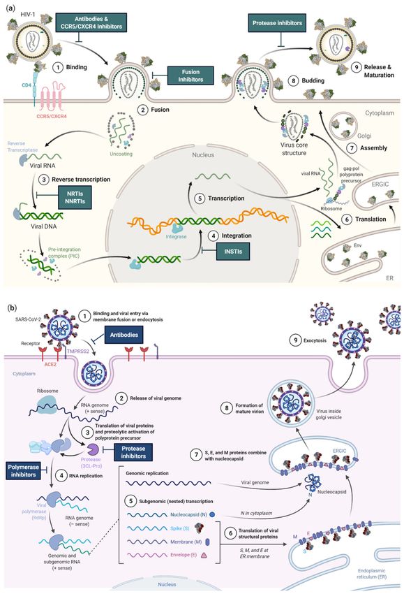
Microorganisms 2021, 9, 1389                                                                                           4 of 61

                               2. Methods
                                     Structural analyses were performed using UCSF Chimera v.1.15rc [88] based on pdb
                               files downloaded from the Research Collaboratory for Structural Bioinformatics Protein
                               Data Bank (RCSB PDB). Protein structures were generated using pdb files 3j5m or 6wpu
                               (prefusion “closed” HIV-1 Env), S.pdb [89] (prefusion “closed” SARS-CoV-2 spike trimer),
                               4l1a (HIV-1 protease in complex with lopinavir), 6wnp (SARS-CoV-2 main protease in
                               complex with Boceprevir), 3v4i/3v81 (HIV-1 reverse transcriptase [RT] in complex with
                               DNA, azathioprine-triphosphate [3v4i; red], and nevirapine [3v81; purple], the latter
                               superimposed after structural overlay of 3v4i and 3v81 RT structures), and 7bv2 (SARS-
                               CoV-2 polymerase RdRp [NSP12] in complex with NSP7, NSP8, template-primer RNA, and
                               Remdesivir triphosphate). Complex entry models were created using structural overlays
                               using MatchMaker in Chimera of pdb structures 6vyb, 6m17, and S_ACE2 [89] (SARS-
                               CoV-2), or 6met and 5vn3 (HIV-1). Models of fusion intermediates were created based
                               on pdb files 6m3w (SARS-CoV-2) and 2zfc (HIV-1). In silico glycosylation of proteins
                               was performed using GlyProt [90], and the composition of oligomannose, hybrid, and
                               complex N-glycans matched with reference literature [91,92]. Viral life cycle schematics
                               were created with BioRender including trimer structures of prefusion “closed” HIV-1 Env
                               (pdb 3j5m) and SARS-CoV-2 spike (pdb 6vxx) as well as activated, “partially open” HIV-1
                               Env (pdb 5vn3) and SARS-CoV-2 spike (pdb 6vyb). Viral sequences were downloaded
                               from the Global initiative on sharing all influenza data (GISAID)-EpiCoV and the Los
                               Alamos National Laboratory (LANL) HIV Databases [30,93]. For better comparability,
                               phylogenetics and genetic diversity analyses were performed using HIV-1 and SARS-CoV-2
                               sequences from one entire year, respectively. For SARS-CoV-2, the first year of the pandemic
                               was studied (mid-December 2019–mid-December 2020), and all full-length sequences with
                               high coverage were downloaded for the studied countries. For HIV-1, we selected a year
                               (January–December) in which a comparable number of HIV-1 unique sequences had been
                               deposited (within 1.5 log difference) relative to the SARS-CoV-2 data set in the same country.
                               Duplicate HIV-1 sequences were removed using the ElimDupes tool from the LANL
                               database. Multiple sequence alignments were performed using Mafft on XSEDE v.7.402
                               as implemented in the CIPRES Science Gateway v. 3.1 [94]. RAxML maximum likelihood
                               trees were generated using RAxML-HPC v.8 on XSEDE with 1000 bootstrap replicates on
                               CIPRES. Phylogenetic trees were visualized using FigTree v.1.4.3 [95]. Highlighter plots
                               were created using ten study sequences covering all major branches of the phylogenetic
                               trees against SARS-CoV-2 or HIV-1 reference sequences Wuhan_Hu_1 (EPI ISL 402125)
                               or HxB2 (K03455), respectively. Genetic distances were calculated using the ape package
                               (“K80” model without pairwise deletion of sites with missing data) and displayed as
                               heatmaps (upper triangle of all pairwise genetic distances) using the complex heatmap
                               package in program R v.4.0.2 x64 and RStudio v.1.3.959 [96,97]. The longitudinal course
                               of clinical and laboratory parameters was modeled based on available data at the time
                               of the manuscript’s completion (June 2021) as summarized and cited in the review’s
                               respective sections.

                               3. Origins of SARS-CoV-2 and HIV-1
                                    HIV-1 and likely SARS-CoV-2 as well, originated from zoonosis as they are both
                               understood to have originally been transmitted to humans from non-human animals.
                               The World Health Organization (WHO) defines zoonosis as a disease or infection that
                               is naturally transmissible from vertebrate animals to humans [98]. HIV is the result of
                               multiple cross-species transmissions of simian immunodeficiency viruses (SIVs) naturally
                               infecting African primates such as African green monkeys, sooty mangabeys, mandrills,
                               chimpanzees, and others [5]. SIVs are largely non-pathogenic in their natural hosts, while
                               HIV-1 causes the acquired immunodeficiency syndrome (AIDS) in humans. There are two
                               major types of human immunodeficiency virus (HIV), type 1 (HIV-1) and type 2 (HIV-2),
                               differing genetically by nearly 55% [99]. HIV-2 origins were confirmed by demonstrating
                               that humans in West Africa harbored HIV-2 strains that resembled a locally circulating
Microorganisms 2021, 9, 1389                                                                                            5 of 61

                               SIV in sooty mangabeys (Cercocebus atys) [100]. This virus has remained largely restricted
                               to West Africa since its discovery in 1989 [101]. On the other hand, HIV-1 disseminated
                               within the human population exhibiting high genetic heterogeneity and giving rise to
                               four distinct groups based on multiple cross-species transmission events: M (major), O
                               (outlier), N (non-M/non-O) and P [102–106]. Group M viruses are responsible for the HIV-1
                               global pandemic, further classified in distinct subtypes (A–D, F–H, and J–L), sub-subtypes
                               (A1–A6, F1, and F2), and circulating and unique recombinant forms [107]. HIV-1 originated
                               from the transmission of a closely related SIV strain from chimpanzees (Pan troglodytes) to
                               humans that was first found in 1989 in two captive chimpanzees in Gabon and isolated
                               from one of them. Sera from these animals cross-reacted with all HIV-1 proteins including
                               envelope proteins [101]. The last common ancestor of HIV-1 group M has been dated to
                               approximately 1910 to 1930, indicating that HIV-1 first emerged in West-Central Africa
                               and spread for some 50 to 70 years before it was recognized [101]. Although the early
                               transmission, dissemination, and establishment of the ape precursors of HIV-1 groups M, N,
                               O, and P in human populations remain unclear, it is believed that transmission of SIV into
                               humans occurred through cutaneous or mucous membrane exposure to infected ape blood
                               and/or body fluids exposures common in the context of bush meat hunting, a longstanding
                               common component of household economies throughout Sub-Saharan Africa [11,101,108].
                               A rapid worldwide spread of HIV-1 has been favored by its enormous genetic variability
                               and rapid evolution, making the virus highly adaptable to new hosts [99].
                                     Since HIV-1, the major driver of the HIV pandemic, originated from great apes such
                               as chimpanzee and gorilla, the initial reservoir and the pathology is mostly restricted
                               to/specific for hominids. In contrast, the reservoir of betacoronaviruses such as severe
                               acute respiratory syndrome coronavirus 1 and 2 (SARS-CoV-1 and SARS-CoV-2) and
                               Middle East respiratory syndrome coronavirus (MERS-CoV) is substantially broader and
                               includes bats, cats, dogs, pangolins, minks, ferrets, and even camelids in the case of MERS-
                               CoV [109–112]. Compared to HIV, coronavirus cross-species transmission events between
                               humans and animals are believed to occur more frequently and back and forth, leading to
                               potentially more diffuse epidemic dynamics that might be harder to control.
                                     SARS-CoV-2 is estimated to have originated between October and December 2019 from
                               zoonosis, but the exact origin remains under extensive scrutiny [113]. The confirmation of
                               a viral pathogen’s zoonotic origin often relies on the isolation of a virus from a non-human
                               animal that shares >99% whole-genome nucleotide identity. In the case of SARS-CoV-2,
                               there has yet to be the isolation of such a >99% similar virus from a non-human animal. The
                               most closely related virus, RaTG13, was isolated in 2013 from a horseshoe bat (Rhinolophus
                               affinis) within the Yunnan province, China, and shares 96% nucleotide identity [114]. While
                               the presence of this sequence in wild bat populations strongly suggests that SARS-CoV-2
                               originated in bats, the sequence divergence of these two viruses represents approximately
                               20 years of evolution, suggesting that RaTG13 can only be regarded as an evolutionary
                               precursor and not a direct progenitor [115]. Moreover, RaTG13 is considerably divergent
                               from SARS-CoV-2 within the receptor-binding domain (RBD) of the spike protein, resulting
                               in an approximately 1,000-fold lower affinity of RaTG13 spike to the human ACE2 receptor
                               than SARS-CoV-2 spike [116]. These findings suggest that there is an intermediate host
                               linking the transmission of SARS-CoV-2 from bats to humans.
                                     The pangolin has been implicated as an intermediate host of SARS-CoV-2 due to the
                               isolation of a coronavirus-denoted Pangolin-CoV (or GD/1/2019) from a sick Malayan pan-
                               golin [117]. Pangolin-CoV is identical to SARS-CoV-2 in all five critical residues for receptor
                               binding of the RBD but has only 92% whole-genome nucleotide identity [117]. Analysis of
                               horizontal gene transfer and recombination lends support for intragenic recombination
                               of the spike genes between RaTG13 and Pangolin-CoV, which could have given rise to
                               the chimeric SARS-CoV-2 [118]. However, neither RaTG13 nor Pangolin-CoV contain the
                               polybasic furin cleavage site at the S1/S2 junction, which is present within the spike protein
                               of SARS-CoV-2 and greatly contributes to the virus’s tropism and pathogenicity [119].
                               Moreover, additional analysis conversely suggests that the presence of identical functional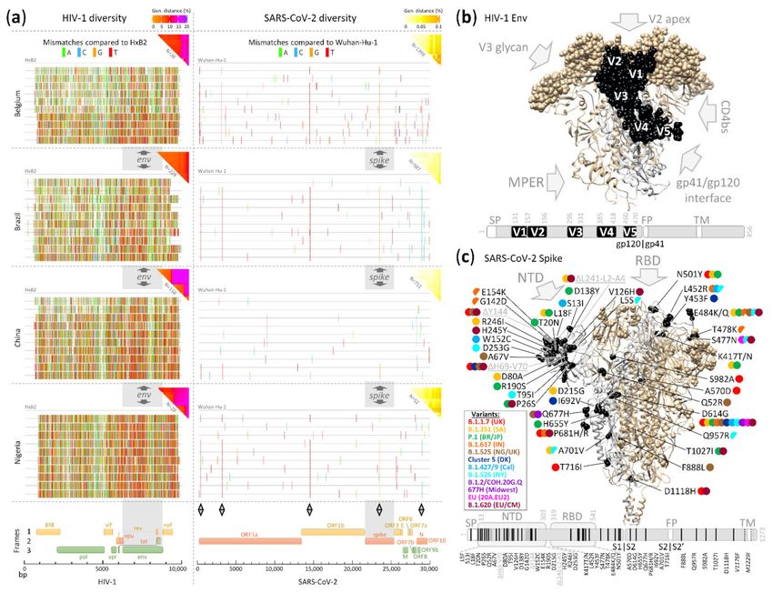
Microorganisms 2021, 9, 1389                                                                                            6 of 61

                               sites within the RBD of SARS-CoV-2 and Pangolin-CoV likely arose independently through
                               random mutations and strong natural selection in addition to recombination, refuting the
                               intermediate transmission of pangolins [120]. Overall, the origin of SARS-CoV-2 remains
                               elusive and in order to clearly identify the natural non-human reservoir of SARS-CoV-2 and
                               confirm its zoonosis, extensive sampling of potential host species is necessary. However,
                               this is likely to be a challenging endeavor as SARS-related coronaviruses are known to be
                               widely distributed across Asia [121]. Additionally, the now widespread distribution of
                               SARS-CoV-2 in humans may result in spillover events to non-human animals, which may
                               undermine any surveillance sampling programs.

                               4. Operating Principles of SARS-CoV-2 and HIV-1
                                    The difference between SARS-CoV-2 and HIV-1extends over their genome organi-
                               zation, repertoire/functioning of viral proteins (see Section 4.1), and viral life cycle (see
                               Section 4.2). The latter includes differences in viral entry, specifically their cellular and
                               receptor tropism (see Section 4.2.1), translation and transcription programs, which, in the
                               case of HIV-1, involve RNA reverse transcription and integration of HIV-1 proviral DNA
                               into the host genome (see Section 4.2.3). Both viruses benefit from their customized annex-
                               ation of the cellular machinery (see Section 4.2.3) and have peculiarities in the proteolytic
                               processing of viral proteins (see Section 4.2.4).

                               4.1. Viral Composition
                                    Both SARS-CoV-2 and HIV-1 are enveloped viruses with a (+)ssRNA genome and
                               a critical set of structural and functional proteins complemented by a broad spectrum
                               of viral co-factors (Figure 1). Despite a ~3-fold difference in genome size, SARS-CoV-2
                               and HIV-1 virions are similarly sized (~100 nm in diameter). According to the Baltimore
                               virus classification, SARS-CoV-2 belongs to group IV viruses, characterized by (+)ssRNA,
                               whereas HIV-1 is listed among group VI viruses, i.e., ssRNA-RT viruses with (+)sense RNA
                               and a DNA intermediate in the life cycle [20,21]. More specifically, SARS-CoV-2 belongs
                               to the family of Coronaviridae (realm: Riboviria, order Nidovirales, genus: Betacoron-
                               aviruses, subgenus: Sarbecoviruses, species: Severe acute respiratory syndrome-related
                               coronaviruses), which are the largest known RNA viruses with genomes approximately
                               30 kb in size (Figure 1, Table 1) [122]. Coronaviruses are named after their crown-like
                               appearance in electron microscopic pictures evoked by their large spike glycoproteins
                               extending from the roughly spherical virions. HIV-1 belongs to the family of Retroviridae
                               (realm: Riboviria, order: Ortervirales, genus: Lentiviruses) [123]. Two copies of HIV-1 RNA
                               are enclosed by a characteristic conical capsid of ~2,000 copies of the viral Gag protein
                               (p24). The capsid is surrounded by a mantle of viral matrix proteins (p17) and a lipid
                               bilayer derived from the infected host cell. The latter is spiked with trimeric viral envelope
                               proteins (Env), the sole viral protein on the HIV-1 surface and the mediator of cellular entry.
                               We refer to this protein also as spike to align it with SARS-CoV-2 nomenclature. While
                               the SARS-CoV-2 spike number is moderate compared to other enveloped viruses such as
                               influenza, it is even lower for HIV-1, with 14 or fewer Env spikes incorporated into the
                               viral membrane (Figure 1, Table 1) [124]. The high plasticity of HIV-1 and SARS-CoV-2,
                               as characteristic for Retroviridae and Coronaviridae, and their potential to tolerate se-
                               quence and structural changes without critical loss of function has warranted their zoonotic
                               transmission and ongoing evolutionary success [125,126].
Microorganisms 2021, 9, 1389 7 of 61
Figure 1. Comparison of HIV-1 and SARS-CoV-2 key viral features. (a) HIV-1 and SARS-CoV-2 are enveloped viruses with
a diameter of ~100 nm. They are decorated with trimeric spike proteins that mediate viral entry, yet SARS-CoV-2 spikesMicroorganisms 2021, 9, 1389 8 of 61
appear in higher numbers than HIV-1 Env (see Table 1). (b) HIV-1 and SARS-CoV-2 possess differently sized, positive-
stranded RNA genomes. The HIV-1 genome is ~10 kb in size, whereas the SARS-CoV-2 genome spans almost 30 kb.
Genomic regions coding for the key functional or structural proteins protease, polymerase, and spike are highlighted in
white, gray, and black and white stripes. (c) Three structural and functional proteins highlighted in (b) are also shown as 3D
structures (ribbon representation), with bound inhibitors shown in red or purple (sphere representation). Protein subunits
are colored differentially. Polymerase-bound RNA/DNA is shown in blue. HIV-1 and SARS-CoV-2 spike proteins are shown
as amino acid backbone structures (left) and glycoproteins (right) with modeled N-glycans (coral; sphere representation).
The HIV-1 genome codes for nine viral genes (gag, pol, env, vif, vpr, vpu, tat, rev,
and nef ) encoding 18 proteins (the Gag proteins MA, CA, SP1, NC, SP2, and P6, the
Pol proteins RT, RNase H, IN, and PR, the Env proteins gp120 and gp41, the regulatory
proteins Tat and Rev, and the accessory proteins Nef, Vpr, Vif, and Vpu). In addition
to the structurally and functionally necessary gag, pol, and env genes, the regulatory
and accessory tat, rev, nef, vif, vpr, and vpu modulate HIV-1 infection, replication, viral
release, and immune recognition [127]. The SARS-CoV-2 genome codes for 14 open-reading
frames (ORFs) encoding 29 proteins [128]. The 50 -end of the genome is dominated by the
large ORF1a and ORF1b genes, comprising more than 2/3 of the entire ~30 kb genome.
They encode polyproteins, which, upon translation, are proteolytically processed into
16 non-structural proteins (NSP1–NSP16) that mostly belong to the replicase–transcriptase
complex. At the 30 -end of the genome, 13 ORFs are expressed from subgenomic RNAs:
in addition to nine accessory proteins, SARS-CoV-2 encodes four structural proteins, as
typical for coronaviruses, i.e., spike, envelope (E), matrix (M), and nucleocapsid (N), the
latter complexing the RNA genome in the absence of a surrounding capsid as is the case
for HIV-1. In contrast to HIV-1, all the three remaining structural SARS-CoV-2 proteins are
incorporated into the viral membrane (spike, E, and M), with spike mediating viral entry.
In addition to the canonical ORFs, numerous discontinuous transcription events make
the SARS-CoV-2 transcriptome highly complex, including transcripts encoding unknown
ORFs with fusions, deletions, and/or frameshifts [129].
SARS-CoV-2 and HIV-1 encode spike glycoproteins, comprised of mainly oligoman-
nose N-glycans in HIV-1 and more balanced complex, oligomannose, and hybrid N-glycans
in SARS-CoV-2 [91,92,130]. The higher number of N-glycans on a smaller spike protein
renders the HIV-1 glycan shield denser than that of SARS-CoV-2, complicating Ab access
to critical entry epitopes with consequences for the development of efficient vaccines
(Figure 1, Table 1).
4.2. Viral Replication
Although HIV-1 and SARS-CoV-2 are both enveloped viruses with a (+)ssRNA
genome, they have evolved different strategies to enter their host cells, replicate, and
release their progeny (Figure 2). Despite engaging different entry receptors and target cells,
HIV-1 and SARS-CoV-2 follow similar principles of class I glycoprotein-mediated viral
fusion and entry (see Section 4.2.1). However, transcription and downstream processes
are critically different (see Section 4.2.2). Maybe the most fundamental difference is that
the SARS-CoV-2 life cycle occurs entirely in the cytoplasm, whereas the HIV-1 life cycle
partially occurs in the nucleus. For this reason, HIV-1 replication takes approximately
double the time of SARS-CoV-2 replication. Specifically, HIV-1 reverse transcription was
shown to initiate at approximately 3 h post infection (h.p.i.), with double-stranded viral
cDNA being detectable 2 h later [44]. In CD4+ T cell lines, integration starts 8.5 h.p.i. and
all viral transcripts are detectable ~15 h.p.i. The viral gene expression peak is reached
between 20 and 23 h.p.i., with ~0.6% of all transcripts in the cell demanded by the virus.
The release of viral particles stretches over several hours and is initiated at 18 h.p.i. and
can continue to 36 h.p.i. in vitro or 60 h.p.i. in vivo (Figure 2a) [44,45]. In contrast, a
SARS-CoV-2 replication cycle takes only ~12 h in A549 cell and in human airway epithelial
cell cultures (HAEC), and time-of-addition experiments showed that initial translation
and viral replication start simultaneously at between 2 and 3 h.p.i. (Figure 2b) [48]. TheMicroorganisms 2021, 9, 1389 9 of 61
following chapter presents the life cycles of both viruses, with a focus on contrasting these
two life cycles.
Figure 2. Replication cycles of (a) HIV-1 and (b) SARS-CoV-2 and major sites of therapeutic intervention. 3CL-pro: 3C-like
protease; ACE2: angiotensin-converting enzyme 2; ER: endoplasmic reticulum; ERGIC: endoplasmic-reticulum-Golgi
intermediate compartment; INSTI: integrase strand transfer inhibitor; NRTI: nucleoside analog reverse transcriptase
inhibitor; NNRTI: non-NRTI; TMPRSS2: Transmembrane protease serine 2.Microorganisms 2021, 9, 1389 10 of 61
4.2.1. Viral Entry
As the first step of the viral replication cycle, cellular entry is one of the most critical,
as it decides the fate of both the virus and the cell. For this reason, the viral glycoproteins
and cellular receptors that facilitate this process are central targets for vaccines, antibody
therapies, and small molecular drugs (Figure 2). For enveloped viruses, including HIV-1
and SARS-CoV-2, entry begins with an attachment step to cellular receptors, followed by
conformational changes of their receptor-binding glycoproteins, and is completed with the
fusion of viral and host membranes.
Attachment of SARS-CoV-2 and HIV-1 is facilitated by the glycoproteins incorporated
within their viral envelope membranes. The glycoproteins of both SARS-CoV-2 and HIV-1
are trimeric class I fusion proteins, named spike or Env (also known as gp160), respectively.
Both are composed of an N-terminal attachment domain (S1; gp120) mediating receptor
binding and a C-terminal fusion domain (S2; gp41) consisting of four critical elements
enabling viral fusion, i.e., fusion peptide (FP), heptad repeat 1 and 2 (HR1, HR2), and
transmembrane domain (Figure 3) [131,132]. To facilitate efficient attachment, both spike
and Env are glycosylated and furin cleaved during viral maturation. Glycosylation aids
immune evasion and is considerably greater on HIV-1 Env than on SARS-CoV-2 spike
(Figure 1c). Cleavage of both glycoproteins by the host protease furin generates non-
covalently bound subunits of their respective N- and C-terminal domains, thereby priming
each glycoprotein for engagement with subsequent receptors or host proteases. The
cellular receptor for SARS-CoV-2 is the widely expressed angiotensin-converting enzyme 2
(ACE2) [133], and it binds via the RBD of spike (Figure 3) [53,134]. Notably, the additional
cell surface receptor neuropilin-1, which is highly expressed in the respiratory and olfactory
epithelium, has been shown to bind exclusively to the furin-cleaved spike, potentiating
SARS-CoV-2 infectivity in these tissues [135,136]. Alternatively, SARS-CoV-2 spike has
also been shown to interact with the host cell receptor CD147 (basigin) to facilitate viral
endocytosis [137]. In contrast to SARS-Cov-2, attachment of HIV-1 occurs via binding of
gp120 to the cell surface immunoglobin glycoprotein CD4 (Figure 3), which is expressed
on subsets of T cells and macrophages [49,138]. Cellular attachment can be initiated or
supported by additional cellular membrane proteins such as integrin α4β7 [139,140].
Upon engagement with their host cell receptors, SARS-CoV-2 spike and HIV-1 Env
undergo conformational changes to facilitate virus–host membrane fusion. The conforma-
tional changes exhibited by both spike and Env enable the extension of their hydrophobic
fusion peptides, which are essential for virus–host membrane fusion and subsequent virus
entry into the host cytoplasm. The molecular triggers for these conformational changes
are different for both viruses. For SARS-CoV-2, extension of the fusion peptide within the
S2 domain is triggered through cleavage by host cell proteases at the S20 site (Figure 3).
The canonical entry occurs through membrane fusion directly at the plasma membrane
and involves S20 cleavage by the host protease TMPRSS2 at the cell surface following
ACE2 engagement [141,142]. Alternatively, SARS-CoV-2 can enter via endocytosis and
membrane-fusion-mediated release from endosomes. In support of this second route
of entry, the endosome-localized host protease cathepsin-L has been shown to partici-
pate in S20 cleavage of SARS-CoV-2 spike at the endosomal membrane, likely following
CD147-mediated endocytosis [137,142]. These alternate mechanisms of SARS-CoV-2 fusion
provide the virus with independent and redundant avenues of entry, which likely con-
tributes to the broad tissue tropism of this virus. In contrast to SARS-CoV-2, the binding
alone of HIV-1 gp120 to its cell surface receptor CD4 is sufficient for triggering confor-
mational changes. These conformational changes expose and stabilize the variable loop
V3-binding site for co-receptor engagement at the cell surface [138]. CCR5 and CXCR4 can
both act as co-receptors for HIV-1. No co-receptors have been identified for SARS-CoV-2.
CCR5 or CXCR4 binding to V3 of gp120 induces further conformational changes to gp120,
which, after dissociation of gp120, releases the fusion peptide of bound gp41 for insertion
into the host cell membrane (Figure 3) [143,144]. Subsequent rearrangement of the heptad
repeat regions of gp41 bring the viral and host cell membranes in close proximity for fusionMicroorganisms 2021, 9, 1389 11 of 61
and release of the viral capsid into the host cell cytoplasm [143]. The released viral capsid
of HIV-1 continues to encapsulate the viral replication components as the pre-integration
complex (PIC), until it delivers the HIV-1 dsDNA to the nucleus. Alternatively, the capsid
of SARS-CoV-2 immediately uncoats the viral RNA upon entry into the cytoplasm.
Figure 3. Spike-mediated cellular entry of HIV-1 (top) and SARS-CoV-2 (bottom). Structural model depicting transi-
tion/activation states of viral spike proteins during viral entry. (1) Prefusion “closed” state, (2) partially “open” state
after interaction of spike proteins with cellular receptors, (3) fusion intermediates after dissociation of cellular attachmentMicroorganisms 2021, 9, 1389 12 of 61
domains gp120 (HIV-1) or S1 (SARS-CoV-2), which exposes fusion peptides for insertion into the target cell membrane.
Schematics of HIV-1 Env and SARS-CoV-2 spike coding genomic regions are shown in the middle with domains colored the
same way as shown in the structural models. ACE2: angiotensin-converting enzyme 2, B0AT1: sodium-dependent neutral
amino acid transporter, RBD: receptor-binding domain, FP: fusion peptide, and HR: heptad repeat.
4.2.2. Translation, Transcription, and Reverse Transcription
The initial stages of SARS-CoV-2 and HIV-1 replication are remarkably different
despite both viruses starting with positive-stranded RNA genomes. Immediately after
uncoating in the cytoplasm, the SARS-CoV-2 genome acts as mRNA for the translation
of two ORFs (Figure 2b). ORF1a and ORF1b produce polyproteins named pp1a and
pp1ab. ORF1a encodes pp1a while the larger pp1ab is the fusion product of ORF1a and
ORF1b, resulting from a -1 ribosome frameshift during translation [47,145]. Following
their proteolytic cleavage into 16 NSP subunits, these polyproteins comprise the complete
SARS-CoV-2 replicase–transcriptase complex (RTC), responsible for the transcription of
the remaining ORFs as well as the full-length gRNA [47]. Conversely, the HIV-1 genome is
reverse-transcribed into dsDNA after entry, a step that is catalyzed by the viral RT prebound
to the viral genome (Figure 2a). Interestingly, this reverse-transcription process, which
lacks a proof-reading step, is much more error-prone than the transcription of the SARS-
CoV-2 genome which is supported by a sophisticated proof-reading mechanism [39,146],
contributing to the comparatively broad genomic diversity of HIV-1 (Table 1, Figure 4).
HIV must transport its nucleic acid to the nucleus via the nuclear pore. Within the nucleus,
the PIC is uncoated and the dsDNA complex is integrated into the host genome by the
viral enzyme Integrase [147–149]. It is only after integration that the HIV-1 genome is
transcribed into mRNAs by host enzymes in the nucleus, which are then transported
out and translated in the cytoplasm. HIV-1 achieves productive infection by preferential
integration of its viral genome in intron regions of highly expressed host genes [147,150].
The chronicity of HIV-1 is caused by latent infection of long-lived memory CD4+ T cells
and constitutes a major barrier towards a cure of HIV-1 infection [151]. Latent infection is
accomplished by integrating HIV-1 into transcriptionally silent regions of the genome of
quiescent CD4+ T cells [152,153].
The transcription of subgenomic (sg) mRNAs and their subsequent translation also
differs considerably between these two viruses. The SARS-CoV-2 RTC forms at lipid
droplet factories within the cytoplasm [47]. There, the NSP12 RNA-dependent RNA
polymerase (RdRp) generates both full-length negative-strand RNA, which acts as the
template for replicating SARS-CoV-2 gRNA and shorter sgRNAs of the accessory and
structural gene-encoding ORFs [47]. Typical of coronaviruses, transcription regulatory
sequences (TRSs) upstream of each of these ORFs prematurely terminate negative-strand
RNA transcription in a process referred to as discontinuous transcription. The resulting
transcripts are a set of structurally polycistronic nested sgRNAs; however, it is assumed
that functionally, these transcripts are monocistronic and that only the 50 -most ORF in
each sgRNA is translated [154,155]. Once translated from these sgRNAs, the structural
proteins spike, E, M, and N package the gRNA to form infectious virions at the ER-to-golgi
intermediate compartment for secretion by exocytosis. In addition to these canonical ORFs
encoding structural proteins, recent ribosomal-profiling has identified 23 translationally
active unannotated SARS-CoV-2 ORFs with currently unknown function [156]. In contrast
to SARS-CoV-2, the transcription of the integrated HIV-1 genome is carried out by cellular
polymerases in the nucleus and, through means of alternate splicing, gives rise to over
50 viral RNA transcripts [157]. Governed by the cellular export machinery, only fully
spliced RNA transcripts are exported from the nucleus for translation. Hence, the HIV-1
gene products can be separated into early and late based on their necessity for intron
retention. The early viral transcripts rev, tat, and nef are encoded by fully spliced transcripts
and consequently, can be immediately exported for translation, while the remaining genes
encoded by partially or unspliced transcripts rely on sufficient levels of Rev to accumulate.
Rev binds HIV-1 intron-containing transcripts and enables their alternate export to theMicroorganisms 2021, 9, 1389 13 of 61
cytoplasm. The structural Gag and Gag-Pol polyproteins are translated from the unspliced
gRNA and consequently are among the last proteins translated. Moreover, Gag is translated
from the gag gene, while Gag-Pol is a fusion protein generated by a ribosomal frameshift
during translation of the gag gene to the alternate pol reading frame [158]. Once all the
HIV-1 structural proteins are translated, virion assembly proceeds at the cell membrane.
Figure 4. Phylogenetic diversity of HIV-1 and SARS-CoV-2. Maximum likelihood phylogenetic trees (RAxML, 1000 boot-
strap replicates) were generated with full length HIV-1 (left) and SARS-CoV-2 (right) genomic sequences from four different
countries/continents. For SARS-CoV-2, all available full-length sequences with high coverage were used, deposited toMicroorganisms 2021, 9, 1389 14 of 61
GISAID within one year since initiation of the outbreak in mid-December 2019. Comparably, HIV-1 sequences from one
entire year were studied, selected based on comparable case numbers (Microorganisms 2021, 9, 1389 15 of 61
in a viral load-dependent manner. In contrast, B cell-specific proteins and neutrophil
chemokines are elevated in individuals with lower viral load. Transcriptional levels of the
SARS-CoV-2 spike-processing host protease TMPRSS2 depend on infection time point and
cell type [171,172]. Time series transcriptome profiling of Calu-3 cells infected in vitro with
a clinical SARS-CoV-2 isolate revealed a strong upregulation of TMPRSS2 mRNA within
the very first few hours post infection [171], whereas at later time points, the levels revert
to baseline or even slightly below [171,172]. Males and older individuals exhibit impaired
transcriptional activity affecting trafficking and/or antiviral responses through the reduced
function of cytotoxic T cells, B cells, and natural killer cells [172]. Proteomic studies further
showed that SARS-CoV-2 reshapes cellular translation, splicing, carbon metabolism, pro-
tein homeostasis, and nucleic acid metabolism [174]. In addition to viral proteins, it was
shown that SARS-CoV-2 RNA directly and specifically binds and/or modulates a broad
network of human proteins in infected human cells [178], and host mitochondria serve as
an organelle platform for anti-SARS-CoV-2 immunity [179]. Approximately one-third of
the cellular RNA-binding proteins (RBPs) are remodeled upon SARS-CoV-2 infection, and
inhibition of these RBPs impairs SARS-CoV-2 infection [180].
4.2.4. Proteolytic Processing of Viral Proteins
A central component of viral replication, including that of SARS-CoV-2 and HIV-1, is
the proteolytic processing of viral proteins, which serves to orchestrate genome replication,
assembly and maturation. This process involves the cleavage of viral proteins mediated by
proteases that are encoded either by the virus or the host. Virally encoded proteases are
typically responsible for the autocatalytic excision from polyproteins in which they reside,
and for subsequent proteolytic processing of the remaining polyprotein components (i.e.,
3CL-pro in pp1a/pp1ab or PR in Gag-Pol). While minimizing coding space within the
viral genome, this polyprotein processing also coordinates synchronized translocation of
tethered viral proteins to assembly sites within the host cell [181]. In addition to processing
by viral proteases, viral proteins, including those of SARS-CoV-2 and HIV-1, are also
cleaved by host proteases (i.e., furin), most notably to mediate viral maturation, which
primes progeny virions for efficient entry into new cells. The critical functional role of
post-translational viral protein cleavage proposes this process as an attractive target for
antiviral therapeutic development.
Characteristic for viruses with a positive-sense RNA genome, SARS-CoV-2 and HIV-1
both encode polyproteins that undergo proteolytic processing by virally encoded proteases.
However, the replication stage at which the polyprotein proteolytic processing occurs
is different between SARS-CoV-2 and HIV-1, reflecting their alternate replication strate-
gies. For SARS-CoV-2, polyprotein processing occurs post viral entry and prior to viral
replication. The two SARS-CoV-2 polyproteins pp1a and pp1ab are translated from the
incoming positive-sense RNA genome and are proteolytically processed by two cysteine
proteases, papain-like protease (PL-pro) and 3-chromtrypsin-like protease (3CL-pro, or
main protease; M-pro), which reside within NSP3 and NSP5, respectively, and are re-
leased auto-catalytically (Figure 1) [182]. PL-pro catalyzes the cleavage of NSP1-3 and the
amino-terminal of NSP4, while 3CL-pro cleaves the carboxyl terminal of NSP4 as well
as the remaining NSP5-16 [183]. These cleavage events are essential for the subsequent
steps of SARS-CoV-2 replication, and consequently, their inhibition through therapeutic
intervention is a topic of ongoing research efforts (Figure 2) [48,184]. In the HIV-1 life cycle,
polyprotein processing occurs at later stages, post viral replication and during viral assem-
bly. Indeed, proteolytic cleavage of HIV-10 s integral structural and replicative proteins Gag
and Gag-Pol typically occurs during virion assembly and maturation at the cell surface
or within the budded virion. Cleavage is performed by the aspartic HIV-1 protease (PR),
itself harbored within the packaged Gag-Pol polyprotein and released auto-catalytically
(Figure 1) [185]. The PR cleavage of Gag generates the main structural proteins MA, CA
and NC, which subsequently rearrange to form the mature, infectious particle [186]. The PR
cleavage of Gag-Pol, while also generating the structural proteins, additionally gives riseMicroorganisms 2021, 9, 1389 16 of 61
to the TFP, PR, RT, RNase H and IN proteins required for initial reverse-transcription and
integration upon entry into a new cell [186]. Incompletely processed HIV-1 polyproteins
fail to covert the assembled virus particle into a mature infectious virion, and HIV-1 PR has
consequently been a target of anti-HIV-1 therapies for numerous years (Figure 2) [186,187].
In addition to polyprotein cleavage by viral proteases, the life cycles of both HIV-1
and SARS-CoV-2 include proteolytic processing of their receptor-binding glycoproteins,
which are performed by host proteases. These events occur late in the life cycle for both
viruses, during a process called viral maturation. Both viruses utilize the host furin-like
proteases for cleavage of their glycosylated receptor-binding proteins within the trans-golgi
network during virion assembly. The SARS-CoV-2 spike protein possesses a furin cleavage
site at the S1/S2 junction [188], which has been demonstrated to be critical for SARS-CoV-2
pathogenicity [119,141,189–191]. It enhances the binding affinity of spike to ACE2 by three
orders of magnitude [116], but is not entirely essential for SARS-CoV-2 infection, possibly
by the secondary spike cleavage event, which can be mediated by proteases other than
furin [28,190,191]. The HIV-1 Env polyprotein cleavage by furin produces the receptor-
binding gp120 and transmembrane gp41 subunits (Figure 3) [32,192–195]. In contrast to
furin-mediated cleavage of SARS-CoV-2 spike, the cleavage of HIV-1 Env appears essential
for HIV-1 entry and infection [196–198].
Unlike HIV-1 and most other viruses, SARS-CoV-2 possesses an additional cleavage
site within its receptor-binding glycoprotein spike, named S20 , which facilitates exposure of
its fusion peptide (Figure 3). This second cleavage is mediated by alternate host proteases
TMPRSS2 and Cathepsin-L. While initial discrepancies in findings disputed the contribu-
tion of either protease, a recent study has determined a spatial delineation underpinning
their alternate contributions [142]. It is now understood that both proteases may cleave
spike to facilitate fusion; however, TMPRSS2 acts to cleave spike at the cell surface, while
Cathepsin-L cleaves spike within the endosome, which likely contributes to the broad
tropism displayed by SARS-CoV-2 [142]. Overall, it is apparent that host proteases are em-
ployed by both viruses during maturation and in the case of SARS-CoV-2 also during host
cell recognition, which primes their receptor-binding glycoproteins for subsequent entry.
5. Humoral Immune Responses
Antibody (Ab) immune responses play a central role in protecting the host from viral
infections [199]. Ab-mediated protection is primarily attributed to the Ab binding and
neutralization capacity (see Section 5.1), complemented by Ab Fc-mediated responses
(see Section 5.2). The Abs’ high specificity to defined viral epitopes imposes a strong
selection pressure, which may favor the selection of viral escape mutations (see Section 5.3).
In all, the relationship between Abs and viruses uniquely shapes their co-evolution in
virus-infected individuals and entire populations.
5.1. Antibody Binding and Neutralization
In natural HIV-1 infection, HIV-1-specific Abs are elicited within the first weeks of in-
fection. These include IgM and subsequently class-switched IgG and IgA, which are mainly
directed against the immunogenic, highly variable Env gp41 and gp120 regions such as V3
at this initial stage. These early non- or weakly neutralizing Abs are narrow, mostly strain
specific, and predisposed to rapid immune evasion [200–202]. Broadly neutralizing Abs
(bnAbs) occur only in a small percentage of HIV-1-infected individuals (~10%), requiring
a few years of continuous antigenic stimulation and maturation, mostly involving high
rates of somatic hypermutation [203]. In HIV-1-infected individuals, the development
of bnAbs, or high neutralization levels in general, are not associated with better clinical
outcome/slow progression, but in turn, correlate with severity of disease and high viral
load (Figure 5) [204–206]. Knowledge gained from the tedious process of natural bnAb
development is currently translated into germline-targeting vaccine strategies with sequen-
tial boosting [207]. Similar to SARS-CoV-2, neutralization is considered the lead effector
function to protect from HIV-1 infection, since passive administration of bnAbs in ani-Microorganisms 2021, 9, 1389 17 of 61
mal models of HIV-1 infection can confer protection against viral challenges [199,208,209].
This strategy is currently tested in human clinical trials [210,211]. However, while vac-
cines can induce sufficiently potent bnAbs against SARS-CoV-2 [212–214], and COVID-19
convalescent individuals acquire protective immunity through natural infection [215],
it has not been possible to induce broadly protective Abs by HIV-1 vaccines [81] and
primary HIV-1 infection does not adequately protect from superinfection [216–218]. Ab
responses induced in participants of HIV-1 human vaccine trials such as RV144 were mostly
non- or weakly neutralizing, waned rapidly, and/or suffered from rapid viral escape (see
Section 5.3) [81,86,219,220].
In contrast to HIV-1 infection, natural SARS-CoV-2 infection rapidly induces neu-
tralizing Abs (nAbs) targeting primarily the spike RBD and N-terminal-domain (NTD)
regions and encompassing a broad range of heavy chain and light chain V genes [221].
Most individuals develop very similar nAb responses with moderate breadth and plasma
neutralization activity that require only low rates of somatic hypermutation [222]. Pro-
longed viral replication in immunocompromised hosts may favor the generation and
selection of nAb escape mutants, which in turn drives Ab affinity maturation and eventu-
ally enhanced neutralization breadth and potency [223]. In humans, previous SARS-CoV-2
infection and anti-spike Ab seropositivity significantly reduce the risk of SARS-CoV-2
reinfection [215,224–229], which is in line with vaccination outcome analyses [230–236]
altogether implying that SARS-CoV-2 binding and/or nAbs exert protective effects. Conse-
quently, neutralization levels have been found highly predictive of immune protection with
seven current vaccines and in convalescent cohorts, and provide an evidence-based model
of SARS-CoV-2 immune protection [237]. Non-human primate models of SARS-CoV-2
infection corroborated these findings and suggest that nAbs play the leading role in protec-
tion from SARS-CoV-2 infection [87,238]. In rhesus macaques, Ab-based protection or their
therapeutic potential is dose dependent. Although low Ab titers were sufficient to protect
rhesus macaques from SARS-CoV-2 infection or reinfection, higher Ab titers were required
to achieve a drop in viral load once infected. Furthermore, cellular immunity contributes to
viral control and may compensate waning or insufficient Ab-mediated responses [87,239]
(see Section 5.1), e.g., CD8+ T cell depletion reduced the protective efficacy of natural
immunity against SARS-CoV-2 reinfection in convalescent animals [87]. In addition to nAb
responses against the spike NTD and RBD regions, phage display screenings for binding
Abs targeting linear epitopes identified common responses in COVID-19 patients against
the fusion peptide and the linker region upstream of the HR2, however with variable
escape mechanisms [240]. These findings are in line with a recent report stating that the Ab
binding response in COVID-19 convalescent individuals converges in >80% to non-RBD
epitopes [241]. The early anti-SARS-CoV-2 binding and nAb response is dominated by
IgM, which wanes rapidly. IgA and IgG peak subsequently, and IgG-mediated neutralizing
responses are most durable and persist over months, mirrored by neutralization half-lives
of a few months in serum but up to >8 months in purified IgG samples [242–245]. The
persisting Ab response can be attributed to a maturing humoral immunity driven by a
sustained SARS-CoV-2 antigenic stimulation in the gut of COVID-19 patients [246]. Similar
to HIV-1, higher anti-SARS-CoV-2 Ab and neutralization levels are preferentially found
in severe cases [55,247–250]. However, COVID-19 survivors exhibit enhanced neutraliza-
tion potency [247] and a more balanced Ab maturation pathway [251] that might even
be critical for survival [252]. More recently, it was shown that severely ill patients do not
necessarily mount higher overall humoral responses than mild cases, but are characterized
by a delayed kinetic of the anti-spike IgG and nAb response [253]. Neutralization is primar-
ily mediated by receptor-blocking nAbs, and they can either inhibit or enhance syncytia
formation [254].Microorganisms 2021, 9, 1389 18 of 61
Figure 5. Courses of natural, untreated HIV-1 and SARS-CoV-2 infection. Estimated models of key clinical, viral, and immune parameters and their longitudinal changes in representative
courses of HIV-1 (a) and SARS-CoV-2 (b) infection. Models of more severe/progressive disease courses are shown on top; mild/slow progressive courses are shown at the bottom. Features
are color-coded according to the legend and key features directly annotated.Microorganisms 2021, 9, 1389 19 of 61
Of note, a recent study showed that some Abs against the spike NTD induce the open
spike conformation and thus enhance the binding capacity to ACE2 and infectivity of SARS-
CoV-2 [255]. Mutational and structural analyses indicated that all infectivity-enhancing
Abs target a common site on the NTD and shar a divalent binding mode. Abs specific for
the infectivity-enhancing site on the NTD were detected at high levels in severe patients.
The identified mechanism of antibody-dependent enhancement (ADE) of viral infection is
Fc receptor-independent. It differs from the Fc receptor-dependent ADE identified with
other viruses in having a lower impact on infection, but affecting a broader range of cells
including ones that do not express Fc receptors [255–257]. Excess amounts of nAbs appear
to suppress ADE in most cases of SARS-CoV-2 infection or vaccination; however, the precise
functional consequences of infectivity-enhancing Abs on SARS-CoV-2 pathogenicity and
vaccines, and their differential impact on variants remain elusive.
In addition to SARS-CoV-2 spike, the N and ORF 3b and 8 proteins are highly im-
munogenic with implications as serological markers [258]. Qualitative differences in early
Ab profiles point to elevated Ab responses to the N protein in deceased individuals [259].
Ab immune responses have mainly been studied in the blood, whereas little data exist
about the responses at the local sites of infection such as the respiratory system. Of interest,
the mucosal immune system comprises the largest part of the immune system. On-site
production of secretory IgA (sIgA) by far exceeds all other immunoglobulin isotypes, which
renders the mucosa, as site of viral entry, prepared for the initial wave of adaptive de-
fense [260]. Consequently, anti-SARS-CoV-2 IgG and IgM levels, which mainly transudate
from the blood into the mucosa, correlate well between both compartments. In contrast,
IgA was found to be more abundant in the mucosa, particularly early during disease, which
supports the hypothesis that SARS-CoV-2 infection triggers local sIgA production [261,262].
Combining immunological and epidemiological analyses on seasonal coronaviruses
has shown that infection-blocking immunity wanes rapidly, but disease-reducing immu-
nity is long-lived, which suggests a model of SARS-CoV-2 transitioning within years to
endemicity with mitigated pathogenicity [263].
5.2. Antibody Fc-Mediated Functions
Antibody Fc-mediated functions complement Ab neutralization functions and provide
a link between Ab- and cell-based immunity (e.g., NK cells and phagocytes) or soluble
effectors (e.g., complement) [264,265]. As such, Fc-mediated Ab functions can act hand in
hand with neutralization or as an additional line of defense before or after neutralization.
In HIV-1 infection, Fc-mediated effector functions have been studied in detail in
recent years, particularly antibody-dependent cellular cytotoxicity (ADCC) and antibody-
dependent cellular phagocytosis (ADCP) [266,267]. Using a quantitative approach in HIV-
1-infected humanized mice and Simian-HIV (SHIV)-infected rhesus macaques, 25–45%
of the total antiviral activity of anti-HIV-1 mAbs was attributed to Fc-mediated effector
functions [268]. In support of that, mAbs with non-functional Fc-receptors had dramat-
ically decreased capacity to protect animal models from SHIV infection [269]. Since the
isolated depletion of complement binding had no impact on the protective activity, Fc-
mediated cellular responses appear to play the dominant role. Indeed, Fc-mediated cellular
responses such as ADCC and ADCP have been associated with protection from HIV-1
disease progression and protection from (S)HIV infection in animal models or in a human
vaccine trial [81,266,270,271]. For example, ADCC responses in the presence of low plasma
IgA/IgG ratios correlated with protection from infection in a large human vaccine trial with
partial efficacy (RV144) [86,220,272–274], yet a complete mechanistic explanation remains
elusive [81,266]. Furthermore, Fcγ phenotyping in vaccinees of the same trial revealed
that distinct single-nucleotide polymorphisms (SNP) in the FCγR2C gene conferred 91%
vaccine efficacy against HIV-1-carrying immunodominant epitopes in Env that experienced
vaccine selection pressure. In contrast, individuals with a different SNP exhibited only 15%
vaccine efficacy [275].Microorganisms 2021, 9, 1389 20 of 61
Many studies have shown a tight linkage between Ab Fc-mediated effector functions
and neutralization in HIV-1 infections [269,276–280]. A recent study showed that the
neutralization activity of an anti-HIV-1 mAb was potentiated >5000-fold in vitro when ex-
pressing the IgG high-affinity Fc receptor FCγRI compared to the same mAb without [278].
Moreover, the antisera from animals immunized with the respective mAb epitope-based
vaccine neutralized diverse HIV-1 clades, including more resistant tier-2 viruses, in an
FCγRI-dependent manner [278]. Nonetheless, the mutual impact between Fc-mediated
functions and neutralization can vary considerably as it was shown, for example, that
Fc-mediated activity was partially redundant for a very potent bnAb [281], and differences
in antibody binding affinity for HIV-1 and SIV Env uncoupled mAb-mediated ADCC
from neutralization [282]. Fc-mediated functions are influenced by the antigenicity and
conformation of the infecting strain/molecular clone, Ab binding levels, Ab specificity, Ab
orientation on the bound antigen, gp120 shedding, capacity to form multivalent antigen–
Ab complexes, degree of internalization of antigen–Ab complexes, and killer cell receptor
ligand expression (e.g., NKG2D) [266,280,283–286].
In SARS-CoV-2 infection, data on the impact of Fc-mediated effector functions are still
unfolding, but similar to HIV-1, these effector functions appear to be critical [239,287,288].
Studies in non-human primates demonstrated that Fc-mediated functions correlated with
protection from SARS-CoV-2 infection [289]. This was confirmed by studies in mice and
hamsters, where nAbs provided better protection when coupled with Fc-receptor func-
tionality [287,290,291]. Fc-effector functions are elicited in symptomatic and asymptomatic
COVID-19 individuals, but they are elevated in severe cases [288]. COVID-19 non-survivors
had a higher incidence of compromised Fc-receptor binding and effector functions, im-
plying a crucial role for Ab Fc-effector functions in limiting severe disease and reducing
patient mortality [251]. An in vitro model of ADCC, using full-length spike proteins ex-
pressed on the surface of a target cell line, and PBMCs from healthy individuals serving as
effector cells, provided additional mechanistic insights. In this model, the ADCC activity of
convalescent plasma decreased only modestly compared to the more pronounced decrease
in neutralization activity. Substantial ADCC activity was maintained in 85% of donors’
plasma up to eight months post symptom onset and strongly correlated with plasma IgG
responses [242]. Notably, three weeks post vaccination with an mRNA vaccine, a time point
at which vaccine efficacy is estimated to be >90%, nAb responses are still mostly absent, but
anti-SARS-CoV-2 ADCC responses well developed [239]. This implies a possible role for
Fc-mediated effector functions and other cellular responses in vaccine-mediated protective
effects. The collected data so far suggest a vital role for Fc-mediated effector functions in
sustained protection from reinfection and vaccine-induced protection.
5.3. Antibody Escape and Mutant Variants
HIV-1 and SARS-CoV-2 Ab escape is based on similar principles of immune pressure
exerted by Abs on their targeted viral epitopes [292]. However, the strength and timing of
the driving immune forces, and the capacities to evade these forces are very different in
both viruses. Important discriminative factors are the acute nature of SARS-CoV-2 infection,
resulting in a small temporal window of active replication and adaptation, combined with
a low mutation rate due to the proof-reading mechanism of the SARS-CoV-2 polymerase
complex [146]. This contrasts with the chronic nature of HIV-1 infection that allows for a
lifelong ongoing viral replication and adaptation with a high mutation rate in the absence
of proof-reading by the HIV-1 polymerase complex. Consequently, HIV-1 immune escape is
a constant factor in almost every HIV-1-infected individual, whereas SARS-CoV-2 immune
escape is rare and seems to occur preferably in immunocompromised individuals with
prolonged viral replication and fostered by treatment with mAbs or convalescent plasma
(blood plasma from a donor who has recovered from COVID-19) (Figure 6) [293–297].You can also read