Sclerosing angiomatoid nodular transformation (SANT) of the spleen: a rare cause of acute abdomen
←
→
Page content transcription
If your browser does not render page correctly, please read the page content below
Journal of Surgical Case Reports, 2021;4, 1–3

                                                                                   doi: 10.1093/jscr/rjab126
                                                                                   Case Report

CASE REPORT

Sclerosing angiomatoid nodular transformation

                                                                                                                                                           Downloaded from https://academic.oup.com/jscr/article/2021/4/rjab126/6217973 by guest on 15 September 2021
(SANT) of the spleen: a rare cause of acute abdomen
Rommel Ojeda1 , Gabriel A. Molina1 ,2 , Galo E. Jiménez1 , Hernán González3 ,
Johanna C. Pinto1 , Andres Jiménez1 and Flor M. Leon4
1
 Department of General Surgery, Hospital IESS Quito Sur, Quito, Ecuador, 2 Universidad San Francisco de Quito
(USFQ), Quito, Ecuador, 3 Hospital de Especialidades Carlos Andrade Marin, Quito, Ecuador, and 4 Instituto
Ecuatoriano de Seguridad Social, Department of Internal Medicine at Hospital IESS Quito Sur, Quito, Ecuador
*Correspondence address. Hospital IESS Quito Sur, Quito, Ecuador, Universidad San Francisco de Quito & Department of General Surgery, Quito 170157,
Ecuador. Tel: +593-998352532; E-mail: gabomolina32@gmail.com

Abstract
Sclerosing angiomatoid nodular transformation (SANT) of the spleen is an extremely rare benign lesion. It originates from
the spleen’s red pulp; however, its pathogenesis is not clearly defined. These tumors are usually asymptomatic or cause
nonspecific abdominal symptoms. Most SANTs are found incidentally on radiographic examination or during surgery for
an unrelated condition. The differential diagnosis from other splenic tumors or malignant lesions can be challenging due
to the risk for a possible malignancy of the suspicious lesion. As more SANTs are being discovered and treated, they should
always be considered in the differential. We present the case of an otherwise healthy 30-year-old female; she presented with
abdominal pain and a mass in her spleen. Surgery was performed, and an SANT was discovered. The patient underwent full
recovery, and on follow-up is doing well.

INTRODUCTION
Sclerosing angiomatoid nodular transformation (SANT) is a rare                     pain became much more severe throughout the year and was
benign disease of the spleen [1, 2]. Clinical symptoms are non-                    accompanied by nausea. Thus, she presented to the emergency
specific, and a suspicious splenic lesion is often suspected, lead-                room. On clinical examination, abdominal pain and tenderness
ing to splenectomy [2, 3]. Diagnosis is usually made histologically                along with a solid mass with severe pain on touch were
after complete resection. We report a 30-year-old female. She                      discovered in her upper abdomen. No fever, vomiting, blood
presented to the emergency room with acute abdomen. A splenic                      in the stool, or other symptoms were found. Due to this, a
mass was identified, and surgery was completed. SANT of the                        computed tomography (CT) revealed an enlarged spleen. It
spleen was discovered; on follow-ups, the patient is doing well.                   measured 15 × 10 × 5.5 cm, and within the spleen, a 5.4 × 5.1 cm
                                                                                   mass and another 5.3 × 2.3 cm mass is seen (Fig. 1A and B).
                                                                                   Laboratory exams revealed leukocytosis with neutrophilia and
CASE REPORT                                                                        an elevated C-reactive protein.
Patient is a 30-year-old female without past medical history.                         With these findings, surgery was needed, and a laparoscopic
She had a 1-year history of mild abdominal pain in her upper                       approach was decided. The enlarged spleen had multiple hemor-
left abdomen; at first, the pain was mild; nonetheless, the                        rhagic foci in its capsule. The larger mass was located mostly on

Received: January 18, 2021. Revised: February 18, 2021. Accepted: March 15, 2021
Published by Oxford University Press and JSCR Publishing Ltd. All rights reserved. © The Author(s) 2021.
This is an Open Access article distributed under the terms of the Creative Commons Attribution Non-Commercial License (http://creativecommons.org/li
censes/by-nc/4.0/), which permits non-commercial re-use, distribution, and reproduction in any medium, provided the original work is properly cited. For
commercial re-use, please contact journals.permissions@oup.com
                                                                                                                                                  1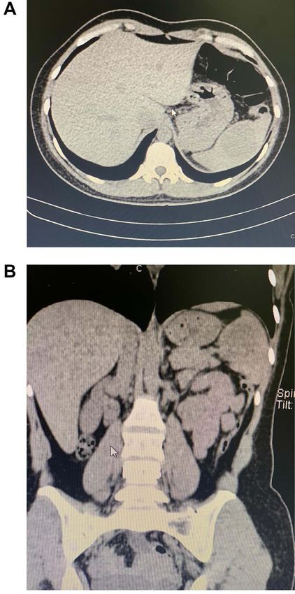
2 R. Ojeda et al.
Downloaded from https://academic.oup.com/jscr/article/2021/4/rjab126/6217973 by guest on 15 September 2021
Figure 2: (A) Spleen completely resected. (B) White SANT masses within the
spleen.
Figure 1: (A) CT, a mass is seen in the spleen. (B) CT, the enlarged spleen with the
mass in the upper pole.
its upper lobe. No other masses, free liquid or lymph nodes were
found. Due to this, a laparoscopic splenectomy was completed.
Pathology revealed a 15 × 10 × 5.5 cm spleen with a 5.4 × 5.1 cm
mass in its upper lobe and a 5.3 × 2.3 mass on its lower lobe;
the masses had a yellowish color and had multiple nodules
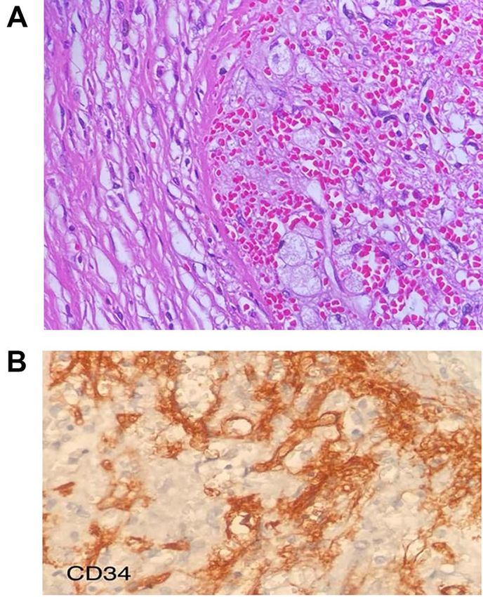
(Fig. 2A and B). Multiple angiomatoid nodules were embedded
in a fibrosclerotic stroma; many regions showed increased vas-
cularity, and strong positivity for CD34 was detected on micro-
scopic evaluation (Fig. 3A and B). SANT was the final diagnosis.
She was discharged on her second postoperative day after
a full diet was initiated. The patient was checked at regular Figure 3: (A) Multiple angiomatoid nodules embedded in a fibrosclerotic stroma.
intervals after surgery. Immunization was given, and no sign of (B) CD34 were detected on microscopic evaluation on the stroma.
recurrence was detected until the first year after surgery.
found during autopsies from cutaneous malignant melanoma
DISCUSSION and breast cancer [1, 2]. The majority of nonlymphoid primary
Tumors of the spleen are rare [1]. Their classification is var- tumors of the spleen are of vascular nature and are usually
ied, but they are usually classified into two main categories: cavernous hemangiomas [2, 3]. Nonetheless, in 2004, Martel
nonlymphoid and lymphoid [1, 2]. The most common primary et al. described a rare primary benign tumor-like lesion of the
malignancies of the spleen are lymphoma and angiosarcoma [1]. spleen with its own pathological features and immunohisto-
Metastasis to the spleen is extremely rare (2.3–7.1%) and usually chemical profile [1, 2]. The name SANT of the spleen appeared [2].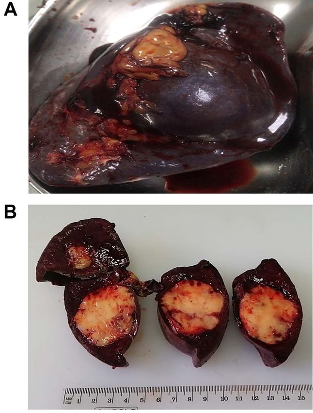
Sclerosing angiomatoid nodular transformation         3

Although it was first reported in 1978 by Silverman and LiVolski    patients should be given in all patients scheduled for elective
and defined as a special kind of hamartoma, Martel et al. were      splenectomy and during the postoperative period for urgent
the ones that defined its characteristics [2, 3].                   or emergent splenectomy patients, as it happened to our
    SANT can be distinguished from other tumors as the              patient [1, 4].
angiomatoid nodules are composed of splenic red pulp elements,         When faced with a splenic lesion, it is critical to recognize the
and its borders are microscopically defined [1, 4]. However,        mass correctly; the relative rarity of splenic metastases and the
its etiology is still under study. It is believed that SANT         uncommonness of SANT combined with the spleen’s fragility
tends to affect middle-aged women as a benign hyperplastic          and the malignant potency of the splenic neoplasms make fast
neoplastic disease, not a neoplasm, but a reactive vascular         and accurate diagnosis a priority. This case highlights the impor-
lesion that can be cured by surgery [1, 5]. Our patient was         tance of timely diagnosis and therapy in any tumor. When faced
a young female without any comorbidity. SANT are sporadic           with a splenic mass, prompt treatment can change the patient’s
tumors with
You can also read