Seasonal shedding patterns of diverse henipavirus related paramyxoviruses in Egyptian rousette bats
←
→
Page content transcription
If your browser does not render page correctly, please read the page content below
www.nature.com/scientificreports
OPEN Seasonal shedding patterns
of diverse henipavirus‑related
paramyxoviruses in Egyptian
rousette bats
Marinda Mortlock1, Marike Geldenhuys1, Muriel Dietrich2, Jonathan H. Epstein1,3,
Jacqueline Weyer1,4,5, Janusz T. Pawęska1,4,5 & Wanda Markotter1*
Bat-borne viruses in the Henipavirus genus have been associated with zoonotic diseases of high
morbidity and mortality in Asia and Australia. In Africa, the Egyptian rousette bat species (Rousettus
aegyptiacus) is an important viral host in which Henipavirus-related viral sequences have previously
been identified. We expanded these findings by assessing the viral dynamics in a southern African
bat population. A longitudinal study of henipavirus diversity and excretion dynamics identified 18
putative viral species circulating in a local population, three with differing seasonal dynamics, and the
winter and spring periods posing a higher risk of virus spillover and transmission. The annual peaks
in virus excretion are most likely driven by subadults and may be linked to the waning of maternal
immunity and recolonization of the roost in early spring. These results provide insightful information
into the bat-host relationship that can be extrapolated to other populations across Africa and be
communicated to at-risk communities as a part of evidence-based public health education and
prevention measures against pathogen spillover threats.
Amidst the global coronavirus disease (COVID-19) pandemic and the detection of possible bat-associated
progenitors of the causative agent, our ability to predict and prevent the future emergence and transmission
of zoonotic pathogens from wildlife is essential for public health security. Not all emerging agents have global
pandemic potential, but even localized epidemics might have severe and devastating impacts on public health,
as exemplifi d by recent outbreaks of Ebola virus disease in Africa and Nipah virus disease in Southeast Asia1,2.
One of the important measures in preventing zoonotic disease spillover is identifying the maintenance popula-
tions and ecosystems where natural reservoirs of potential zoonotic agents occur. Bats have been identified as
reservoirs for many zoonotic viruses and predicted to host a signifi antly higher proportion than other mammals,
although this is still under debate with a recent study suggesting a homogenous risk among mammalian taxo-
nomic orders3,4. As such, biosurveillance for bat-borne pathogens has seen a marked increase over the last few
decades and has pointedly improved our knowledge of chiropteran hosts and their associated viral diversity5–11.
The Egyptian rousette bat (Rousettus aegyptiacus) is the known natural reservoir for zoonotic viruses such as
Marburg and Sosuga v irus12–15. Th s species has a broad geographical distribution spanning sub-Saharan Africa
and parts of North Africa, the Middle East, and Southwest Asia16–19. In recent years, it has been associated with
various viruses belonging to at least 16 viral families, including paramyxoviruses related to the Henipavirus
genus5,6,9,19–25. Bat-borne henipaviruses, Hendra and Nipah viruses, are signifi ant human and animal pathogens
due to their high morbidity and mortality rates26. Nipah virus has also been shown to be capable of sustained
human-to-human transmission chains27. Although these viruses have mainly been associated with Pteropus bat
species, recent evidence suggesting an association of Nipah virus with Rousettus leschenaultii from India has also
been reported28. The detection of related henipaviruses in African bat species and serological evidence of human
exposure to these viruses could indicate that spillover is occurring undetected, which raises concern given the
pervasive lack of public health resources in many African countries29.
1
Centre for Viral Zoonoses, Department of Medical Virology, University of Pretoria, Pretoria 0001, South
Africa. 2UMR Processus Infectieux en Milieu Insulaire Tropical, 97490 Sainte‑Clotilde, Reunion Island,
France. 3EcoHealth Alliance, New York, NY 10001, USA. 4Centre for Emerging Zoonotic and Parasitic Diseases,
National Institute for Communicable Diseases of the National Health Laboratory Services, Johannesburg 2131,
South Africa. 5Department of Microbiology and Infectious Diseases, School of Pathology, University of
Witwatersrand, Johannesburg 2131, South Africa. *email: wanda.markotter@up.ac.za
Scientific Reports | (2021) 11:24262 | https://doi.org/10.1038/s41598-021-03641-w 1
Vol.:(0123456789)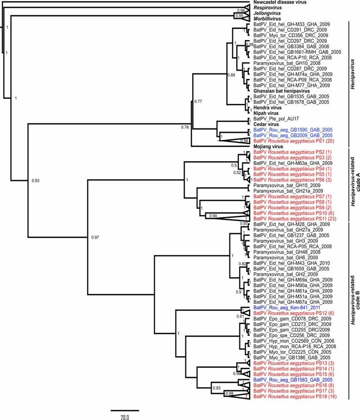
www.nature.com/scientificreports/
Henipavirus dynamics in their pteropid hosts and drivers of disease emergence have been studied in more
detail for Hendra and Nipah virus in Pteropus spp. from Australia and South Asia, respectively. Definitive sea-
sonality and magnitude of Hendra virus excretion pulses coinciding with disease spillover and outbreaks have
been reported, although viral excretion has been documented year-round30–33. Excretion pulses were shown to
be signifi antly higher during the dry winter s eason31; however, variations across latitudes were observed towards
Australia’s northern parts, where excretion was reported in dry and wet winter r egions32. Winter spillover varia-
tion was found to be related to winter temperatures, which affect bat behaviors and physiology, as well as human
behavior34. For Nipah virus, viral infections in bats were not seasonal but rather were driven by waning humoral
immunity in adult bat populations. Introduction of virus to a colony where seroprevalence was below a threshold
of herd immunity was a likely determinant of an outbreak within b ats35.
Investigations into African bat-borne henipaviruses have mostly been limited to the straw-colored fruit bat
(Eidolon helvum) with a diversity of henipa- and related viruses described—one of which, Ghanaian bat henipa-
virus, has been classifi d into the Henipavirus genus5,29,36–38. A longitudinal assessment of henipavirus antibodies
in a captive colony of E. helvum bats found pups to lose their maternal antibody protection between four and
twelve months following b irth39. Evidence suggests virus maintenance at a population level and horizontal virus
transmission during the pregnancy and lactation stages of the reproductive period. These findings were more
recently supported through molecular assessment of viral persistence and excretion in the same captive colony
of E. helvum bats. The results suggested viral shedding throughout the year; however, a signifi ant seasonal
pattern was detected, with peaks observed in July and J anuary40,41. However, limited follow-up studies have
been performed to gain information on the viral dynamics, pathogenicity, and host ecology of newly described
henipaviruses and related viruses in other bat species, such as R. aegyptiacus.
To address the role of R. aegyptiacus as a natural reservoir for henipavirus and related viruses on the Afri-
can continent, we performed initial biosurveillance followed by longitudinal viral excretion studies in a South
African bat population. Here, we report the detection of 18 henipa- and related paramyxoviruses, seasonal
excretion patterns of three putative species, and the influence of age on infection status. These fi dings can have
implications for paramyxovirus biosurveillance and risk assessments across the vast geographical distribution
of this bat species.
Materials and methods
Study site and regulatory requirements. Th s study targeted a population of R. aegyptiacus roosting
in Matlapitsi cave in Limpopo Province, South Africa (GPS: − 24.11487, 30.12151)42. These bats are present at
this site year-round, although population size varies depending on the time of the year, i.e., higher numbers
during the reproductive season (September to February) and lower numbers over the colder months (May to
August). Sampling of bats was conducted longitudinally on a near-monthly basis over several years as part of a
more extensive study on zoonotic virus biosurveillance. The cave is situated in the Matlapitsi valley, close to a
rural community with free-roaming livestock and fruit trees planted between human dwellings. Humans have
previously frequented the cave for religious practices.
Ethical clearance for the study was granted by the University of Pretoria Animal Ethics Committee, Faculty
of Veterinary Science, Onderstepoort, Pretoria, South Africa (Nos. 058-14 and 054-14) and the National Health
Laboratory Service (NHLS) Animal Ethics Committee, Sandringham, Johannesburg, South Africa (No. 137/12).
Permits for sample collection were obtained from the Department of Economic Development, Environment
and Tourism of the Limpopo Provincial Government in South Africa (Nos. CPM006806, ZA/LP/91509 and
ZA/LP/100499). Permission to conduct research on animals under section 20 of the Animal Disease Act (No.
35 of 1984) was granted by the Department of Agriculture, Land Reform and Rural Development (DALRRD)
of South Africa (No. 12/11/1/1/8). All methods were performed in accordance with the relevant guidelines and
regulations as stipulated by the institutional ethical approval boards.
Sample collection was performed using personal protective equipment, including Tyvek coveralls, powered
air-purifying respirators, fluid-resistant boots, double-layer nitrile gloves, and leather gloves, when handling
bats. Single-use items were decontaminated with sodium hypochlorite (5 g/L prepared with a 1:10 dilution of
domestic bleach), double-bagged into biohazard bags and transported to the BSL4 NHLS facility for autoclaving
and incineration. All reusable equipment was decontaminated with a sodium hypochlorite (5 g/L) solution and
sprayed down with 70% ethanol.
Weather data were obtained from the South African Weather Service at a daily resolution for the longitudi-
nal sampling period. The nearest weather station (Tzaneen-Westfalia Estate; ~ 42 km) and rain station (Wolk-
berg; ~ 14 km) were selected to calculate monthly temperature and rainfall averages (Supplementary Dataset 1).
Sample collection and sample sets. Several sample types were used (Table 1), with initial biosurveil-
lance conducted on archival spleen and urine samples opportunistically collected over several years (Supple-
mentary Fig. S1). These sample types were selected based on previous fi dings reporting successful detection of
henipavirus-related viral sequences in fruit bats5,36,37. Collection of these tissues and urine samples from indi-
vidual bats was performed as previously described whereby bats were sedated with a mixture of ketamine and
xylazine (1:2; a volume of 0.05 to 0.1 mg/g body mass) and euthanized through cardiac e xsanguination9. As a
noninvasive approach, population-level urine and fecal samples were collected monthly over more than two
years to assess paramyxovirus excretion over time. Population-level urine was collected as previously described,
and care was taken to collect clear urine samples not containing fecal m atter9. Briefly, urine droplets were col-
lected using cotton-tipped swabs or by pipetting from plastic trays placed in the cave underneath roosting bats.
For the collection of population-level fecal samples, fresh droppings were collected from the cave fl or under-
neath roosting bats. Th ee fecal deposits were collected using cotton-tipped swabs and pooled. Rectal swabs
Scientific Reports | (2021) 11:24262 | https://doi.org/10.1038/s41598-021-03641-w 2
Vol:.(1234567890)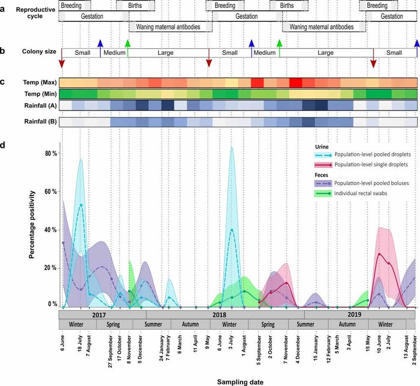
www.nature.com/scientificreports/
Sample set Sample type Number Description Collection date
1 Tissuea 302 Spleen samples from individual batsc 2012–2018
2 Urinea 58 Collected from individual bats 2013–2018
b Collected from sheets placed under-
3a Population-level urine (pooled) 260 June 2017–August 2018
neath roosting bats (10 per pool)
Collected from sheets placed under-
3b Population-level urine (not-pooled)b 475 September 2018–September 2019
neath roosting bats
Collected from the cave fl or under-
4 Population-level fecal (pooled)b 871 June 2017–September 2019
neath roosting bats (3 per pool)
5 Rectal swabsb 712 Collected from individual bats November 2017–May 2019d
Table 1. Sample selection for assessing the diversity and excretion dynamics of henipa- and related viruses
in Egyptian rousette bats. a Opportunistically collected. b Longitudinal monthly collection. c Kidney, liver, lung
and intestinal tissue tested for spleen-positive individuals. d Collection of rectal swabs was not performed for
January, March or December 2018.
collected from individual bats were included in the study to determine whether specific host characteristics such
as age, sex, or forearm mass index (FMI) can be correlated with infection status. The swabs were collected under
anesthetics during catch-and-release sampling using a mixture of ketamine and xylazine on 1:2 ratio (a volume
of 0.05 to 0.1 mg/g body mass) as previously described9. Sterile cotton swabs (VWR Critical Swab) were dipped
in 1 × phosphate-buffered saline (PBS) before being inserted into the rectum of bats and rotated three times. All
biological samples were immediately transferred into a liquid nitrogen-charged dryshipper for transport to the
laboratory and stored at -80 °C until further processing.
Morphological measurements of all bats were taken, including weight, forearm length, age, and sex. The age of
the bats was determined based on forearm measurements, where > 89 mm was classifi d as adults and ≤ 89 mm as
subadults42–44. Each bat was marked by a 3-letter-3-digit tattoo code on the propatagium of the left ventral wing
of the bat. Th s procedure was performed while the bats were sedated and allowed for analyses of recaptured bats.
Briefly, this involved cleaning of the wing surface with AIMS animal tissue prep (AIMS™), after which the code
was tattooed onto the wing. Tattoo ink (AIMS Permanent black pigment; AIMS™, USA) was injected into the
skin using a seven-point round liner (7RL) needle and a Bold Monk Ordinary Tattoo Machine (GetInked, South
Africa). Bats recaptured during the same sampling event as when initial marking was done were released. Upon
recapture during a subsequent monthly sampling event, sample collection was repeated, and the tattoo number
was documented. Detailed sample information is recorded in Supplementary Dataset 2 available online. During
sampling, collection of proportional numbers representing both sexes and age groups was attempted as much
as possible. However, colony structure varied over time, particularly from February to April, where noticeably
higher numbers of subadults were sampled.
Sample preparation and molecular testing. Sample processing was performed in a biosafety level 3
facility (BSL3) at the Prinshof campus, University of Pretoria, South Africa. RNA was extracted from tissue and
urine samples using TRIzol® reagent (Invitrogen) according to the manufacturer’s instructions. Rectal swabs and
fecal material were resuspended in 800 µl and 400 µl 1 × PBS, respectively, and vortexed briefly to ensure proper
resuspension. Two hundred microliters of the suspension was inactivated with the addition of an equal volume
of 2 × DNA/RNA shield solution (Zymo Research), and RNA extraction was performed using the Zymogen
Quick-RNA MiniPrep Plus kit (Zymo Research) according to the manufacturer’s specifi ations.
Reverse transcription was performed using either the SuperScript III or IV™ reverse transcriptase enzyme
(Invitrogen) with 100 ng of purifi d random hexamers and 5 µl extracted RNA according to the manufacturer’s
recommendations. Incubation was performed at 25 °C for 5 min, 60 °C for 50 min and 85 °C for 5 min. Nucleic
acid amplifi ation was performed using Res-Mor-Hen (RMH) primers targeting the polymerase gene in combina-
tion with an in-house optimized two-step heminested RT-PCR a ssay45. For the first round of PCR amplification,
each reaction consisted of 5 µl of cDNA, 0.2 mM RMH-F1 primer, 0.3 mM RMH-R primer, 0.4 mM dNTP mix
(Thermo Scientific), 2 mM MgCl2, 1 × DreamTaq™ buffer, 1.25 U DreamTaq™ polymerase (5 U/µl) and nuclease-
free water to a fi al volume of 50 µl. Amplifi ation was performed at 94 °C for 2 min; 40 cycles of 94 °C for 15 s,
48 °C for 30 s and 72 °C for 30 s; and 72 °C for 7 min. Hemi-nested reaction setup and cycling conditions were
performed as described for the fi st round of amplifi ation using 0.3 mM of the RMH-F2 and RMH-R primers,
excluding MgCl2 and nuclease-water adapted accordingly.
Products were analyzed on a 1.5% agarose gel, and positive samples were subjected to Sanger sequencing on
an ABI 3500xl instrument using the BigDye Terminator v3.1 Cycle Sequencing Kit (Thermo-Fisher Scientifi ).
For all spleen-positive individuals, tissue from the kidney, liver, lung and intestine was also tested for paramyxo-
virus RNA.
Statistical analyses. Prevalence of excretion: effect of sample type and pooling. An overall detection fre-
quency was calculated as the number of PCR-positive samples over the number of tested samples for each type
of sample: pooled swabs, individual rectal swabs, pooled urine droplets and single droplets. Because pooling can
induce an upward bias of the prevalence, we also estimated a corrected overall prevalence for pooled samples
using the Prevalence package and truePrevPools function in R (v.4.0.2) as previously described46,47.
Scientific Reports | (2021) 11:24262 | https://doi.org/10.1038/s41598-021-03641-w 3
Vol.:(0123456789)www.nature.com/scientificreports/
Sample type Number tested Number positive Positivity Corrected positivity Variation over time
p < 0.001
Population-level urine (pooled) 260 16 6.2 ± 2.9% 3.4 ± 1.3%
(GLM: χ214 = 52.479)
p < 0.001
Population-level urine (not-pooled) 475 25 5.3 ± 2.0% n/a
(GLM: χ212 = 64.442)
p < 0.001
Population-level fecal (pooled) 871 46 5.3 ± 1.5% 2.3 ± 0.6%
(GLM: χ227 = 81.541)
p = 0.024
Rectal swabs 712 12 1.7 ± 0.9% n/a
(GLM: χ215 = 27.648)
Table 2. Overall detection rates of henipavirus and related viral RNA in different sample types across the
sampling period.
For urine, we tested the effect of the sampling approach (pooled vs not pooled) on the percentage positiv-
ity over the period for which the two collection methods were applied (November 2017 to May 2019). We also
compared the prevalence between urine and feces to assess the effect of sample type, fi st using pooled urine and
pooled feces samples (from June 2017 to August 2018) and then single urine and pooled feces samples (from
September 2018 to May 2019). Comparisons were performed using Chi-squared tests in R.
Temporal excretion dynamics. Data collected from the different sample types (sample sets 3, 4 and 5; Table 1)
were considered separately to investigate the excretion dynamics of henipa- and related viruses. We used gen-
eralized linear models (GLMs) in R, with the sampling date (coded as different sessions) used as an explanatory
variable and a binomial error and a logit link function. For individual rectal swabs, host sex, age and FMI, and
their interactions were also included in the model. The forearm mass index was determined with forearm meas-
urements, and the weight of the bats was expressed in kg/m2. ANOVA of the GLMs and a chi-squared test were
performed to assess the signifi ance of the variables.
Estimating viral diversity. To estimate viral diversity, all sequences were aligned in BioEdit (v7.2.5)
and trimmed to equal length48. The corresponding gene regions for Hendra and Nipah viruses were obtained
from complete genomes available through the National Centre for Biotechnology for Information (NCBI) and
included in the alignment (GenBank accession numbers NC_001906 and NC_002728, respectively). The align-
ment was subsequently translated to amino acid sequences, and a similarity matrix was generated. An amino
acid sequence identity of 94% shared between Hendra and Nipah virus for this region was used as a conservative
cut-off value to identify putative viral species. Th s cut-off was used to further group sequences together for a
fi al count of putative paramyxovirus species.
Phylogenetic analyses of detected viral sequences. Sequence alignments of detected viral sequences
and representatives from the literature were generated using BioEdit (Supplementary Tables S1 and S2)48. The
substitution model selection for the nucleotide alignment was performed using jModelTest (v2.1.10)49. Bayesian
phylogenetic analyses were performed using the predetermined best fit model in BEAST (v2.5) with 10,000,000
iterations sampling every 1000 trees and was checked for an effective sample size (ESS) value of > 200 to ensure
convergence50. The fi al phylogeny was generated using FigTree (v1.4.2) (2006–2012 Andrew Rambaut, Institute
of Evolutionary Biology, University of Edinburgh). Clades representing the same putative henipavirus or related
virus species were collapsed for improved visualization.
Results
Paramyxoviral detection rate and temporal excretion dynamics. To investigate the detection
rates, diversity, and viral excretion dynamics of henipa- and related viruses associated with R. aegyptiacus bats,
we sampled and tested a total of 2678 samples collected between 2012 and 2019. Although the assay also targeted
viruses from the Respiro- and Morbillivirus genera, only viral sequences related to the Henipavirus genus were
detected. Initial biosurveillance performed using opportunistically collected spleen and urine samples from
individual bats between 2012 and 2018 (Table 1) indicated the presence of henipa- and related viruses in this bat
population. Low detection frequencies of 1.66% (n = 5) and 1.72% (n = 1) were found in the two sample types,
respectively. Of the five bats with viral RNA present in splenic tissue, one additionally tested positive for RNA in
the renal tissue with an identical RNA sequence reported in both.
Further investigation into the preliminary fi dings detected viral RNA in 4.35% of pooled roost urine samples
and in 2% of fecal samples across the entire sampling period (Table 2). We found no difference in the estimated
detection rates when using pooled fecal samples from beneath the colony or rectal swabs from individual bats
(Chi-squared test: χ21 = 0.520, p = 0.819). Finally, comparison of viral RNA excretion in pooled feces and urine
showed a signifi antly higher excretion in urine, both using pooled urine samples from June 2017 to August
2018 (Chi-squared test: χ21 = 7.380, p = 0.007) and single urine droplets from September 2018 to May 2019
(χ21 = 11.115, p < 0.001). None of the 77 recaptured bats tested positive for paramyxovirus RNA in their rectal
swabs, and inferences on changes in infection status of individual bats throughout the sampling period could
not be made.
Assessment of the temporal excretion dynamics of the henipavirus and related viruses indicated a strong
variation over time regardless of sample type (Table 2). Overall, viral RNA excretion was detected throughout
Scientific Reports | (2021) 11:24262 | https://doi.org/10.1038/s41598-021-03641-w 4
Vol:.(1234567890)www.nature.com/scientificreports/
most of the year, with a peak in viral excretion observed around June/July for three consecutive years (Fig. 1).
Lower excretion levels were reported from September to November and seemingly decreased towards the end
of each year. Notably, a cyclic absence of excretion was observed in autumn between February and May 2018
and 2019. Peak viral excretion is believed to follow in line with the waning of maternal antibodies. The viral
excretion peaks also fell within the winter months for the region characterized by a cold, dry climate and limited
food availability (Fig. 1). However, at a lower detection frequency, the continued excretion during the spring
(September to November) coincides with the recolonization of the roost (Fig. 1).
Analyses of bat morphometric and morphological data of individuals from which rectal swabs were included
indicated no effect of sex (GLM: χ21 = 0.519, p = 0.471) or FMI (GLM: χ21 = 1.761, p = 0.184) on infection status.
A correlation between age and infection status was found, with subadults being more infected than adults (GLM:
χ21 = 6.199, p = 0.013). Although the proportion of subadults sampled was much higher in the period following
the annual birth pulse (February to May), viral positives were detected during times where age classes were not
skewed (Supplementary Fig. S2). This suggests that the correlation between subadults and infection status was
not affected by the seasonal abundance of subadults.
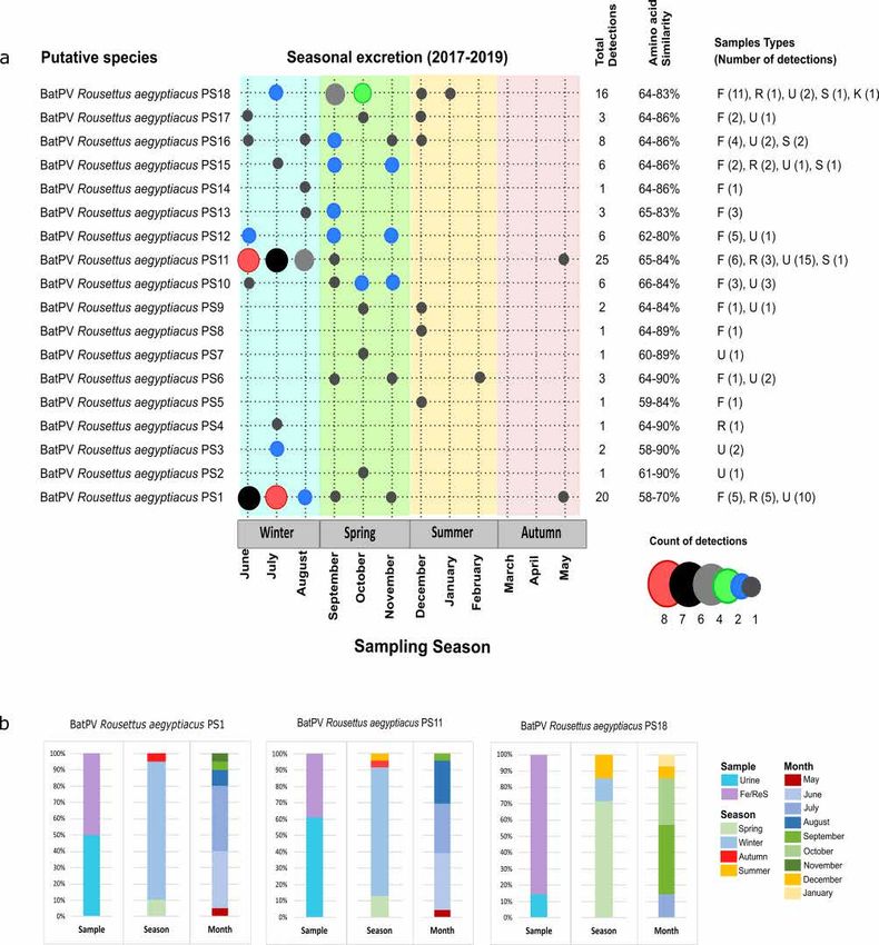
Viral diversity and phylogenetic analyses. The henipa- and related viral diversity in R. aegyptiacus
have not been considered extensively in previous biosurveillance studies. Assessment of the amino acid similar-
ity between 106 viral sequences detected in this study indicated the presence of 18 putative viral species (PS)
using a conservative estimate of 94% similarity at the amino acid level (Fig. 2). The overall similarity shared
between putative viral species ranged from 58 to 90%, with Bat Rousettus aegyptiacus PS1 being most divergent
at 58–70% similarity shared. Sequences highly similar (up to 99.3% amino acid similarity) to Bat Rousettus
aegyptiacus PS1, PS11, PS12 and PS16 were previously reported in the same bat species from Kenya, Ghana and
Rwanda5,6,21. The remaining 14 putative viral species are newly described and contribute to paramyxovirus diver-
sity in Egyptian rousette bats. Most putative viral species were observed at a very low frequency and detected on
less than 10 occasions throughout the study.
Th ee of the putative henipavirus and related viral species, BatPV Rousettus aegyptiacus PS1, PS11 and PS18,
were the most frequently detected in bat excretions, being recorded 20, 23 and 14 times, respectively (Fig. 2).
When considering the sample type and time of sampling, the profiles of excretion were similar for BatPV Rouset-
tus aegyptiacus PS1 and PS11. These two species were detected near equally in both urine and feces/rectal swabs
and predominantly in the winter months of June and July. In contrast, BatPV Rousettus aegyptiacus PS18 was
more frequently detected in feces/rectal swabs and appeared later in the year during the spring months. These
three species drive the excretion peaks observed in winter and spring, respectively (Fig. 1).
All sequences detected phylogenetically grouped with the Henipavirus genus or in closely related sister clades,
and none with the Respiro- or Morbillivirus genera which was also targeted by the assay. Analysis indicated that
BatPV Rousettus aegyptiacus PS1, which is most diverse from the other detected sequences, groups among
viruses within the Henipavirus genus (Fig. 3). Putative species BatPV Rousettus aegyptiacus PS2 to PS11 group
together in the Henipavirus-related clade A and PS12 to PS18 in Henipavirus-related clade B. Viruses grouping in
Henipavirus-related clade B were overall more prevalent in gastrointestinal excretions—as described for BatPV
Rousettus aegyptiacus PS18. However, a definite association with this route of excretion cannot be made across
the clade due to the overall low detection of the associated putative viral species (Fig. 2). As reported for the
putative species analyses, some of the viral sequences grouped closely with those detected in the same host spe-
cies from different African countries. The clustering of viruses specific to certain bat species/genera corresponds
to previous hypotheses of host specificity of bat-borne p aramyxoviruses5,6.
Discussion
Biosurveillance studies in wildlife signifi antly contribute to the identifi ation of viral diversity and unrecognized
host species. Cross-sectional studies are often limited due to sample type bias, time of sampling and sample sizes
that are not optimal for detecting and assessing viral diversity. As such, there is a need for more targeted longitu-
dinal biosurveillance to adequately address research questions related to bat-borne viral diversity, maintenance,
and dynamics within bat populations. Findings associating R. aegyptiacus bats with henipaviruses have been
limited, with only seven detections reported from populations around Equatorial Africa5,6,20,21. By focusing our
research on this bat species through maximizing our sample sizes and adding a temporal component, we were
able to obtain high-resolution data and insight into the dynamics of henipa- and related virus diversity.
Findings from our biosurveillance support and supplement previous detections of henipa- and related viruses
and expand the known virus diversity with at least 14 putative henipa- and related virus species. Th s is consider-
ably higher than that reported for viruses belonging to other viral families, such as the Filo-, Adeno-, Herpes- and
Coronaviridae (Geldenhuys et al. in preparation), detected in the same target p opulation14,51,52. High viral diver-
sity and strain detection rates increase the probability of strains with zoonotic potential present in these popula-
tions, as exemplifi d by henipaviruses and Sosuga pararubulavirus, which are observed at a higher frequency in
their respective bat reservoirs3,12,30. In conjunction, the repeat detection of viral RNA across years could be an
indication of the establishment and maintenance of these viruses within their natural reservoir populations and
increased intraspecies transmissibility. The risk of human and livestock exposure or disease spillover might be
considered much lower for the diversity documented at a very low detection rate; however, this diversity should
not be disregarded, as their zoonotic potential remains unknown. The three dominant putative species presenting
with much higher detection frequencies provide the opportunity for increased exposure and potential spillover,
posing a higher risk for the local community. Moreover, the detection of these viruses in bat excreta highlights
the potential for environmental contamination not only in the cave where these bats roost but also across their
foraging range, as previously demonstrated for Nipah virus53.
Scientific Reports | (2021) 11:24262 | https://doi.org/10.1038/s41598-021-03641-w 5
Vol.:(0123456789)www.nature.com/scientificreports/
Figure 1. Detection of paramyxoviral RNA in a population of Egyptian rousette bats and descriptive host
and environmental considerations from Matlapitsi cave, Limpopo Province, South Africa, 2017–2019. (a)
Estimated stages over the reproductive cycle of the Egyptian rousette bat populations in South Africa (based
on unpublished observations made during catch-and-release sampling and previous fi dings) 42. Shaded areas
represent an early and late estimate for each stage; (b) Colony size variation across the sampling period defi ed
as small—when the population size is at its lowest (~ 3000), medium—when the roost is recolonized before the
birthing season (~ 7000), and large—following the birthing pulse (> 9000)42. Red arrows indicate the periods
of mass departure from the roost near the end of May, the blue arrows represent recolonization of the roost
in spring, and the green arrows indicate the influx of naïve individuals during the birthing pulse; (c) Monthly
climate data (maximum and minimum temperatures and rainfall (A) from the closest weather station to the
roost and additional rainfall (B) data from a closely situated rain station. The maximum temperature color scale
bar ranges from light red to dark red (22–32.6 °C), minimum temperatures from green to light orange (8.4–
19 °C), and rainfall from light to dark blue (0–168 mm); (d) Percentage positivity of urine, fecal and rectal swab
samples for henipa- and related viral RNA detected monthly over the longitudinal sampling period calculated
per monthly sampling event. Colored dots represent raw point data, with dashed lines showing values predicted
by a loess function in R. Shaded areas represent 95% confide ce intervals.
Serological investigations into henipavirus dynamics in E. helvum bats in Africa provided initial insight
into the association of these viruses with African fruit bat species39,54. These studies primarily relied on the
cross-reactivity of African henipaviruses with Nipah virus and reported the limitation of not working with a
Scientific Reports | (2021) 11:24262 | https://doi.org/10.1038/s41598-021-03641-w 6
Vol:.(1234567890)www.nature.com/scientificreports/
Figure 2. Putative henipavirus and related virus species detected in an Egyptian rousette bat population at
Matlapitsi cave, Limpopo Province, South Africa. a. Seasonal excretion of putative viral species indicated in
different color bars per season (sampling period June 2017 to September 2019). The number of detections is
represented by dots of different sizes and colors. The total number of detections represents both the initial
tissue-based biosurveillance and longitudinal excretion data. Amino acid similarities were based on a 150
amino acid sequence and indicate the similarity shared between the putative species. Sample types are F—fecal,
R—rectal swabs, U—urine, S—spleen, and K—kidney. b. Bar charts represent the proportion of RNA-positive
samples for putative species (PS) 1 (n = 20), PS11 (n = 23), and PS18 (n = 14) detected in bat excreta specific o
sample type, season, and month of detection. For sample type, detections for fecal (Fe) and rectal swabs (ReS)
were combined, as they form part of the same excretion pathway.
fully characterized host–pathogen s ystem39. Studies considering the specifi drivers of the dynamics of unclas-
sifi d bat-borne viral diversity in their associated bat species are lacking. Although some drivers are considered
Scientific Reports | (2021) 11:24262 | https://doi.org/10.1038/s41598-021-03641-w 7
Vol.:(0123456789)www.nature.com/scientificreports/
Figure 3. Bayesian phylogenetic analyses of partial paramyxovirus polymerase (L) gene sequences detected
in Egyptian rousette bats. The proportional phylogeny was based on an alignment of sequences with 439
nucleotide lengths. Bayesian phylogenetic analysis was performed using the TIM3 + I + G substitution model
with a resulting effective sample size (ESS) value of 594.3. Sequence names in red represent sequences detected
in this study, blue—sequences detected in the same bat species from other countries, and black—representative
sequences from the literature. Numbers in brackets at the end of red sequence names indicate the number of
detections per sequence. Posterior probabilities of more than 0.5 are indicated at internal nodes, and large clades
were collapsed. Newcastle disease virus was selected as an outgroup, and other Orthoparamyxovirinae genera
are indicated in bold italics. GenBank accession numbers for all sequences used in the phylogenetic analyses are
provided in Supplementary Tables S1 and S2.
Scientific Reports | (2021) 11:24262 | https://doi.org/10.1038/s41598-021-03641-w 8
Vol:.(1234567890)www.nature.com/scientificreports/
common for many viruses, such as the effect of season and reproduction, regional specific drivers can additionally
interplay and affect virus dynamics and spillover potential. Th s is exemplifi d by the presence of Marburg virus
in South African populations of R. aegyptiacus without local disease emergence—possibly due to the absence of
extrinsic factors driving outbreaks in other parts of Africa such as hunting for and consumption of bat bushmeat
or entering of caves for guano m ining14,51. With our longitudinal molecular approach, we were able to study the
dynamics of African henipa- and related viruses in a single host species. Compared to previous reports, synchro-
nized shedding of multiple putative viral species was observed yearly, although dominant putative viral species
displaying variable seasonality were detected9,41,55. Similar fi dings have been reported from a captive population
of E. helvum bats in Ghana, whereby distinct paramyxoviral sequences display various shedding patterns41. Th s
suggests the involvement of different local drivers for various paramyxovirus taxa, highlighting the limitations
of lower resolution studies where such differences might be overlooked.
The temporal dynamics of BatPV Rousettus aegyptiacus PS1 and PS11 are in line with data previously reported
for Hendra virus with a characteristic winter seasonality for peak viral excretion31. During this time, the bat
population at Matlapitsi cave is at its lowest (Fig. 1), with most of the population exiting the roost to overwinter
in other locations with a warmer climate and where food is likely more readily available42. For the remaining
individuals, low food availability and dependance on alternate food sources, such as cultivated fruit, could result
in nutritional stress. Th s has previously been suggested to have a physiological impact on bats and reduced
immunocompetence, making them susceptible to i nfection30,56. Intraspecies transmission and exposure of local
humans and livestock populations to bat-borne paramyxoviruses are more likely to occur in winter due to the
aggregation of R. aegyptiacus bats around limited food sources, and these bats potentially feeding on cultivated
fruiting trees planted throughout human settlements, as observed over the course of field studies in the area.
Although colder winter temperatures and nutritional stress may intensify pulses of viral shedding, the obser-
vation of a period of no detectable excretion before the observed peak in the winter months coincides with the
influx of naïve individuals (subadults) into the colony following the birth pulse and suggests the involvement of
maternal antibody protection (Fig. 1). Waning of maternal immunity provides a synchronized influx of naïve
individuals who could be exposed to these viruses, resulting in large-scale horizontal transmission, infection, and
virus detection during these pulses. The birthing pulse for the targeted South African population of R. aegyptiacus
in this study was documented in late spring (October/November), which is approximately six months before
the peak in viral e xcretion42. These data correspond to serological findings of the loss of henipavirus maternal
antibodies in E. helvum bats at four to 12 months (averaged at six months) following p arturition39,54. While our
study did not include a serological aspect to confirm the influence of maternal immunity on infection acquisition
during the study period, our molecular fi dings seem to support a greater risk of infection with henipavirus and
related viruses following the loss of maternal immunity.
A difference in seasonality was described for BatPV Rousettus aegyptiacus PS18, with excretion predominantly
detected in the spring, coinciding with the recolonization of the roost. The detection of this putative viral spe-
cies predominantly upon roost colonization could represent the viral species diversity in other roosts that are
reintroduced here with the influx of infected individuals into the c olony57. However, since this is an open popu-
lation with regular movement in and out of the colony to one or more different roosts, the possibility of a large
metapopulation across southern Africa should be considered before such inferences can be made42. It becomes
increasingly important to study bat movement patterns and collect spatiotemporal virological and serological
data across the distribution of this bat species. Viral interspecies interactions could also account for a shift in
the seasonality of excretion between the virus taxa, as previously demonstrated for rhinovirus and parainfluenza
virus coinfections in humans58.
Our study provides some considerations on how enzootic henipavirus and related viruses in R. aegyptiacus
circulate within a southern population and the associated exposure risk in this area. At a local scale, the period
with the highest risk for human and livestock exposure to henipavirus and related viruses was determined to be
winter through spring. When the natural wild fig food source of R. aegyptiacus is limited during winter, increased
human contact is possible when bats seek other food sources, such as the cultivated fruit trees found between
human dwellings within the area. In addition, R. aegyptiacus has been documented to expand their home ranges
in winter and forage further away from the roosts, which results in increased dispersion to other settlements
within the greater area17. The Tzaneen region northeast of the cave is known for its tropical and subtropical
agriculture with cultivated fruits as one of their main lines of produce—which could serve as a secondary food
source for these bats. Therefore, risk assessments for winter months should include settlements within a wider
radius around the cave roost. Overwintering roosts also warrant consideration for risk assessments due to the
observed increase in viral shedding during this time and the associated infection status of subadults participat-
ing in the movement between roosts. Th s highlights the need to incorporate bat tracking in larger-scale studies
to identify secondary roosts. During spring, when the roost is recolonized and the fig trees start to fruit, the
now larger population of bats will remain mostly localized and feed and within the Matlapitsi valley, increasing
contact rates with humans and free-roaming livestock. Consequently, the landscape structure, bat movement
and feeding behaviour, proximity of the human settlement to a roosting cave, and human behaviour might be
the most important risk factors to consider when performing risk assessments and providing recommendations
to the local community.
Extrapolating these conclusions to other populations of R. aegyptiacus bats across its geographical distribu-
tion should be made with caution, as the viral dynamics might be markedly different due to differences in life-
history traits and environmental conditions. Egyptian rousette populations closer to the equator, such as those
in Uganda, have bi-annual birthing pulses, which is likely due to bimodal rainfall and the year-round availability
of food in these tropical r egions59,60. As such, there will be two periods where an influx of naïve individuals into
the population is observed. If the waning of maternal immunity is a driver of henipavirus and related virus
dynamics within this bat species, two peaks in viral excretion would be expected within a short succession.
Scientific Reports | (2021) 11:24262 | https://doi.org/10.1038/s41598-021-03641-w 9
Vol.:(0123456789)www.nature.com/scientificreports/
However, if climatic conditions such as dry winter periods and nutritional stress are determining factors, then
the expected excretion peaks would likely not be as pronounced. In contrast, the most southern population of
R. aegyptiacus, documented on Table Mountain in South Africa, occurs in a region characterized by a wet-cold
winter climate. Th s population is not believed to migrate to other roosts and represents a more closed popula-
tion structure61. Assessing these intrinsic and extrinsic factors at a local scale will ultimately provide the data
necessary to understand henipavirus and related virus dynamics in R. aegyptiacus bats across their distribution
and aid in the movement towards more spatiotemporal predictions of disease emergence hotspots.
Received: 17 September 2021; Accepted: 6 December 2021
References
1. IlungaKalenga, O. et al. The Ongoing Ebola Epidemic in the Democratic Republic of Congo, 2018–2019. N. Engl. J. Med. 381,
373–383 (2019).
2. Arunkumar, G. et al. Outbreak investigation of nipah virus disease in Kerala, India, 2018. J. Infect. Dis. 219, 1867–1878 (2019).
3. Olival, K. J. et al. Host and viral traits predict zoonotic spillover from mammals. Nature 546, 646–650 (2017).
4. Mollentze, N. & Streicker, D. G. Viral zoonotic risk is homogenous among taxonomic orders of mammalian and avian reservoir
hosts. Proc. Natl. Acad. Sci. USA. 117, 9423–9430 (2020).
5. Drexler, J. F. et al. Bats host major mammalian paramyxoviruses. Nat. Commun. 3, 1–12 (2012).
6. Mortlock, M. et al. Novel paramyxoviruses in bats from Sub-Saharan Africa, 2007–2012. Emerg. Infect. Dis. 21, 1840–1843 (2015).
7. Wilkinson, D. A. et al. Highly diverse morbillivirus-related paramyxoviruses in wild fauna of the southwestern Indian Ocean
Islands: Evidence of exchange between introduced and endemic small mammals. J. Virol. 88, 8268–8277 (2014).
8. Mélade, J. et al. An eco-epidemiological study of Morbilli-related paramyxovirus infection in Madagascar bats reveals host-
switching as the dominant macro-evolutionary mechanism. Sci. Rep. 6, 1–12 (2016).
9. Mortlock, M., Dietrich, M., Weyer, J., Paweska, J. T. & Markotter, W. Co-circulation and excretion dynamics of diverse Rubula:
And related viruses in Egyptian rousette bats from South Africa. Viruses 11, 1–22 (2019).
10. Anthony, S. J. et al. Global patterns in coronavirus diversity. Virus Evol. 3, 1–15 (2017).
11. Geldenhuys, M. et al. A metagenomic viral discovery approach identifies potential zoonotic and novel mammalian viruses in
Neoromicia bats within South Africa. PLoS ONE 13, 1–27 (2018).
12. Amman, B. R. et al. A recently discovered pathogenic paramyxovirus, Sosuga Virus, is present in Rousettus aegyptiacus fruit bats
at multiple locations in Uganda. J. Wildl. Dis. 51, 774–779 (2015).
13. Amman, B. R. et al. Isolation of Angola-like Marburg virus from Egyptian rousette bats from West Africa. Nat. Commun. 11, 1–9
(2020).
14. Pawęska, J. T. et al. Shedding of marburg virus in naturally infected egyptian rousette bats, South Africa, 2017. Emerg. Infect. Dis.
26, 3051–3055 (2020).
15. Albariño, C. G. et al. Novel paramyxovirus associated with severe acute febrile disease, South Sudan and Uganda, 2012. Emerg.
Infect. Dis. 20, 211–216 (2014).
16. Benda, P., Vallo, P., Hulva, P. & Horáček, I. The Egyptian fruit bat Rousettus aegyptiacus (Chiroptera: Pteropodidae) in the Palae-
arctic: Geographical variation and taxonomic status. Biol. 67, 1230–1244 (2012).
17. Luĉan, R. K. et al. Spatial activity and feeding ecology of the endangered northern population of the Egyptian fruit bat (Rousettus
aegyptiacus). J. Mammal. 97, 815–822 (2016).
18. Strachinis, I., Kalaentzis, K., Katsiyiannis, P. & Kazilas, C. First record of the Egyptian fruit bat, Rousettus aegyptiacus (Pteropo-
didae), from Kastellorizo island, Greece. Mammalia 82, 611–613 (2018).
19. ACR. African Chiroptera Report 2020. (2020).
20. Conrardy, C. et al. Molecular detection of adenoviruses, rhabdoviruses, and paramyxoviruses in bats from Kenya. Am. J. Trop.
Med. Hyg. 91, 258–266 (2014).
21. Markotter, W. et al. Paramyxo- and coronaviruses in Rwandan bats. Trop. Med. Infect. Dis. 4, 1–11 (2019).
22. van Vuren, P. J. et al. A novel adenovirus isolated from the Egyptian fruit bat in South Africa is closely related to recent isolates
from China. Sci. Rep. 8, 9584 (2018).
23. Sasaki, M. et al. Identifi ation of group A rotaviruses from Zambian fruit bats provides evidence for long-distance dispersal events
in Africa. Infect. Genet. Evol. 63, 104–109 (2018).
24. Kandeil, A. et al. Isolation and characterization of a distinct influenza A virus from Egyptian bats. J. Virol. 93, 18 (2018).
25. Waruhiu, C. et al. Molecular detection of viruses in Kenyan bats and discovery of novel astroviruses, caliciviruses and rotaviruses.
Virol. Sin. 32, 101–114 (2017).
26. Halpin, K. & Rota, P. A Review of Hendra Virus and Nipah Virus Infections in Man and Other Animals. In Zoonoses: Infections
Affecting Humans and Animals: Focus on Public Health Aspects (ed. Sing, A.) 997–1012 (Springer, 2015).
27. Gurley, E. S. et al. Person-to-person transmission of Nipah Virus in a Bangladeshi community. Emerg. Infect. Dis. 13, 1031–1037
(2007).
28. Gokhale, M. D. et al. Detection of possible Nipah virus infection in Rousettus leschenaultii and Pipistrellus Pipistrellus bats in
Maharashtra, India. J. Infect. Public Health 14, 1010–1012 (2021).
29. Pernet, O. et al. Evidence for henipavirus spillover into human populations in Africa. Nat. Commun. 5, 1–10 (2014).
30. Paez, D. J. et al. Conditions affecting the timing and magnitude of Hendra virus shedding across pteropodid bat populations in
Australia. Epidemiol. Infect. 145, 3143–3153 (2017).
31. Field, H. et al. Spatiotemporal aspects of Hendra virus infection in pteropid bats (flying-foxes) in Eastern Australia. PLoS ONE
10, 1–14 (2015).
32. Martin, G. et al. Hendra virus spillover is a bimodal system driven by climatic factors. EcoHealth 15, 526–542 (2018).
33. Field, H. et al. Hendra virus infection dynamics in Australian fruit bats. PLoS ONE 6, 1–6 (2011).
34. McKee, C. D. et al. The ecology of nipah virus in bangladesh: A nexus of land-use change and opportunistic feeding behavior in
bats. Viruses 13, 1–10 (2021).
35. Epstein, J. H. et al. Nipah virus dynamics in bats and implications for spillover to humans. Proc. Natl. Acad. Sci. USA. 117,
29190–29201 (2020).
36. Baker, K. S. et al. Co-circulation of diverse paramyxoviruses in an urban African fruit bat population. J. Gen. Virol. 93, 850–856
(2012).
37. Muleya, W. et al. Molecular epidemiology of paramyxoviruses in frugivorous Eidolon helvum bats in Zambia. J. Vet. Med. Sci. 76,
611–614 (2014).
38. Weiss, S. et al. Henipavirus-related sequences in fruit bat bushmeat, Republic of Congo. Emerg. Infect. Dis. 18, 1536–1537 (2012).
39. Baker, K. S. et al. Viral antibody dynamics in a chiropteran host. J. Anim. Ecol. 83, 415–428 (2014).
Scientific Reports | (2021) 11:24262 | https://doi.org/10.1038/s41598-021-03641-w 10
Vol:.(1234567890)www.nature.com/scientificreports/
40. Gibson, L. et al. Persistence of multiple paramyxoviruses in a closed captive colony of fruit bats (Eidolon helvum). Viruses 13, 1659
(2021).
41. Jolma, E. R. et al. Longitudinal secretion of paramyxovirus RNA in the urine of straw-coloured fruit bats (Eidolon helvum). Viruses
13, 1654 (2021).
42. Jacobsen, N. H. G. & Du Plessis, E. Observations on the ecology and biology of the Cape fruit bat Rousettus aegyptiacus leachi in
the Eastern Transvaal. S. Afr. J. Sci. 72, 270–273 (1976).
43. Lučan, R. K. et al. Reproductive seasonality of the Egyptian fruit bat (Rousettus aegyptiacus) at the northern limits of its distribu-
tion. J. Mammal. 95, 1036–1042 (2014).
44. Mutere, F. A. The breeding biology of the fruit bat Rousettus aegyptiacus E. Geoffroy living at 0°22’S. Acta Trop. 25, 97–108 (1968).
45. Tong, S., Chern, S.-W.W., Li, Y., Pallansch, M. A. & Anderson, L. J. Sensitive and broadly reactive reverse transcription-PCR assays
to detect novel paramyxoviruses. J. Clin. Microbiol. 46, 2652–2658 (2008).
46. R Core Team. R: A Language and Environment for Statistical Computing. (R Foundation for Statistical Computing, 2018). https://
www.R-project.org/.
47. Devleesschauwer, B. et al. The prevalence package. 1–28 (2014).
48. Hall, T. A. BioEdit: A user-friendly biological sequence alignment editor and analysis program for Windows 95/98/NT. Nucleic
Acids Symp. Ser. 41, 95–98 (1999).
49. Darriba, D., Taboada, G. L., Doallo, R. & Posada, D. jModelTest 2: More models, new heuristics and high-performance computing.
Nat. Methods 9, 1–4 (2015).
50. Bouckaert, R. et al. BEAST 2.5: An advanced software platform for Bayesian evolutionary analysis. BioRxiv (2018).
51. Paweska, J. T. et al. Marburg virus infection in Egyptian rousette bats, South Africa, 2013–2014. Emerg. Infect. Dis. 24, 1134–1137
(2018).
52. Dietrich, M., Kearney, T., Seamark, E. C. J., Paweska, J. T. & Markotter, W. Synchronized shift of oral, faecal and urinary microbiotas
in bats and natural infection dynamics during seasonal reproduction. R. Soc. Open Sci. 5, 180021 (2018).
53. Chua, K. B. et al. Isolation of Nipah virus from Malaysian Island flying-foxes. Microbes Infect. 4, 145–151 (2002).
54. Peel, A. J. et al. Support for viral persistence in bats from age-specific serology and models of maternal immunity. Sci. Rep. 8, 1–11
(2018).
55. Peel, A. J. et al. Synchronous shedding of multiple bat paramyxoviruses coincides with peak periods of Hendra virus spillover.
Emerg. Microbes Infect. 8, 1314–1323 (2019).
56. Plowright, R. K. et al. Reproduction and nutritional stress are risk factors for Hendra virus infection in little red flying foxes
(Pteropus scapulatus). Proc. R. Soc. B Biol. Sci. 275, 861–869 (2008).
57. Epstein, J. H. et al. Pteropus vampyrus, a hunted migratory species with a multinational home-range and a need for regional
management. J. Appl. Ecol. 46, 991–1002 (2009).
58. Pinky, L. & Dobrovolny, H. M. Coinfections of the respiratory tract: Viral competition for resources. PLoS ONE 11, 1–19 (2016).
59. Cumming, G. S. & Bernard, R. T. F. Rainfall, food abundance and timing of parturition in African bats. Oecologia 111, 309–317
(1997).
60. Thomas, D. W. & Marshall, A. G. Reproduction and growth in three species of West African fruit bats. J. Zool. Lond. 202, 265–281
(1984).
61. Barclay, R. M. R. & Jacobs, D. S. Differences in the foraging behaviour of male and female Egyptian fruit bats (Rousettus aegyptia-
cus). Can. J. Zool. 89, 466–473 (2011).
Acknowledgements
We would like to thank all the staff and students at the Biosurveillance and Ecology of Emerging Zoonoses
Research group at the University of Pretoria and the Centre for Emerging Zoonotic and Parasitic Diseases at
the National Institute for Communicable Diseases for their assistance with the monthly collection of samples.
Additionally, we thank the South African Weather Service for providing us with weather data. We acknowledge
Marinda Mortlock and Marike Geldenhuys for the images used in the manuscript. Th s work was fi ancially sup-
ported in part by the National Research Foundation (NRF) of South Africa: the DSI-NRF South African Research
Chair held by W.M. Grant No. 98339 (including postdoctoral fellowship funding), an NRF Doctoral bursary
(Grant No: 82938). The NRF is also thanked for funding the equipment based at the DNA Sanger sequencing
facility in the Faculty of Natural and Agricultural Sciences, University of Pretoria (UID:78566), which was used
to generate Sanger sequencing data presented in this work. Opinions expressed and conclusions arrived at are
those of the author and are not necessarily to be attributed to the NRF. Research and student support were also
provided by the Poliomyelitis Research Foundation through a research grant (No: 14/16) and student support
grant (No: 13/47). The project or effort depicted was or is sponsored by the Department of the Defense, Defense
Th eat Reduction Agency (HDTRA1-20-1-0025). The content of the information does not necessarily refl ct
the position or the policy of the federal government, and no offi al endorsement should be inferred. Additional
funding was provided by the South African Medical Research Council (SA-MRC)—Self-Initiated Research
(SIR) Grant in support of this research and publication. M.G. and M.M. were also supported by the University
of Pretoria’s postdoctoral funding program.
Author contributions
Contributions per author are as follow: M.M. conceptualization, methodology, investigation, formal analyses,
preparation of figu es and writing of the original draft manuscript; M.G. investigation, methodology, manuscript
review and editing; M.D. data analyses, statistics and manuscript review and editing; J.W. conceptualization,
supervision, preparation of figures and manuscript review and editing; J.H.E. funding acquisition, manuscript
review and editing; J.T.P. resources, manuscript review and editing; and W.M. supervision, resources, funding
acquisition, conceptualization, manuscript review and editing. All authors reviewed and accepted the manuscript.
Competing interests
The authors declare no competing interests.
Additional information
Supplementary Information The online version contains supplementary material available at https://doi.org/
10.1038/s41598-021-03641-w.
Scientific Reports | (2021) 11:24262 | https://doi.org/10.1038/s41598-021-03641-w 11
Vol.:(0123456789)www.nature.com/scientificreports/
Correspondence and requests for materials should be addressed to W.M.
Reprints and permissions information is available at www.nature.com/reprints.
Publisher’s note Springer Nature remains neutral with regard to jurisdictional claims in published maps and
institutional affiliations.
Open Access Th s article is licensed under a Creative Commons Attribution 4.0 International
License, which permits use, sharing, adaptation, distribution and reproduction in any medium or
format, as long as you give appropriate credit to the original author(s) and the source, provide a link to the
Creative Commons licence, and indicate if changes were made. The images or other third party material in this
article are included in the article’s Creative Commons licence, unless indicated otherwise in a credit line to the
material. If material is not included in the article’s Creative Commons licence and your intended use is not
permitted by statutory regulation or exceeds the permitted use, you will need to obtain permission directly from
the copyright holder. To view a copy of this licence, visit http://creativecommons.org/licenses/by/4.0/.
© The Author(s) 2021
Scientific Reports | (2021) 11:24262 | https://doi.org/10.1038/s41598-021-03641-w 12
Vol:.(1234567890)You can also read