SINGLE AMINO ACID VARIANT (SAV) PERCENTAGE AND MONOMER MODELING OF SPIKE PROTEIN OF SARS-COV-2 IN JORDAN - PREPRINTS.ORG
←
→
Page content transcription
If your browser does not render page correctly, please read the page content below
Preprints (www.preprints.org) | NOT PEER-REVIEWED | Posted: 14 June 2020                   doi:10.20944/preprints202006.0184.v1

              Article

              Single Amino Acid Variant (SAV) percentage and
              monomer modeling of spike protein of SARS-CoV-2 in
              Jordan
              Walid Al-Zyoud 1*, Hazem Haddad 2 & Ramzi Foudeh 3
              1  Department of Biomedical Engineering, School of Applied Medical Sciences, German Jordanian University,
              Amman, Jordan; walid.alzyoud@gju.edu.jo
              2 Princess Haya Biotechnology Centre, Jordan University of Science and Technology, Irbid, Jordan;

                  hazem_haddad1981@just.edu.jo
              3 Jordan Society of Genetic Engineers (JSGE), Amman, Jordan; r.foudeh1312@gmail.com

                * Correspondence: walid.alzyoud@gju.edu.jo;

              Abstract:
              Spike protein is the surface glycoprotein of the severe acute respiratory syndrome-coronavirus-2 (SARS-
              CoV-2) necessary for the entry of the virus via the transmembrane receptors of the human endothelial
              cells of the respiratoty system for the virus to be engulfed causing COVID-19 disease after priming by
              type II transmembrane protease TMPRSS2 and then binding with the angiotensin-converting enzyme 2
              (ACE2). Therefore, mutations and amino acid variants analysis are essential in understanding the
              mechanism of binding of spike protein with its receptor to have an insights on possibilities to design a
              peptide or nucleotide-based vaccine for COVID-19. Here, we employed Iterative Threading Assembly
              Refinement (I-TASSER) and Multiple Alignment using Fast Fourier Transform (MAFFT) to predict the
              three-dimensional monomer structure of spike protein of SARS-CoV-2 and to analyze the amino acid
              variants for protein sequences from GISAID database for samples collected from Jordan in a try to find
              an explanation for the low confirmed number of COVID-19 in Jordan. Our Protein Homology/analogY
              Recognition Engine V 2.0 (Phyre2) findings showed four single amino acid variants (SAV) found in 20
              samples of SARS-CoV-2. What is equal to 5% of samples showed tyrosine deletion at Y144 located in the
              SARS-CoV-like_Spike_S1_NTD (N terminal domain), 62% showed aspartate substitution to glycine at
              D614G located in the SARS-CoV-2_Spike_S1_RBD (spike recognition binding site), 5% showed aspartate
              substitution to tyrosine at D1139Y and 5% showed glycine substitution to serine at G1167S both located
              in the Corona_S2 domain. The findings have shown lower mutational sensitivity in all variants that might
              not affect the function of spike glycoprotein except for D614G, which has the highest mutational
              sensitivity score (5 out of 9) indicating a higher likelihood to affect the function of the spike protein. This
              might suggest, in general, a reduced transmitability of SARS-CoV-2 in Jordan.

              Key words: COVID-19, SARS, spike, variants, & structure.

            © 2020 by the author(s). Distributed under a Creative Commons CC BY license.
Preprints (www.preprints.org) | NOT PEER-REVIEWED | Posted: 14 June 2020                doi:10.20944/preprints202006.0184.v1

                                                                                                               2 of 10

               1. Introduction:
              Severe acute respiratory syndrome coronavirus 2 (SARS-CoV-2) caused an outbreak in Wuhan city,
              China, at the beginning of December 2019 that rapidly spread across the country and to other nations
              around the world and characterized as a pandemic by the World Health Organization WHO [1]. The first
              case of SARS-CoV-2 was reported the ministry of health in Jordan on March 2, 2020 for a Jordanian citizen
              who returned from Italy. To the date of this report, there are 953 confirmed cases, 678 recovered and 9
              deaths of COVID-19 in Jordan, according to the official web site launched by the Jordanian Ministry of
              health as a unified source of information about coronavirus (https://corona.moh.gov.jo/en).

              SARS-CoV-2 has a positive, single-strand RNA genome that is over 29 kilobases in length, which belongs
              to one of the four genera of Orthocoronaviridae, the beta-coronavirus [2]. Moreover, SARS-CoV-2 encodes
              four major structural proteins, the envelope (E), membrane (M), nucleocapsid (N), and spike (S) proteins.
              Spike protein (approx. 180 kDa) is the surface glycoprotein of the severe acute respiratory syndrome-
              coronavirus-2 (SARS-CoV-2)[3]. Spike glycoprotein is necessary for the interaction of the virus with
              human cell receptors for a sequential combination of the viral encompass with the cell membrane to be
              engulfed and permit COVID-19 disease by binding with the angiotensin-converting enzyme 2 (ACE2)
              [4][5] after an evident activation by type II transmembrane protease TMPRSS2 [6].

              Here, to understand the early steps of COVID-19 infection, we predicted a three-dimensional structure
              of the spike glycoprotein of SARS-CoV-2 from positive nasopharyngeal specimens collected in Jordan
              and sequenced by Biolab Diagnostic Laboratories (Jordan) & Andersen lab at Scripps Research (USA) who
              published sequences were retrieved from GISAID, a maintained global database based in Germany. The
              insight in this work is helpful for scientists to understand different molecular and cytological approaches
              involved in vaccine development for COVID-19.

               2. Materials and Methods:
              Genomic sequence retrieval
              A total of 20 whole-genome sequences of SARS-CoV-2 collected from Jordan were retrieved from GISAID
              database and analyzed at the amino sequence level of the spike glycoprotein. The database showed that
              the nasopharyngeal specimens were collected through March 2020 only with GISAID sequential
              accession number from EPI_ISL_429992 to EPI_ISL_4300015.

              Iterative Threading ASSEmbly Refinement (I-TASSER)
              To produce a predicted three-dimensional structure for the S-protein of SARS-CoV-2 collected in Jordan
              as a PDB file, a hierarchical approach to protein structure and function prediction known as I-TASSER
              server was used. The I-TASSER pipeline consists of three steps: 1) identification of models, 2) assembly
              of full-length structures, and 3) annotation of structure-based functions.

              Submitting sequence in FASTA format and Multiple Alignment using Fast Fourier Transform
              The FASTA formats of the spike gene were aligned (Appendix A), isolated and translated into 1273 amino
              acids from the whole genome 20 (Jordan) sequences plus 1 reference sequence (accession number
              YP_009724390.1) of the SARS-CoV-2 by using an open-source functions by the The University of Alcalá,
              Madrid, Spain at (http://biomodel.uah.es/en/lab/cybertory/analysis/trans.htm) and the BLAST
              function at the NCBI, a web-based service, in addition to Multiple Alignment using Fast Fourier
              Transform (MAFFT) [7] and viewed by Jalview [8] of Dundee University Scotland. Then the FASTA
              format of an amino acid sequence of spike protein was submitted to I-TASSER server to get protein
              structure and function prediction (see appendix for the submitted Sequence in FASTA format).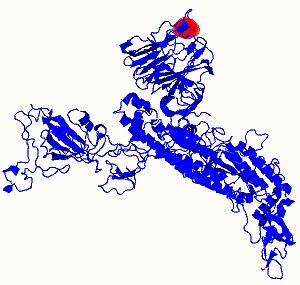
Preprints (www.preprints.org) | NOT PEER-REVIEWED | Posted: 14 June 2020                 doi:10.20944/preprints202006.0184.v1

                                                                                                                3 of 10

              Single Amino Acid Variant (SAV) Phenotype, protein modeling, and mutation analysis
              Four Amino Acid Variant (SAV) found from 20 spike glycoprotein submitted and retrieved by Phyre2
              server to predict mutational sensitivity [9–11].

              Nomenclature sequence Amino Acid Variant (SAV) and annotation used the accession number
              Surface glycoprotein [Severe acute respiratory syndrome coronavirus 2] with accession number
              YP_009724390.1 was used as a reference sequence to compare with, and it was downloaded from
              https://www.ncbi.nlm.nih.gov/protein/YP_009724390.1?report=fasta.

               3. Results:
              Predicted Secondary Structure
              Initially, the I-TASSER was utilized to recognize the basic templates from the PDB by multiple threading
              approach LOMETS, with full-length atomic models produced by iterative fragment assembly simulations
              based on templates. Function insights of the targeted molecule are then obtained by rethreading the
              three-dimensional models via the BioLiP database of protein functions. Figure 1 shows the predicted
              secondary structure of the SARS-CoV-2 spike glycoprotein tested in Jordan.

              Figure 1: Spike glycoprotein predicted secondary structure included SARS-CoV-like_Spike_S1_NTD (N terminal
              domain), SARS-CoV-2_Spike_S1_RBD (spike recognition binding site) and Corona_S2 doamin.

              The final predicted model of the monomer of spike protein by I-TASSER
              For each target, I-TASSER simulations called decoys generate an extensive collection of structural
              conformations. I-TASSER uses the SPICKER to cluster all architectural structures based on the pair-sided
              similarity and records up to 5 models corresponding to the five largest structural clusters. The reliability
              of each model is evaluated quantitatively by a C-score based on the value of threaded prototype
              alignments and the parameters of convergence of structural mounting simulations. C-score is usually [-
              5, 2], where a higher-value C-score means a more positive and vice versa scale. Following the association
              observed between these attributes, the TM-score and RMSD are calculated using the C and the protein
              frequency. Since the group size classes the top 5 models, in some situations, a higher C-score is possible
              for the lower-ranking models. While the first model is better in most cases, as seen in our research (Fig.
              4), lower-level models can also be better than higher-level models. If the I-TASSER simulations converge,
              less than 5 clusters can have been generated; it usually shows that because of the converged simulations,
              the models have good quality.

              Very recently, the experimental cryo-EM structures of the glycoprotein of SARS-CoV-2 have been
              reported by two groups; Wrapp et al. and Walls et al. and the structures are available in the Protein Data
              Bank, e.g., 6vsb, 6vxx [12] & [3]. While the deposited structures are not full-length and not solved by
              crystallography but cryo-EM, the resolution of those structures is close to the crystal structures. Zhang’s
              group (zhanglab.ccmb.med.umich.edu/COVID-19/) has modeled other structural models of SARS-CoV-
              2, including the surface glycoprotein, with I-TASSAR. We found no difference between the recently
              reported experimental structures and the in silico models we generated, even with the new single amino
              variants.
Preprints (www.preprints.org) | NOT PEER-REVIEWED | Posted: 14 June 2020 doi:10.20944/preprints202006.0184.v1
4 of 10
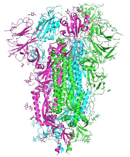
(a) (b)
Figure 4 : (a) The final I-TASSER predicted spike glycoprotein spike protein monomer model, the PDB file is
provided in the supplementary data, and (b) the trimer spike glycoprotein as elucidated by Cryo-EM; PDB:6vxx [12].
The top two proteins structurally close to the spike glycoprotein in the Protein Data Bank (as identified
by TM-align) are listed in Table 1. In Table 2 the top five hits of closest Enzyme Commission (EC) numbers
and active sites are listed.
Table 1: Proteins structurally close to the spike glycoprotein in the Protein Data Bank (as identified by TM-align).
Protein rankings are based on the structural alignment TM score in the PDB library between the query
template and known structures. RMSDa the RMSD among structurally aligned residues of TM-align;
IDENa is the structurally related region's percentage sequence identity; Cov reflects the alignment range
of the TM-alignment and is proportional to the sum by the length of query protein of structurally aligned
residues. 5x58A: Prefusion structure of SARS-CoV spike glycoprotein, conformation 1 (viral protein);
6nzkA: Structural basis for human coronavirus attachment to sialic acid receptors (viral protein).
Table 2: Enzyme Commission (EC) numbers and active sites
1ileA: Isoleucyl-tRNA synthetase (aminoacyl-tRNA synthetase): 1k32A: Crystal structure of the tricorn protease
(hydrolase); 3eqlM: Crystal structure of the T. Thermophilus RNA polymerase holoenzyme in complex with antibiotic
myxopyronin (transferase); 2pdaA: Crystal structure of the complex between pyruvate-ferredoxin oxidoreductase
from Desulfovibrio africanus and pyruvate (oxidoreductase); 1ej6A: Reovirus core (virus).Preprints (www.preprints.org) | NOT PEER-REVIEWED | Posted: 14 June 2020 doi:10.20944/preprints202006.0184.v1
5 of 10
One powerful way of multiple sequence alignment is the Multiple Alignment using Fast Fourier
Transform (MAFFT) as shown in figure 5 below [8]. Figure 6 (a) shows the location of single amino acid
variants del Y144 & D614G on spike protein as they have the highest mutation sensitivity scores and (b)
shows the mutation sensitivity histograms for all four single amino acid variants on spike protein; Y 144
D 614, D 1139, and G 1167 calculated according to [10].
Figure 5 : Multiple Sequence Alignment showing Amino Acid Variant (SAV) viewed by Jalview. The blue
arrow and the red box are just cursers; the white columns are the location of the SAV along the full length of
amino acid sequence of spike protein for 20 SARS-CoV-2 samples from Jordan. YP-009724390 is the reference
sequence of spike protein of from Wuhan. The number 1273 is the total number of amino acid in the
sequence.Preprints (www.preprints.org) | NOT PEER-REVIEWED | Posted: 14 June 2020 doi:10.20944/preprints202006.0184.v1
6 of 10
(a)
del Y144 D614G
(b)
Y 144 D 614 D 1139 G 1167
Figure 6: (Phyre2) Mutational sensitivity (a) the location of single amino acid variants del Y144 & D614G on spike
protein as they have the highest mutation sensitivity scores. (b) The mutation sensitivity histograms for all four
single amino acid variants on spike protein.
Table 3 shows that the D614G has the highest percentages and mutation sensitivity in the spike protein
in the Jordanian population. This mutation showed the substitution of aspartate; a bulky amino acid to
glycine; the simplest amino acid ever.
Table 3: SAV in spike-protein of SRAS-CoV-2; percentage and mutational sensitivity scores in Jordanian population
SAV in spike-protein of SAV Percentage in (Phyre2) Mutational sensitivity Score out of
SRAS-CoV-2 Jordanian population (9=high mutation sensitivity, 0=low)
Deletion at Y144 5% 3
D614G 62% 5
D1139Y 5% 1
G1167S 5% 1
4. Discussion:
In this study, we used the spike gene sequences from 20 whole-genome sequences of SARS-CoV-2
collected from Jordan retrieved from GISAID database and analyzed at the amino sequence level of the
spike glycoprotein versus a reference sequence of the surface glycoprotein [Severe acute respiratory
syndrome coronavirus 2; (SARS-CoV-2)] own the accession number YP_009724390.1 of Wuhan. Our
findings showed that, the molecules which were structurally close to the spike glycoprotein from the
Enzyme Commission (EC) numbers and active sites included Isoleucyl-tRNA synthetase which provide
the protein the ability to synthesize tRNA, Crystal structure of the tricorn protease (hydrolase) which
provide the protein the ability to hydrolyse the host proteins need for viral entry; Crystal structure of the
T. Thermophilus RNA polymerase holoenzyme (transferase) which provide the protein the ability to
synthesize the viral RNA; Crystal structure of the complex between pyruvate-ferredoxin oxidoreductasePreprints (www.preprints.org) | NOT PEER-REVIEWED | Posted: 14 June 2020 doi:10.20944/preprints202006.0184.v1
7 of 10
from Desulfovibrio africanus and pyruvate (oxidoreductase); and Reovirus core (virus) all might explain
the ability of SARS-CoV-2 in getting inside the human host cells.
Four Amino Acid Variants (SAV) founded in 20 samples of SARS-CoV-2 were discovered in this study
with 5% of samples showed tyrosine deletion at Y144 located in the SARS-CoV-like_Spike_S1_NTD (N
terminal domain), 62% showed aspartate substitution to glycine at D614G located in the SARS-CoV-
2_Spike_S1_RBD (spike recognition binding site), 5% showed aspartate substitution to tyrosine at
D1139Y and 5% showed glycine substitution to serine at G1167S both located in the Corona_S2 domain.
From a biochemistry point of view, the D614G mutation showed the substitution of aspartate; a bulky
amino acid to glycine; the simplest amino acid ever, which might suggest a reduced new affinity between
spike protein and its receptors. The findings have shown lower mutational sensitivity in all variants that
might not affect the function of spike glycoprotein except for D614G, which has the highest mutational
sensitivity score (5 out of 9) indicating a higher likelihood to affect the function of the spike protein. This
might suggest, in general, a reduced transmitability of SARS-CoV-2 in Jordan. The D614G substitution
was previously reported as a dominant mutation in Europe [9]. The I-TASSER predicted monomer three-
dimensional structures of spike protein of SARS-CoV-2 had similar stability structures for all of the four
Amino Acid Variant (SAV) when we compared the reference sequence of the spike glycoprotein
YP_009724390.1 (SARS-CoV-2) with FASTA sequences of spike glycoproteins from Jordanian population;
no change on the three-dimensional structure was noticed.
The generated three-dimensional monomer structure of the spike protein of SARS-CoV-2 is consistent
with a perfusion conformation structure reported in the literature [3]. Like any in silico study, this study
is limited with the capabilities of utilized servers and algorithms to it is highly dependent on the initial
templates used for calculations, so if the initial template scoring is not good enough then this might affect
the final output files.
5. Conclusion:
This is the first study of its kind in the Middle East to predict the monomer three-dimensional structure
of the spike glycoprotein from SARS-CoV-2 of Jordanian specimens. In addition, we reported four amino
acid variants, which might explain the current low number of COVID-19 cases, 953 confirmed cases, and
678 recovered and 9 deaths. However, the highest frequency mutation in our study, with 62% of samples
showed aspartate substitution to glycine at D614G is consistent with other reports for samples collected
in Europe at the same time of our samples collection, March 2020. In this study, we consider the mutation
D614G as the dominant local mutation in Jordan. We believe that the reported four amino acid variants
especially the tyrosine deletion at Y144 located in the SARS-CoV-like_Spike_S1_NTD (N terminal
domain) and the aspartate substitution to glycine at D614G located in the SARS-CoV-2_Spike_S1_RBD
(spike recognition binding site) have collectively reduced the spike protein affinity of SARS-CoV-2 with
ACE2 receptors in the Jordanian population. It is highly recommended to keep monitoring the mutation
rate of SARS-CoV-2 in Jordan in monthly bases with higher number of samples to fulfil a statistical power.
Some of the low percentage appeared mutations e.g. 5% might be increased if the population size is
higher.
Author Contributions: “Conceptualization, W.A-Z., R.F & H.H.; methodology, W.A-Z & H.H.; software, W.A-Z &
H.H.; validation, W.A-Z & H.H. and R.F.; formal analysis, W.A-Z & H.H.; investigation, W.A-Z & H.H.; resources,
R.F.; data curation, W.A-Z & H.H.; writing—original draft preparation, W.A-Z.; writing—review and editing,
W.A-Z & H.H.; visualization, W.A-Z & H.H.; supervision, R.F.; project administration, R.F.; funding acquisition,
W.A-Z & H.H. All authors have read and agreed to the published version of the manuscript.”
Funding: “This research received no external funding”Preprints (www.preprints.org) | NOT PEER-REVIEWED | Posted: 14 June 2020 doi:10.20944/preprints202006.0184.v1
8 of 10
Acknowledgments: The authors acknowledge Biolab Diagnostic Laboratories (Jordan) & Andersen lab at Scripps
Research (USA) who published sequences were retrieved from GISAID, a maintained global database based in
Germany.
Conflicts of Interest: “The authors declare no conflict of interest.”
Appendix A:
Submitted Sequence in FASTA format
del. Y144 Sample 23
>hCoV-19/Jordan/SR-042/2020|EPI_ISL_430000|2020-03-30
MFVFLVLLPLVSSQCVNLTTRTQLPPAYTNSFTRGVYYPDKVFRSSVLHSTQDLFLPFFSNVTWFHAIHVSGTNGTKRFDNPVLPF
NDGVYFASTEKSNIIRGWIFGTTLDSKTQSLLIVNNATNVVIKVCEFQFCNDPFLGVYHKNNKSWMESEFRVYSSANNCTFEYVS
QPFLMDLEGKQGNFKNLREFVFKNIDGYFKIYSKHTPINLVRDLPQGFSALEPLVDLPIGINITRFQTLLALHRSYLTPGDSSSGWT
AGAAAYYVGYLQPRTFLLKYNENGTITDAVDCALDPLSETKCTLKSFTVEKGIYQTSNFRVQPTESIVRFPNITNLCPFGEVFNAT
RFASVYAWNRKRISNCVADYSVLYNSASFSTFKCYGVSPTKLNDLCFTNVYADSFVIRGDEVRQIAPGQTGKIADYNYKLPDDFT
GCVIAWNSNNLDSKVGGNYNYLYRLFRKSNLKPFERDISTEIYQAGSTPCNGVEGFNCYFPLQSYGFQPTNGVGYQPYRVVVLSF
ELLHAPATVCGPKKSTNLVKNKCVNFNFNGLTGTGVLTESNKKFLPFQQFGRDIADTTDAVRDPQTLEILDITPCSFGGVSVITP
GTNTSNQVAVLYQDVNCTEVPVAIHADQLTPTWRVYSTGSNVFQTRAGCLIGAEHVNNSYECDIPIGAGICASYQTQTNSPRRA
RSVASQSIIAYTMSLGAENSVAYSNNSIAIPTNFTISVTTEILPVSMTKTSVDCTMYICGDSTECSNLLLQYGSFCTQLNRALTGIAV
EQDKNTQEVFAQVKQIYKTPPIKDFGGFNFSQILPDPSKPSKRSFIEDLLFNKVTLADAGFIKQYGDCLGDIAARDLICAQKFNGL
TVLPPLLTDEMIAQYTSALLAGTITSGWTFGAGAALQIPFAMQMAYRFNGIGVTQNVLYENQKLIANQFNSAIGKIQDSLSSTAS
ALGKLQDVVNQNAQALNTLVKQLSSNFGAISSVLNDILSRLDKVEAEVQIDRLITGRLQSLQTYVTQQLIRAAEIRASANLAATK
MSECVLGQSKRVDFCGKGYHLMSFPQSAPHGVVFLHVTYVPAQEKNFTTAPAICHDGKAHFPREGVFVSNGTHWFVTQRNFY
EPQIITTDNTFVSGNCDVVIGIVNNTVYDPLQPELDSFKEELDKYFKNHTSPDVDLGDISGINASVVNIQKEIDRLNEVAKNLNES
LIDLQELGKYEQYIKWPWYIWLGFIAGLIAIVMVTIMLCCMTSCCSCLKGCCSCGSCCKFDEDDSEPVLKGVKLHYT
D1139Y Sample 21
>hCoV-19/Jordan/SR-033/2020|EPI_ISL_429993|2020-03-16
MFVFLVLLPLVSSQCVNLTTRTQLPPAYTNSFTRGVYYPDKVFRSSVLHSTQDLFLPFFSNVTWFHAIHVSGTNGTKRFDNPVLPF
NDGVYFASTEKSNIIRGWIFGTTLDSKTQSLLIVNNATNVVIKVCEFQFCNDPFLGVYYHKNNKSWMESEFRVYSSANNCTFEYV
SQPFLMDLEGKQGNFKNLREFVFKNIDGYFKIYSKHTPINLVRDLPQGFSALEPLVDLPIGINITRFQTLLALHRSYLTPGDSSSGW
TAGAAAYYVGYLQPRTFLLKYNENGTITDAVDCALDPLSETKCTLKSFTVEKGIYQTSNFRVQPTESIVRFPNITNLCPFGEVFNA
TRFASVYAWNRKRISNCVADYSVLYNSASFSTFKCYGVSPTKLNDLCFTNVYADSFVIRGDEVRQIAPGQTGKIADYNYKLPDDF
TGCVIAWNSNNLDSKVGGNYNYLYRLFRKSNLKPFERDISTEIYQAGSTPCNGVEGFNCYFPLQSYGFQPTNGVGYQPYRVVVL
SFELLHAPATVCGPKKSTNLVKNKCVNFNFNGLTGTGVLTESNKKFLPFQQFGRDIADTTDAVRDPQTLEILDITPCSFGGVSVIT
PGTNTSNQVAVLYQGVNCTEVPVAIHADQLTPTWRVYSTGSNVFQTRAGCLIGAEHVNNSYECDIPIGAGICASYQTQTNSPRR
ARSVASQSIIAYTMSLGAENSVAYSNNSIAIPTNFTISVTTEILPVSMTKTSVDCTMYICGDSTECSNLLLQYGSFCTQLNRALTGIA
VEQDKNTQEVFAQVKQIYKTPPIKDFGGFNFSQILPDPSKPSKRSFIEDLLFNKVTLADAGFIKQYGDCLGDIAARDLICAQKFNG
LTVLPPLLTDEMIAQYTSALLAGTITSGWTFGAGAALQIPFAMQMAYRFNGIGVTQNVLYENQKLIANQFNSAIGKIQDSLSSTA
SALGKLQDVVNQNAQALNTLVKQLSSNFGAISSVLNDILSRLDKVEAEVQIDRLITGRLQSLQTYVTQQLIRAAEIRASANLAAT
KMSECVLGQSKRVDFCGKGYHLMSFPQSAPHGVVFLHVTYVPAQEKNFTTAPAICHDGKAHFPREGVFVSNGTHWFVTQRNF
YEPQIITTDNTFVSGNCDVVIGIVNNTVYYPLQPELDSFKEELDKYFKNHTSPDVDLGDISGINASVVNIQKEIDRLNEVAKNLNE
SLIDLQELGKYEQYIKWPWYIWLGFIAGLIAIVMVTIMLCCMTSCCSCLKGCCSCGSCCKFDEDDSEPVLKGVKLHYT
G1167S Sample 16
>hCoV-19/Jordan/SR-039/2020|EPI_ISL_429998|2020-03-28
MFVFLVLLPLVSSQCVNLTTRTQLPPAYTNSFTRGVYYPDKVFRSSVLHSTQDLFLPFFSNVTWFHAIHVSGTNGTKRFDNPVLPF
NDGVYFASTEKSNIIRGWIFGTTLDSKTQSLLIVNNATNVVIKVCEFQFCNDPFLGVYYHKNNKSWMESEFRVYSSANNCTFEYV
SQPFLMDLEGKQGNFKNLREFVFKNIDGYFKIYSKHTPINLVRDLPQGFSALEPLVDLPIGINITRFQTLLALHRSYLTPGDSSSGW
TAGAAAYYVGYLQPRTFLLKYNENGTITDAVDCALDPLSETKCTLKSFTVEKGIYQTSNFRVQPTESIVRFPNITNLCPFGEVFNA
TRFASVYAWNRKRISNCVADYSVLYNSASFSTFKCYGVSPTKLNDLCFTNVYADSFVIRGDEVRQIAPGQTGKIADYNYKLPDDF
TGCVIAWNSNNLDSKVGGNYNYLYRLFRKSNLKPFERDISTEIYQAGSTPCNGVEGFNCYFPLQSYGFQPTNGVGYQPYRVVVL
SFELLHAPATVCGPKKSTNLVKNKCVNFNFNGLTGTGVLTESNKKFLPFQQFGRDIADTTDAVRDPQTLEILDITPCSFGGVSVIT
PGTNTSNQVAVLYQDVNCTEVPVAIHADQLTPTWRVYSTGSNVFQTRAGCLIGAEHVNNSYECDIPIGAGICASYQTQTNSPRR
ARSVASQSIIAYTMSLGAENSVAYSNNSIAIPTNFTISVTTEILPVSMTKTSVDCTMYICGDSTECSNLLLQYGSFCTQLNRALTGIA
VEQDKNTQEVFAQVKQIYKTPPIKDFGGFNFSQILPDPSKPSKRSFIEDLLFNKVTLADAGFIKQYGDCLGDIAARDLICAQKFNG
LTVLPPLLTDEMIAQYTSALLAGTITSGWTFGAGAALQIPFAMQMAYRFNGIGVTQNVLYENQKLIANQFNSAIGKIQDSLSSTA
SALGKLQDVVNQNAQALNTLVKQLSSNFGAISSVLNDILSRLDKVEAEVQIDRLITGRLQSLQTYVTQQLIRAAEIRASANLAAT
KMSECVLGQSKRVDFCGKGYHLMSFPQSAPHGVVFLHVTYVPAQEKNFTTAPAICHDGKAHFPREGVFVSNGTHWFVTQRNF
YEPQIITTDNTFVSGNCDVVIGIVNNTVYDPLQPELDSFKEELDKYFKNHTSPDVDLSDISGINASVVNIQKEIDRLNEVAKNLNES
LIDLQELGKYEQYIKWPWYIWLGFIAGLIAIVMVTIMLCCMTSCCSCLKGCCSCGSCCKFDEDDSEPVLKGVKLHYTPreprints (www.preprints.org) | NOT PEER-REVIEWED | Posted: 14 June 2020 doi:10.20944/preprints202006.0184.v1
9 of 10
D614G Sample 3,5,7,8,9,10,11,12,13,15,18,20 and 21
Example:
>hCoV-19/Jordan/SR-036/2020|EPI_ISL_429996|2020-03-23
MFVFLVLLPLVSSQCVNLTTRTQLPPAYTNSFTRGVYYPDKVFRSSVLHSTQDLFLPFFSNVTWFHAIHVSGTNGTKRFDNPVLPF
NDGVYFASTEKSNIIRGWIFGTTLDSKTQSLLIVNNATNVVIKVCEFQFCNDPFLGVYYHKNNKSWMESEFRVYSSANNCTFEYV
SQPFLMDLEGKQGNFKNLREFVFKNIDGYFKIYSKHTPINLVRDLPQGFSALEPLVDLPIGINITRFQTLLALHRSYLTPGDSSSGW
TAGAAAYYVGYLQPRTFLLKYNENGTITDAVDCALDPLSETKCTLKSFTVEKGIYQTSNFRVQPTESIVRFPNITNLCPFGEVFNA
TRFASVYAWNRKRISNCVADYSVLYNSASFSTFKCYGVSPTKLNDLCFTNVYADSFVIRGDEVRQIAPGQTGKIADYNYKLPDDF
TGCVIAWNSNNLDSKVGGNYNYLYRLFRKSNLKPFERDISTEIYQAGSTPCNGVEGFNCYFPLQSYGFQPTNGVGYQPYRVVVL
SFELLHAPATVCGPKKSTNLVKNKCVNFNFNGLTGTGVLTESNKKFLPFQQFGRDIADTTDAVRDPQTLEILDITPCSFGGVSVIT
PGTNTSNQVAVLYQGVNCTEVPVAIHADQLTPTWRVYSTGSNVFQTRAGCLIGAEHVNNSYECDIPIGAGICASYQTQTNSPRR
ARSVASQSIIAYTMSLGAENSVAYSNNSIAIPTNFTISVTTEILPVSMTKTSVDCTMYICGDSTECSNLLLQYGSFCTQLNRALTGIA
VEQDKNTQEVFAQVKQIYKTPPIKDFGGFNFSQILPDPSKPSKRSFIEDLLFNKVTLADAGFIKQYGDCLGDIAARDLICAQKFNG
LTVLPPLLTDEMIAQYTSALLAGTITSGWTFGAGAALQIPFAMQMAYRFNGIGVTQNVLYENQKLIANQFNSAIGKIQDSLSSTA
SALGKLQDVVNQNAQALNTLVKQLSSNFGAISSVLNDILSRLDKVEAEVQIDRLITGRLQSLQTYVTQQLIRAAEIRASANLAAT
KMSECVLGQSKRVDFCGKGYHLMSFPQSAPHGVVFLHVTYVPAQEKNFTTAPAICHDGKAHFPREGVFVSNGTHWFVTQRNF
YEPQIITTDNTFVSGNCDVVIGIVNNTVYDPLQPELDSFKEELDKYFKNHTSPDVDLGDISGINASVVNIQKEIDRLNEVAKNLNE
SLIDLQELGKYEQYIKWPWYIWLGFIAGLIAIVMVTIMLCCMTSCCSCLKGCCSCGSCCKFDEDDSEPVLKGVKLHYTPreprints (www.preprints.org) | NOT PEER-REVIEWED | Posted: 14 June 2020 doi:10.20944/preprints202006.0184.v1
10 of 10
References:
1. Wu F, Zhao S, Yu B, Chen YM, Wang W, Song ZG, et al. A new coronavirus associated with
human respiratory disease in China. Nature. 2020 Mar 12;579(7798):265–9.
2. Lu R, Zhao X, Li J, Niu P, Yang B, Wu H, et al. Genomic characterisation and epidemiology of
2019 novel coronavirus: implications for virus origins and receptor binding. Lancet. 2020 Feb
22;395(10224):565–74.
3. Daniel Wrapp, Nianshuang Wang, Kizzmekia S. Corbett, Jory A. Goldsmith C-LH, Olubukola
Abiona, Barney S. Graham JSM. Cryo-EM structure of the 2019-nCoV spike in the prefusion
conformation. - PubMed - NCBI. Science. 2020.
4. Lan J, Ge J, Yu J, Shan S, Zhou H, Fan S, et al. Structure of the SARS-CoV-2 spike receptor-
binding domain bound to the ACE2 receptor. Nature. 2020 Mar 30;1–6.
5. Zhang H, Penninger JM, Li Y, Zhong N, Slutsky AS. Angiotensin-converting enzyme 2 (ACE2)
as a SARS-CoV-2 receptor: molecular mechanisms and potential therapeutic target. Intensive
Care Med. 2020 Apr 1;46(4):586–90.
6. Glowacka I, Bertram S, Muller MA, Allen P, Soilleux E, Pfefferle S, et al. Evidence that TMPRSS2
Activates the Severe Acute Respiratory Syndrome Coronavirus Spike Protein for Membrane
Fusion and Reduces Viral Control by the Humoral Immune Response. J Virol. 2011 May
1;85(9):4122–34.
7. Abio Madeira F´, Mi Park Y, Lee J, Buso N, Gur T, Madhusoodanan N, et al. The EMBL-EBI
search and sequence analysis tools APIs in 2019. Web Serv issue Publ online. 2019;47.
8. Waterhouse AM, Procter JB, Martin DMA, Clamp M, Barton GJ. Jalview Version 2-A multiple
sequence alignment editor and analysis workbench. Bioinformatics. 2009;25(9):1189–91.
9. Angyal A, Brown RL, Carrilero L, Green LR, Groves DC, Johnson KJ, et al. Spike mutation
pipeline reveals the emergence of a more transmissible form of SARS-CoV-2 on behalf of the
Sheffield COVID-19 Genomics Group#, LaBranche CC2, and Montefiori DC2.
10. Yates CM, Filippis I, Kelley LA, Sternberg MJE. SuSPect: Enhanced prediction of single amino
acid variant (SAV) phenotype using network features. J Mol Biol. 2014 Jul 15;426(14):2692–701.
11. Kelley LA, Mezulis S, Yates CM, Wass MN, Sternberg MJE. The Phyre2 web portal for protein
modeling, prediction and analysis. Nat Protoc. 2015 Jun 30;10(6):845–58.
12. Walls AC, Park YJ, Tortorici MA, Wall A, McGuire AT, Veesler D. Structure, Function, and
Antigenicity of the SARS-CoV-2 Spike Glycoprotein. Cell. 2020 Apr 16;181(2):281-292.e6.You can also read