Splenic masses in dogs - Part 1: Epidemiologic, clinical characteristics as well as histopathologic diagnosis in 249 cases (2000-2011)
←
→
Page content transcription
If your browser does not render page correctly, please read the page content below
Original Article                                                                                                                                       © Schattauer 2012   1

Splenic masses in dogs
Part 1: Epidemiologic, clinical characteristics as well as histopathologic diagnosis
in 249 cases (2000–2011)
N. Eberle1; V. von Babo1; I. Nolte1; W. Baumgärtner2; D. Betz1
1Small Animal   Hospital, University of Veterinary Medicine Hannover, Foundation; 2Department for Pathology, University of Veterinary Medicine Hannover, Foundation

 Key words                                                                                 Schlüsselwörter
 Canine, splenic mass, staging, hemoperitoneum                                             Hund, Umfangsvermehrung Milz, Staging, Hämaskos

 Summary                                                                                   Zusammenfassung
 Objective: Splenic masses have a high prevalence and are more com-                        Gegenstand: Umfangsvermehrungen der Milz haben beim Hund eine
 mon than diffuse splenic enlargement in dogs. It was the aim of the                       hohe Prävalenz und kommen häufiger vor als eine diffuse Vergrößerung
 present study to retrospectively describe clinical aspects and histopa-                   des Organs. Ziel der Studie war die retrospektive Beschreibung der kli-
 thologic characteristics of dogs with splenic masses. Material and                        nischen Aspekte und die histopathologische Charakteristika von Um-
 methods: Records of patients with a histologically diagnosed splenic                      fangsvermehrungen der Milz beim Hund. Material und Methoden:
 mass between January 2000 and March 2011 were reviewed. Results:                          Die Auswertung erfasste die Daten von Hunden mit der histopathologi-
 249 dogs met the inclusion criteria and could be included in the study.                   schen Diagnose einer Umfangsvermehrung der Milz zwischen Januar
 Splenic masses were diagnosed histologically as non-malignant dis-                        2000 und März 2011. Ergebnisse: 249 Hunde erfüllten die Einschluss-
 ease (n = 117; 47%) and malignant splenic disease (n = 132; 53%).                         kriterien und gingen in die Studie ein. Die Umfangsvermehrungen wur-
 Hemangiosarcoma was the most common histological diagnosis                                den histologisch als nichtmaligne Erkrankung (n = 117; 47%) und ma-
 (n = 97; 73.5%). Other malignant tumors included sarcoma (n = 14), fi-                    ligne Milzerkrankung (n = 132; 53%) klassifiziert. Das Hämangiosar-
 brohistiocytic nodules (n = 9) as well as lymphoma, blastoma and ade-                     kom war der häufigste maligne Tumor (n = 97; 73.5%). Als weitere ma-
 nocarcinoma. The non-malignant masses consisted of nodular hyper-                         ligne Tumoren wurden Sarkome (n = 14), fibrohistiozytäre Umfangs-
 plasia (n = 60), splenic hematoma (n = 41), and splenitis (n = 6). Dogs                   vermehrungen (n = 9), Lymphome, Blastome und Adenokarzinome dia-
 with hemoperitoneum had a higher frequency of splenic neoplasia.                          gnostiziert. Die nichtmalignen Umfangsvermehrungen umfassten no-
 Conclusion: The results corroborate previous findings that hemangio-                      duläre Hyperplasien (n = 60), Milzhämatome (n = 41) und Splenitiden
 sarcoma is the most frequent neoplasm of the canine spleen. However,                      (n = 6). Bei Hunden mit Hämoabdomen bestand eine höhere Wahr-
 in approximately half of the cases benign lesions were histologically                     scheinlichkeit für eine Milzneoplasie. Schlussfolgerung: Die Ergebnis-
 diagnosed. Clinical relevance: It is essential that a frank discussion is                 se der Studie bestätigen bisherige Resultate, dass das Hämangiosar-
 held with owners regarding the prognosis associated with the treat-                       kom beim Hund den häufigsten Milztumor darstellt. Allerdings wurden
 ment of dogs with a splenic mass associated with hemoperitoneum.                          bei annähernd der Hälfte der histologisch untersuchen Milzen nichtma-
                                                                                           ligne Veränderungen diagnostiziert. Klinische Relevanz: Eine ausführ-
                                                                                           liche Information der Besitzer hinsichtlich der Prognose und Therapie
                                                                                           von Hunden mit einer Umfangsvermehrung der Milz in Verbindung mit
                                                                                           einem Hämoabdomen ist bedeutsam.

 Correspondence to                                                                         Umfangsvermehrungen der Milz beim Hund. Teil 1: Epidemiologische Aspek-
 Nina Eberle, Dr. med. vet., Diplomate ECVIM-CA (Oncology)                                 te, klinische Charakteristika sowie histopathologische Befunde von 249 Fäl-
 Small Animal Hospital                                                                     len (2000–2011)
 University of Veterinary Medicine Hannover, Foundation                                    (English version of) Tierärztl Prax 2012; 40 (K): 250–260
 Bünteweg 9, D-30559 Hannover                                                              Received: December 23, 2011
 Germany                                                                                   Accepted after revision: June 5, 2012
 Email: Nina.Eberle@tiho-hannover.de

Introduction                                                                               evidence against this (10). According to this study, non-malignant
                                                                                           masses, such as hematoma and nodular hyperplasia even account
The high prevalence of splenic masses in dogs, along with the sur-                         for the majority of focal canine splenic masses (10).
gical accessibility of the spleen results in a relatively large number                         Hemangiosarcoma is a malignant tumor of vascular endothe-
of splenectomies performed in veterinary medicine (1, 13). Splenic                         lial origin (2). In dogs, the most common site of hemangiosarcoma
nodular masses are widely considered to be indicative of hem-                              is the spleen. Because of the high potential of splenic hemangiosar-
angiosarcoma (5, 17, 26). Recent publications, however, provide                            coma to develop metastasis, the prognosis is poor (2, 29). Com-

                                                                                                                Tierärztliche Praxis Kleintiere 4/2012; 40(K): 250–260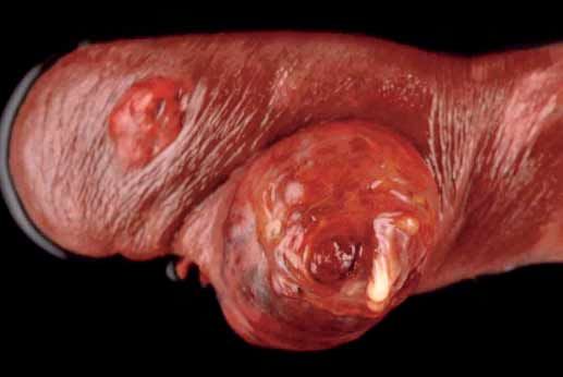
2   N. Eberle et al.: Characteristics of canine splenic masses

    mon sites for metastasis via hematogenous or transabdominal            nal effusion. The size of a mass (largest dimension) was evaluated
    routes are the liver, the omentum, the lung and the heart (2). The     and recorded. All images were viewed by use of a standard com-
    two thirds rule has been applied to canine splenic masses. Appro-      puter workstation with Digital Imaging and Communications in
    ximately two thirds of all splenic masses will have a malignant        Medicine (DICOM) viewing softwarea and 3-megapixel grayscale
    tumor, and two thirds of these malignancies will have a hem-           monitorsb. For the determination of the dimension the largest dia-
    angiosarcoma of the spleen (4, 16, 24).                                meter was measured in DICOM.
       Presenting complaints vary and can range from vague, non-               With abdominal ultrasonography the appearance of the splenic
    specific illness, asymptomatic swelling of the abdomen to acute        and hepatic lesions was evaluated. The splenic mass was categor-
    death secondary to hypotensive shock (1, 12). Clinical signs com-      ized as being cavernous, inhomogeneous, or homogenous. Mass
    monly associated in dogs with ruptured splenic masses are related      size (largest dimension) was evaluated and recorded. The appear-
    to severe anemia (12).                                                 ance of the liver parenchyma was categorized as homogenous, in-
       Abdominal radiographs and ultrasonography are used to evalu-        homogeneous or containing a cavernous mass. The abdominal
    ate the spleen. The ultrasonographic appearance of splenic hema-       cavity was also reviewed for the presence of effusion. Cardiac ultra-
    tomas and hemangiosarcomas has been described, but both tend           sound was performed in order to evaluate for the evidence of
    to vary in echogenicity and appearance (30, 31). Based on radio-       lesions compatible with metastasis. The echocardiographic exam-
    graphic findings, ultrasonographic findings and gross inspection,      ination and the abdominal ultrasound were performed by clini-
    malignant and non-malignant splenic masses have non-specific           cians with long-standing academic experience in cardiac and ab-
    characteristics (1, 8, 13, 30).                                        dominal ultrasound.
       It was the aim of the present study to retrospectively describe
    clinical aspects and histopathological characteristics of dogs with    Tissue sampling and histopathologic examination
    splenic masses and, based on this, to verify previously described
    histopathologic distributions for the own hospital population.         The entire spleen was surgically removed. An open liver biopsy was
                                                                           obtained with suture fracture on the periphery of the liver lobe and
                                                                           fixed in 10% buffered formalin. In case of focal lesions or other
    Material and methods                                                   changes of the liver parenchyma an additional wedge biopsy was
                                                                           performed.
    Patients                                                                   Following splenectomy or euthanasia, each spleen was fixed in
                                                                           10% buffered formalin. Tissue samples from at least two represen-
    Dogs with a histopathologically confirmed splenic mass were in-        tative areas of the margin between the splenic masses and adjacent
    cluded in the retrospective study. Exclusion criteria were splenic     spleen and one sample of the centre of the mass plus a minimum of
    enlargement without a splenic mass, sole medical management            two grossly appearing normal sections of the spleen were sampled.
    and lack of histopathologic examination.                               In case of multiple splenic masses, every nodule was examined. The
        The routine diagnostic workup included general examination,        samples were dehydrated and embedded in paraffin, and 5-μm
    complete blood count, serum biochemistry, thoracic and abdo-           sections were prepared and stained with hematoxylin and eosin. A
    minal radiographs, transabdominal ultrasound to describe the           specific histopathologic diagnosis was made for each mass and the
    splenic mass and the liver and to evaluate for abdominal effu-         presumed normal section of spleen. Each mass was classified as
    sion, as well as an echocardiographic examination. Information re-     malignant or nonmalignant by a board-certified pathologist of
    trieved from medical records included signalment and presenting        the Department for Pathology. The examination of the spleen was
    complaint (weakness, lethargy, collapse, unspecific symptoms, fe-      a routine diagnostic work-up so that different board-certified pa-
    ver, abdominal distension, weight loss).                               thologists were involved in the diagnosis. Immunhistochemical
        Clinical staging in dogs with hemangiosarcoma was performed        staining procedures were conducted in cases of indifferent find-
    according to the WHO clinical staging system for canine hem-           ings in the hematoxylin and eosin stained sections. The antibodies
    angiosarcoma (24). Dogs that are classified as stage I have a pri-     used in immunhistochemistry included CD 117, CD 3, CD 79a,
    mary tumor only, whereas dogs classified as stage II have a primary    MAC 387, lysozym, vimentin, desmin, smooth muscle actin, factor
    tumor with splenic rupture or lymph node involvement, and dogs         VII-antigen, MHC-II, melan-A, S-100 protein and cytokeratin.
    classified as stage III have a primary tumor with splenic rupture or
    lymph node involvement and evidence of distant metastasis.

    Diagnostic imaging
                                                                           a   Digital Imaging and Communications in Medicine (DICOM) viewing soft-
    Abdominal radiographs were evaluated by one board-certified                ware dicom PACS Version 5; Oehm & Rehbein GmbH, Rostock, Germany
    oncologist (NE) for the presence of a mass in the mid or cranial       b
                                                                               EIZO RadiForce RX 211 grayscale monitor; EIZO NANAO corporation, Is-
    abdomen, and generalized loss of detail associated with abdomi-            hikawa, Japan

    Tierärztliche Praxis Kleintiere 4/2012; 40(K): 250–260                                                                     © Schattauer 2012
N. Eberle et al.: Characteristics of canine splenic masses 3
Table 1 Patient characteristics (weight, sex distribution, age) in dogs with Tab. 1 Patientendaten (Gewicht, Geschlecht, Alter) bei Hunden mit Um-
splenic mass with versus without hemoperitoneum (HP) and with malignant fangsvermehrung der Milz mit versus ohne Hämoperitoneum (HP) sowie mit
(MT) versus non-malignant tumor (NMT) (p < 0.05 indicates a significant dif- malignem (MT) versus nichtmalignem Tumor (NMT) (p < 0,05 bezeichnet ei-
ference). nen signifikanten Unterschied)
Dogs with HP Dogs without HP p Dogs with MT Dogs with NMT p
n = 145 (58%) n = 104 (42%) n = 134 n = 115
Median body weight (kg) 31 26 0.002 30.5 28 0.008
Range body weight (kg) 6–60 2–85 # 6–85 2–55 #
Male 63 (43%) 41 (39%) 0.602 55 (42%) 49 (43%) 1.000
Male castrated 28 (19%) 12 (12%) 0.117 23 (17%) 17 (15%) 0.605
Female 18 (13%) 33 (32%) 0.000 20 (15%) 31 (26%) 0.029
Female spayed 36 (25%) 18 (17%) 0.164 34 (26%) 20 (16%) 0.123
Median age (years) 10 10 0.334 10 11 0.758
Range age (years) 2–16 5–18 # 2–18 5–11 #
Statistics dy weight than those with no evidence of abdominal effusion
(n = 104) (p = 0.002). Furthermore, there was a higher frequency
Descriptive statistics (median, minimum, maximum) of the con- of female dogs in the non-hemoperitoneum group. There was no
tinuous variables were calculated separately for the nonmalig- other difference in population characteristics between dogs with
nant and malignant splenic masses. The data were not normally and without hemoperitoneum (씰Table 1).
distributed, so parametric methods were used to analyze for statis-
tical significance. In order to verify the similarity or difference be- 3. Comparison of dogs with malignant and non-malignant
tween groups with malignant versus non-malignant masses as tumor
well as those with hemoperitoneum and without hemoperito- Dogs with malignant tumor (n = 134) had a higher median body
neum, a Mann Whitney U-test was performed for continuous vari- weight than those without malignant tumor (n = 115) (p = 0.008).
ables and Fischer’s exact test for categorical variables. A p-value of There was a higher frequency of female dogs in the non-malignant
< 0.05 was considered significant. All statistical analyses were per- group (p < 0.029). No other difference in population characteris-
formed using SPSS 19.0 statistics Software.c tics between dogs with malignant and non-malignant tumor was
found (씰Table 1).
Results Presenting complaint
Patient characteristics 1. Complete patient population
The dogs were presented with clinical signs of apathy/listlessness
1. Complete patient population (n = 114; 46%), collapse (n = 26; 10%), abdominal distension
A total of 249 with a splenic mass were included into the study be- (n = 15; 6%), no appetite (n = 16; 7%), fever (n = 6; 2%), and un-
tween January 2000 and March 2011. Eighty-five dogs were mixed- specific symptoms (n = 22; 9%). In 50 dogs (20%), the diagnosis
breed, with the remaining 164 dogs belonging to 57 breeds. Breeds was an incidental finding. The general condition was assessed by
represented by more than five dogs included the German Shepherd the veterinarian as unremarkable (n = 49; 20%), slightly reduced
(n = 25), Golden Retriever (n = 13), Labrador Retriever (n = 12), (n = 34; 14%), moderately reduced (n = 96; 39%), and profoundly
Dachshund (n = 8) and Boxer (n = 5). The median body weight reduced (n = 70; 28%).
was 30 kg (range 2–85 kg). Gender distribution was as follows: 104
male dogs (40 male castrated), 51 female dogs (54 female spayed) 2. Comparison of dogs with and without hemoperitoneum
amounting to a male : female ratio of 2 : 1. Median age at the time of Apathy/listlessness and collapse were significantly (p < 0.000;
diagnosis was 10 years (range 2–18 years). p = 0.005) more common in the hemoperitoneum group
(씰Table 2). Also, the general condition in dogs with hemoperito-
2. Comparison of dogs with and without hemoperitoneum neum was more frequently profoundly reduced (n = 56; p < 0.000).
Dogs with hemoperitoneum (n = 145) had a higher median bo- In the dogs with a splenic mass without hemoperitoneum, it was
c SPSS 19.0; SPSS Inc., Chicago, IL, USA
Tierärztliche Praxis Kleintiere 4/2012; 40(K): 250–2604 N. Eberle et al.: Characteristics of canine splenic masses
more often an incidental finding (n = 40; p < 0.000) than in dogs Ultrasound findings
with hemoperitoneum (n = 10).
1. Complete patient population
3. Comparison of dogs with malignant and non-malignant The appearance of the splenic mass was characterized with ab-
tumor dominal ultrasonography in 158 dogs (63%). The mass appeared
Dogs presented with clinical signs of apathy/listlessness more fre- inhomogeneous in 91 dogs (58%) and cavernous in 67 cases
quently had a malignant splenic mass (p < 0.000). In cases in which (42%). The median largest dimension of the splenic mass was 6 cm
the splenic mass was an incidental finding, 32% had a non-malig- (range 2–15 cm). The appearance of the liver parenchyma was
nant splenic lesion and 10% had a malignant tumor (씰Table 2) characterized in 167 dogs (67%), presenting as homogenous in 124
(p < 0.000). dogs (74%), inhomogeneous in 37 dogs (22%), or cavitated in six
cases (4%).
Diagnostic imaging In 151 dogs (61%) echocardiography was performed. A right
atrial mass could be seen in four cases (3%).
Radiographic appearance
2. Comparison of dogs with and without hemoperitoneum
Abdominal radiographs were performed in 223 dogs and revealed The splenic mass in dogs without hemoperitoneum was more
a mid or cranial abdominal mass in 137 cases (56.4%). The medi- commonly inhomogeneous (p = 0.002) than in dogs with hemo-
an largest dimension of the mass was 11 cm (range 3–28 cm). A peritoneum. The liver was more commonly homogenous in dogs
generalized loss of detail associated with abdominal effusion was without hemoperiteoneum (p = 0.012; 씰Table 4).
obvious in 37 dogs (15.2%). Abdominal radiographs of 19 dogs
(8%) showed no intraabdominal mass or signs of hemoabdomen. 3. Comparison of dogs with malignant and non-malignant
In six dogs, no radiographs were performed. Because of gastric di- tumor
latation (n = 5), pyometra (n = 1) and a large intraabdominal lipo- Dogs with a malignant tumor more commonly had a cavitated
ma (n = 1) the radiographic evaluation of the spleen was not pos- mass than dogs with a non-malignant tumor (p = 0.003). A cavi-
sible in some patients. There was a significant difference between tated appearance of the liver was associated with a malignant
the size of non-malignant splenic masses (median 12 cm; range tumor in all cases (p = 0.031) (씰Table 4).
4–28 cm) and malignant splenic masses (median 10 cm; range
3–24 cm) (p = 0.001) (씰Table 3). Furthermore, loss of detail was Laboratory findings
significantly more common in dogs with a malignant tumor
(p < 0.000). 1. Complete patient population
The median hematocrit was 34% (range 9–70%; reference range
Table 2 Presenting complaint in dogs with splenic mass with verus with- Tab. 2 Symptomatik bei Hunden mit Umfangsvermehrung der Milz mit
out hemoperitoneum (HP) and with malignant (MT) versus non-malignant versus ohne Hämoperitoneum (HP) sowie mit malignem (MT) versus nicht-
tumor (NMT) (p < 0.05 indicates a significant difference). malignem Tumor (NMT) (p < 0,05 bezeichnet einen signifikanten Unter-
schied)
Dogs with HP Dogs without HP p Dogs with MT Dogs with NMT p
n = 145 n = 104 n = 134 n = 115
Apathy/listlessness 86 (59%) 28 (27%) 0.000 76 (59%) 38 (33%) 0.000
Collapse 22 (15%) 4 (4%) 0.005 16 (12%) 10 (9%) 0.421
Abdominal distension 10 (7%) 5 (5%) 0.496 6 (4%) 9 (8%) 0.424
No appetite 7 (5%) 9 (9%) 0.421 11 (8%) 5 (4%) 0.606
Fever 2 (1%) 4 (4%) 0.239 2 (1%) 4 (3%) 0.424
Unspecific symptoms 8 (6%) 14 (13%) 0.040 10 (7%) 12 (10%) 0.507
Incidental finding 10 (7%) 40 (38%) 0.000 13 (10%) 37 (32%) 0.000
Unremarkable 12 (8%) 37 (36%) 0.000 17 (13%) 32 (28%) 0.006
Slightly reduced 12 (8%) 22 (21%) 0.008 17 (13%) 17 (15%) 0.357
Moderately reduced 65 (45%) 31 (30%) 0.018 58 (43%) 38 (33%) 0.069
Profoundly reduced 56 (39%) 14 (13%) 0.000 42 (31%) 28 (24%) 0.323
Tierärztliche Praxis Kleintiere 4/2012; 40(K): 250–260 © Schattauer 2012N. Eberle et al.: Characteristics of canine splenic masses 5
40–55%); the median platelet count was 199,500/μL (range count (p = 0.000), white blood count (p = 0.046) and the PTT
700–1,007,000/μL; reference range 150.000–500.000/μL); and (p = 0.012) (씰Fig. 1).
the median white blood count (WBC) was 15 × 103/μL (range
1–127 ×103/μL; reference range 6.0–12.0 ×10³/μL). The prothrom- 3. Comparison of dogs with malignant and non-malignant
bin time (PT) was recorded in 148 dogs, and the activated partial tumor
thromboplastin time (aPTT) was documented in 149 dogs. The In the group of patients with malignant tumor, the prothrombin
median PT was 85% (range 12–150%; reference range 75–130%), time (PT) was recorded in 85 dogs, and the activated partial
and the median aPTT was 14 s (range 5–130 s; reference range thromboplastin time (aPTT) was recorded in 86 dogs. In dogs with
10.0–13.1 s). non-malignant tumor the prothrombin time (PT) was recorded in
63 dogs, and the activated partial thromboplastin time (aPTT) was
2. Comparison of dogs with and without hemoperitoneum recorded in 63 dogs. There was a significant difference in the he-
The prothrombin time (PT) was recorded in 92 dogs with hemo- matocrit (p = 0.000), platelet count (p = 0.002), white blood count
peritoneum and in 56 dogs without hemoperitoneum. The acti- (p = 0.009), and activated PTT (p = 0.001) between the dogs with
vated partial thromboplastin time (aPTT) was recorded in 93 dogs malignant and with non-malignant tumor of the spleen (씰Fig. 1).
with hemoperitoneum and 56 dogs without. There was significant
difference in the laboratory findings between the dogs with and
without hemoperitoneum in the hematocrit (p = 0.000), platelet
Table 3 Radiographic findings of dogs with splenic mass with versus Tab. 3 Röntgenologische Befunde bei Hunden mit Umfangsvermehrung
without hemoperitoneum (HP) and with malignant (MT) versus non-malig- der Milz mit versus ohne Hämoperitoneum (HP) sowie mit malignem (MT)
nant tumor (NMT) (p < 0.05 indicates a significant difference). versus nichtmalignem Tumor (NMT) (p < 0,05 bezeichnet einen signifikanten
Unterschied)
Dogs with HP Dogs without HP p Dogs with MT Dogs with NMT p
n = 130 n = 93 n = 123 n = 100
Abdominal mass 75 62 0.246 64 73 0.031
Diameter (cm) 11 10,5 0.536 10 12 0.001
Range (cm) 3–23 3–28 # 3–24 4–24 #
Loss of detail 35 2 0.000 32 5 0.000
Splenomegaly 14 16 0.236 21 9 0.122
Unremarkable 6 13 0.027 6 13 0.059
Table 4 Ultrasound findings of spleen and liver in dogs with splenic mass Tab. 4 Sonographische Befunde von Milz und Leber bei Hunden mit Um-
with versus without hemoperitoneum (HP) and with malignant (MT) versus fangsvermehrung der Milz mit versus ohne Hämoperitoneum (HP) sowie mit
non-malignant tumor (NMT) (p < 0.05 indicates a significant difference). malignem (MT) versus nichtmalignem Tumor (NMT) (p < 0,05 bezeichnet ei-
nen signifikanten Unterschied)
Dogs with HP Dogs without HP p Dogs with MT Dogs with NMT p
n = 82 n = 76 n = 90 n = 68
Spleen
Inhomogeneous 41 (50)% 50 (66%) 0.002 44 (49%) 47 (69%) 0.293
Cavitated 41 (50%) 26 (34%) 0.664 46 (51%) 21 (31%) 0.003
Diameter (cm) 7 5 0.575 6 5 0.574
Range (cm) 2–15 1–15 # 1–12 2–15 #
Liver n = 92 n = 75 n = 91 n = 76
Inhomogeneous 24 (26%) 13 (18%) 0.471 23 (25%) 14 (19%) 0.285
Homogeneous 63 (69%) 61 (81%) 0.012 62 (68%) 62 (81%) 0.339
Cavitated 5 (5%) 1 (1%) 0.405 6 (7%) 0 0.031
Tierärztliche Praxis Kleintiere 4/2012; 40(K): 250–2606 N. Eberle et al.: Characteristics of canine splenic masses
a) b)
c) d)
Fig. 1 Hematocrit (a), white blood count (b), platelet count (c), pro-
thrombin time (d), and activated partial thromboplastin time (e) in dogs
with splenic mass with versus without hemoperitoneum (HP) and with
malignant (MT) versus non-malignant tumor (NMT) (asterisks indicate
significant differences between the groups [p < 0.05]; º outliers).
Abb. 1 Hämatokrit (a), Leukozytenzahl (b), Thrombozytenzahl (c), Pro-
thrombinzeit (d) und aktivierte partielle Thromboplastinzeit (e) bei
Hunden mit Umfangsvermehrung der Milz mit versus ohne Hämoperito-
neum (HP) sowie mit malignem (MT) versus nichtmalignem Tumor (NMT)
(Stern bezeichnet signifikante Unterschiede zwischen den Gruppen
e) [p < 0,05]; º Ausreißer).
Tierärztliche Praxis Kleintiere 4/2012; 40(K): 250–260 © Schattauer 2012N. Eberle et al.: Characteristics of canine splenic masses 7
Histopathologic diagnoses of splenic masses Other non-malignant findings in the spleen were a myelolipoma, a
fibrosis of the splenic parenchyma and one spleen with fibrous
1. Complete patient population granulation tissue (씰Table 5).
The entire spleen was submitted to the Department of Pathology
so that multiple tissue specimens could be selected to ensure an ac- 2. Comparison of dogs with and without hemoperitoneum
curate diagnosis. In 44 dogs, a necropsy was performed because the In patients with hemoperitoneum, a malignant tumor within
owner elected euthanasia at the time of diagnosis. the spleen was found in 67%, and a non-malignant tumor in
Based upon histopathologic examination, 132 of 249 (53%) 33%. The most common malignant tumor in dogs with hemo-
dogs had a malignant neoplasm diagnosed within the spleen. Of peritoneum was hemangiosarcoma with 82 cases (56%). In the
these, 97 (39%) were diagnosed with hemangiosarcoma and 14 group without hemoperitoneum, 14% (n = 15) had a hemangio-
(5%) were diagnosed with sarcoma. Two of the sarcomas were a sarcoma (p < 0.000). Splenic lymphoma was detected in three pa-
fibrosarcoma and one was a leiomyosarcoma. Other malignant tients with a hemoperitoneum. Nodular hyperplasia was more
diagnoses comprised fibrohistiocytic nodules (n = 9; 3%), malig- common in the group of patients without hemoperitoneum
nant lymphoma (n = 3), blastoma (n = 7), myeloproliferative dis- (p = 0.000). The incidence of hematoma was nearly equally dis-
ease suspected myeloid leukemia (n = 1), and adenocarcinoma tributed (씰Table 6).
(n = 1) (씰Table 5). Immunohistochemistry was performed in nine
tissue samples of splenic masses. These further investigations led to WHO clinical staging
the diagnosis of four fibrohistiocytic nodules, two sarcomas, one
hematoma, and one lymphoma. One mass was inconclusive even Dogs with splenic hemangiosarcoma were classified as stage I in
with immunohistochemistry. 12 cases (12%), whereas 54 dogs were classified as stage II (56%),
117 of 249 (47%) dogs were diagnosed with non-malignant dis- and 31 dogs were classified as stage III (32%).
ease. 60 dogs (24%) were diagnosed with lymphocytic nodular hy-
perplasia, 41 dogs (16%) with splenic hematoma, six (3%) with
splenitis, four dogs (2%) with hemangioma, and three dogs
showed extramedullary hematopoiesis within the spleen (1%).
Table 6 Histopathological distribution of splenic masses in dogs with or
without hemoperitoneum (HP) (p < 0.05 indicates a significant difference).
Tab. 6 Histopathologische Charakterisierung von Umfangsvermehrungen
Table 5 Histopathological distribution of splenic masses in dogs. der Milz bei Hunden mit versus ohne Hämoperitoneum (HP) (p < 0,05 be-
Tab. 5 Histopathologische Charakterisierung von Umfangsvermehrungen zeichnet einen signifikanten Unterschied)
der Milz bei Hunden
Dogs with HP Dogs without HP p
Dogs with splenic mass n = 145 n = 104
n = 249
Malignant tumor 97 (67%) 35 (34%) 0.000
Malignant tumor 132 (53%)
Hemangiosarcoma 82 (56%) 15 (14%) 0.000
Hemangiosarcoma 97 (39%)
Sarcoma 5 (3%) 9 (9%) 0.242
Sarcoma 14 (5%)
Fibrohistiocytic nodules 3 (2%) 6 (6%) 0.723
Fibrohistiocytic nodules 9 (3%)
Lymphoma 3 (2%) 0 #
Lymphoma 3 (1%)
Blastoma 4 (3%) 3 (3%) #
Blastoma 7 (3%)
Adenocarcinoma 0 1 (1%) #
Adenocarcinoma 1 (1%)
Myeloid leukemia 0 1 (1%) #
Myeloproliferative disease suspected 1 (1%)
Non-malignant 48 (33%) 69 (66%) 0.000
myeloid leukemia
tumor
Non-malignant tumor 117 (47%)
Hematoma 23 (16%) 18 (17%) 0.863
Nodular hyperplasia 60 (24%)
Nodular hyperplasia 19 (13%) 41 (39%) 0.000
Hematoma 41 (16%)
Hemangioma 3 (2%) 1 (1%) #
Splenitis 6 (3%)
Splenitis 2 (1%) 4 (4%) 0.239
Hemangioma 4 (2%)
Extramedullary 1 (1%) 2 (2%) #
Extramedullary hematopoeisis 3 (1%) hematopoeisis
Other 3 (1%) Other 0 3 (3%) #
Tierärztliche Praxis Kleintiere 4/2012; 40(K): 250–2608 N. Eberle et al.: Characteristics of canine splenic masses
Histopathologic examination of liver biopsies In those cases, surgical intervention could result in long-term sur-
vival times. Differentiating between malignant and benign splenic
A liver biopsy specimen was obtained in 173 of the 249 dogs (69%). masses before splenectomy would be helpful when treatment deci-
Of the dogs with splenic hemangiosarcoma, 26 (15%) had histo- sions are being made to better advise owners on the extent of the
pathologic confirmation of metastases within the liver at the time disease.
of examination. In two dogs with splenic sarcoma metastases with- In contrast to the presented results, the two thirds rule has been
in the liver were diagnosed (1.7%). Five dogs had metastases of applied to canine splenic masses. Approximately two thirds of all
other malignant tumors within the liver (blastoma n = 3, adeno- splenic masses will have a malignant tumor, and two thirds of these
carcinoma n = 1, bile duct carcinoma n = 1). No evidence of malig- malignancies will have a hemangiosarcoma of the spleen (4, 16,
nant hepatic neoplasia based upon histopathologic examination 24). According to one study non-malignant masses, such as hema-
was detected in 140 of 173 (81%) dogs (씰Table 7). toma and nodular hyperplasia even account for the majority of
focal canine splenic masses (10). The majority of the malignant
splenic masses in the present study were hemangiosarcomas
Discussion (n = 97; 73.5%), which is consistent with the results of other
studies (4, 21). Other malignant tumors in the present study were
The goal of the study was to retrospectively describe clinical as- sarcomas of different origin, fibrohistiocytic nodules, malignant
pects and histopathological characteristics of dogs with splenic lymphoma, blastoma, myeloid leukemia, and adenocarcinoma.
masses. Dogs included had a splenic mass either with or without The nonmalignant splenic masses comprised of nearly similar
hemoperitoneum. The distribution of malignant and non-malig- numbers of splenic nodular hyperplasia (n = 60; 24%) and splenic
nant tumors was nearly equal with 53% malignant and 47% be- hematomas (n = 41; 16%). Previous studies evaluated the preva-
nign masses. In this study population, patients with a splenic mass lence and histological classification of disease of the canine spleen
had a chance of nearly one half of having a non-malignant tumor. (4, 24, 25). The frequency of splenic hematoma amounted to
Table 7 Histopathological distribution of hepatic lesions in dogs with or Tab. 7 Histopathologische Charakterisierung von Umfangsvermehrungen
without hemoperitoneum (HP) and with malignant (MT) or non-malignant der Milz bei Hunden mit versus ohne Hämoperitoneum (HP) sowie mit mali-
tumor (NMT) (p < 0.05 indicates a significant difference). gnem (MT) versus nichtmalignem Tumor (NMT) (p < 0,05 bezeichnet einen
signifikanten Unterschied)
Dogs with HP Dogs without HP p Dogs with MT Dogs with NMT p
n = 114 n = 59 n = 103 n = 70
Hemangiosarcoma 20 6 0.057 26 0 0.000
Sarcoma 0 2 0.072 2 0 0.250
Malignant, other than 3 2 0.642 5 0 0.624
sarcoma
Non-malignant 91 49 0.020 70 70 0.444
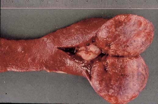
a) b)
Fig. 2 Macroscopic image of a spleen with a hemangiosarcoma (a) and with a nodular hyperplasia (b).
Abb. 2 Makroskopisches Bild einer Milz mit einem Hämangiosarkom (a) und einer nodulären Hyperplasie
Tierärztliche Praxis Kleintiere 4/2012; 40(K): 250–260 © Schattauer 2012N. Eberle et al.: Characteristics of canine splenic masses 9
10–20% of all examined spleens, which is comparable with the re- as body weight demonstrated no difference between the dogs with
sults of the present study. malignant and those with non-malignant splenic masses.
Hematomas in humans are almost always the result of abdomi- In a previous retrospective study the biological behaviour of
nal trauma. Splenic hematomas in dogs often occur secondary to splenic hemangiosarcoma was characterized (27). Metastatic pat-
an underlying splenic disorder, such as primary nodular hyperpla- tern data of 25 dogs presented with clinical signs related to splenic
sia, and are uncommonly a result of blunt trauma (24). An associ- mass that had undergone necropsy were analysed. Six of 25 dogs
ation between splenic hematomas and underlying splenic nodular had right atrial hemangiosarcoma. Disease confined to the peri-
hyperplasia has been proposed. It has been suggested that the pres- toneal cavity was present in 79% of dogs without right atrial in-
ence of nodular hyperplasia may disrupt the normal blood supply volvement. The most common metastatic sites in these dogs were
of the canine spleen with secondary regional blood accumulation liver, omentum and mesentery (27). In the present study a liver
and hematoma formation (24). Nontraumatic hemoabdomen is biopsy specimen was obtained from 173 of 249 dogs. Histology re-
considered to be a poor prognostic indicator, and dogs with hemo- vealed hemangiosarcoma in 26 (15%) dogs and seven dogs (4%)
peritoneum have been shown to be significantly more likely to have had evidence of metastasis of other malignant neoplasias. On the
hemangiosarcoma than splenic nodular hyperplasia/hematoma other hand, in 70 cases of liver biopsy with a malignant splenic
complex (1, 16, 21). In the present study, a higher proportion tumor the histopathologic examination showed no evidence for
(67%) of dogs with peritoneal effusion based on ultrasound or la- malignant neoplasia. Therefore, hepatic nodules identified during
parotomy had a malignant splenic mass. The presence of hemo- celiotomy could represent a nonmalignant process, such as vacuo-
peritoneum has been indicated as a predictor of malignancy in lar change or hyperplasia and should not automatically be inter-
studies of canine splenic disease (1, 16, 21). In the study from Ham- preted as being malignant. On the other hand, biopsy sites may not
mond et al. (12), the prevalence of malignant splenic neoplasia was have been representative. Incidence of malignancy in liver biopsies
76% in dogs with hemoperitoneum, whereas only 24% dogs had a therefore may have been underestimated in the present study. As
benign splenic lesion. In the present study the distribution of macroscopic changes within the liver detected during surgery can-
splenic masses was comparable. not be distinguished from benign lesions such as hyperplastic no-
Some of the splenic masses included in this study (n = 50; 20%) dules, it is imperative not to recommend euthanasia without histo-
were identified incidentally with abdominal ultrasound or celio- pathologic confirmation.
tomy. These patients were presented for various clinical signs not Hemangiosarcoma and splenic hematoma have been reported
directly related to the presence of a splenic mass. Of these dogs, to be macroscopically indistinguishable from each other in most
26% (n = 13) had a malignant tumor and 37 (74%) had a non-ma- cases (1, 16, 21, 24). Abdominal radiography and ultrasonography
lignant tumor. This result is of interest, because the decision are traditionally used to evaluate the spleen (10). Based on radio-
whether or not to perform a splenectomy in dogs without hemo- graphic findings, ultrasonographic findings and gross inspection,
peritoneum must be carefully considered. Splenectomized dogs malignant and nonmalignant splenic masses have nonspecific
may have some reduction of functional immune surveillance. This characteristics (10). In the present study, there was no difference
may have negative consequences such as increased susceptibility to between the size (largest diameter) of nonmalignant (median
microbial infection and erythrocyte parasitism (6). However, the 5 cm) and malignant splenic masses (median 6 cm) in the ultra-
results of the present study pointed out a risk of one fourth to have sound examination. Even though, there was a difference between
a malignant tumor in incidental splenic masses. Furthermore, the the size of nonmalignant (median 12 cm) and malignant splenic
risk of a rupture of the splenic mass resulting in a life threatening masses (median 10 cm) in the radiographic estimation of the lar-
condition should be taken into account in the process of decision gest diameter (p = 0.001), it was not possible to predict malignan-
making. cy. Both findings are in agreement with prior studies (16, 25). An
The literature shows that German Shepherd dogs, Labrador Re- additional finding of the study is the diverse median largest size of
trievers, and Golden Retrievers are at higher risk for the devel- the splenic masses in the radiographic and ultrasound examina-
opment of abdominal hemangiosarcoma and splenic hematoma tion. An explanation could be the difficulty to measure very large
(1, 5, 19, 20, 23–25). The findings of the present study support the splenic masses appropriately. The results of the present study em-
breed predisposition data from the previous reports. Because they phasizes the inability to distinguish between malignant and non-
are common breeds in the general population of the hospital, it is malignant splenic masses based on size or other gross findings (16,
difficult to conclude whether these breeds were overrepresented. 24). Ivancic et al. (15) showed that even contrast harmonic ultra-
The results from the present study were consistent with previous sonography of splenic masses cannot distinguish between splenic
reports that older and larger dogs are predisposed to splenic mass- hemangiosarcoma and hematoma. Due to the retrospective nature
es (16, 20). There is not a consistent sex predilection in the litera- of the study, a more detailed description of the sonographic char-
ture (16, 21). In contrast to the literature however the present study acteristics of liver and spleen was not possible.
showed overrepresentation of male dogs (104 dogs [40 male cas- Analysis of a recent study suggested that anemic dogs with clini-
trated] versus 51 female dogs [54 female spayed]). Furthermore, cal signs, a splenic mass, and hemoperitoneum that required a
comparison of epidemiologic data and patient characteristics such transfusion without a history of trauma or coagulopathy had a
Tierärztliche Praxis Kleintiere 4/2012; 40(K): 250–26010 N. Eberle et al.: Characteristics of canine splenic masses
76% risk of having malignant neoplasia and a 70% risk of having ranted. Studies evaluating hepatic parenchyma, using helical CT
hemangiosarcoma (12). In the present study, the presence of ane- while infusing contrast material (wash in/wash out) may improve
mia showed a significant difference between dogs with malignant the sensitivity for detecting hepatic neoplasia (9, 26).
and non-malignant tumor (p < 0.000). Cytological examination of fine-needle aspirates could be an
A limitation of the present study lies in its retrospective nature aid in the diagnosis of splenic masses. In human medicine, an as-
and in that not all of the above-mentioned parameters were evalu- sociation between target lesions in the liver and malignancy has
ated in every case. Since the majority of dogs with splenic masses been described (28). The presence of a target lesion had a positive
with hemoperitoneum in the present study were presented as predictive value for malignancy of 86% (28).
emergency cases at times when an experienced radiologist was not In veterinary medicine, the finding of one or more target lesions
available, ultrasonography was only performed in 158 cases. Addi- in the liver or spleen had a positive predictive value for malignan-
tionally, a possible bias due to including patients with splenic cy of 74% (3). In case of multiple target lesions within one organ,
masses that did not undergo surgery needs to be considered. An- the positive predictive value for malignancy was 81% (3). In one
other limitation of the study is that description of gross inspection, other study, the accuracy rate of splenic cytological examination
number of splenic masses and macroscopic size of the spleen was was only 38% (7). A reason for the poor accuracy rate for splenic
not performed due to the retrospective nature of the study. A more masses can lie in the highly cavernous composition of some mass-
detailed characterization of the macroscopic findings of the spleen es. Even when the fine-needle aspirate is obtained from a visibly
could hold additional information in comparison to ultrasound solid part of the mass, the specimen can be contaminated with
examination. blood and contain unrepresentative cellular material. Because of
Future studies in dogs with splenic masses may involve new the inconsistent use of fine-needle aspirations in the present study,
diagnostic imaging modalities such as contrast-enhanced ultra- this technique could not be evaluated any further and should be
sound and computed tomography (14). The use of ultrasound, considered for future investigations of splenic masses in the dog.
computed tomography, and magnetic resonance imaging of the
spleen and liver for detection of mass lesions in humans has been Conflict of interest
extensively described (7, 9, 22, 26). Computed tomography of ca- The authors confirm not to have any conflict of interest.
nine splenic masses showed that malignant masses had lower
Houndsfield values than nonmalignant masses pre- and post-con- Acknowledgments
trast medium administration (10). Further investigation of pre- The authors thank members of the oncology service, as well as the
operative abdominal CT evaluation of patients is therefore war- emergency and surgery staff.
Conclusion for practice References
Distinguishing between benign or malignant splenic masses based on
1. Aronsohn MG, Dubiel B, Roberts B, Powers BE. Prognosis for acute nontrau-
history, signalement, laboratory findings, radiographs, and ultrasound matic hemoperitoneum in the dogs: a retrospective analysis of 60 cases
examination was not possible in the dogs in this study. The prognosis (2003–2006). J Am Anim Hosp Assoc 2009; 45: 72–77.
for splenic masses however varies depending upon whether the mass 2. Brown NO, Patnaik AK, MacEwan EG. Canine hemangiosarcoma: retrospec-
tive analysis of 104 cases. J Am Vet Med Assoc 1985; 186: 56–58.
is benign or malignant. Preoperative diagnostic methods to identify
3. Cuccovillo A, Lamb CR. Cellular featues of sonographic target lesions of the
dogs with malignant disease would be helpful when treatment deci- liver and spleen in 21 dogs and cats. Vet Radiol Ultrasound 2002; 43:
sions are being made in dogs with splenic masses to better advise 275–278.
owners on the extent of the disease. Veterinarians are obligated to en- 4. Day MJ, Lucke VM, Pearson H. A review of pathological diagnosis made from
87 canine splenic biopsies. J Small Anim Pract 1995; 36: 426–433.
sure that owners of these dogs are informed about the prevalence of
5. De Morais, HA, Argyle DJ, O’Brian RT. Disease of the spleen. In: Textbook
hemangiosarcomas, treatment options, and the overall poor progno- of Veterinary Internal Medicine, 7th edn. Ettinger SJ, Feldman EC, eds. St.
sis associated with hemangiosarcoma before committing to treat- Louis, Missouri: Saunders 2010; 810–819.
ment. Due to this prognosis many owners opt to have their dog eutha- 6. Dietrich CF, Kratzer W, Stroberl D Danse E, Fessl R, Bunk A, Vossas U,
Hauenstein K, Koch W, Blank W, Oudkerk M, Hahn D, Greis C. Assessment
nized if evidence of a malignant mass or metastatic lesions are found
of metastatic liver disease in patients with primary extrahepatic tumours
preoperatively or intraoperatively. On the other hand, patients with by contrast-enhanced sonography versus CT and MRI. World J Gastroente-
splenic mass and evidence of hemoperitoneum have a chance of one rol 2006; 12: 1699–1705.
third to have a non-malignant tumor, and surgical intervention can re- 7. Eich CS, Whitehair JG, Moroff SD, Heeb LA. The accuracy of intraoperative
cytopathological diagnosis compared with conventional histopathological
sult in long-term survival times. It is furthermore important that dogs
diagnosis. J Am Anim Hosp Assoc 2000; 36: 16–18.
with liver nodules at the time of surgery may have benign hepatic hy- 8. Feeney DA, Johnston GR, Hardy RM. Two-dimensional, gray-scale ultraso-
perplastic nodules and that euthanasia at the time of surgery should nography for assessment of hepatic and splenic neoplasia in the dog and cat.
not be recommended to owners solely on the basis of macroscopic J Am Vet Med Assoc 1984; 184: 68–81.
9. Ferrozzi F, Bova D, Draghi F, Garlaschi G. CT findings in primary vascular tu-
liver nodules.
mors of the spleen. Am J Roentgenol 1996; 166:1097–1101.
Tierärztliche Praxis Kleintiere 4/2012; 40(K): 250–260 © Schattauer 2012N. Eberle et al.: Characteristics of canine splenic masses 11
10. Fife WD, Samii VF, Drost WT, Mattoon JS, Hoshaw-Woodard S. Comparison 21. Pyrmak C, McKee LJ, Goldschmidt MH, Glickman LT. Epidemiologic, clini-
between malignant and non-malignant splenic masses in dogs using con- cal, pathologic, and prognostic chararcteristics of splenic hemangiosarcoma
trast-enhanced computed tomography. Vet Radiol Ultrasound 2004; 45: and splenic hematoma in dogs: 217 cases (1985). J Am Vet Med Assoc 1988;
289–297. 193: 706–712.
11. Hammer A, Couto CG. Diagnosing and treating canine hemangiosarcoma. 22. Quaia E, D’Onofrio M, Palumbo A, Comparison of contrast-enhanced ultra-
Vet Med 1992; 3: 188–201. sonography verses baseline ultrasound and contrast-enhanced computed to-
12. Hammond TN, Pesillo-Crosby SA. Prevalence of hemangiosarcoma in mography in metastatic disease of the liver: Diagnostic performance and
anemic dogs with splenic mass and hemoperitoneum requiring a transfu- confidence. Eur Radiol 2006; 16: 1599–1609.
sion: 71 cases (2003–2005) J Am Vet Med Assoc 2008; 232: 553–558. 23. Srebrenik N, Appelby RC. Breed prevalence and sites of hemangioma and
13. Hanson JA, Pennick DG. Ultrasonographic evaluation of traumatic splenic hemangiosarcoma in dogs. Vet Rec 1991; 129: 408–409.
hematoma and literature review. Vet Radiol Ultrasound 1994; 35: 463–466. 24. Spangler WL, Culbertson MR. Prevalence, type, and importance of splenic
14. Irausquin RA, Scavelli TD, Corti L, Stefanacci JD, DeMarco J, Flood S, Rohr- disease in dogs: 1480 cases (1985–1989). J Am Vet Med Assoc 1992; 200:
bach BW. Comparative evaluation of the liver in dogs with a splenic mass by 829–834.
using ultrasonography and contrast-enhanced computed tomography. Can 25. Spangler WL, Kass PH. Pathologic factors affecting postsplenectomy survival
Vet J 2008; 49: 46–52. in dogs. J Vet Intern Med 1997; 11: 166–171.
15. Ivancic M, Long F, Seiler GS. Contrast harmonic ultrasonography of splenic 26. Stark DD, Wittenberg J, Butch RJ, Ferrucci JT Jr. Hepatic metastases: Ran-
masses and associated liver nodules in dogs. J Am Vet Med Assoc 2009; 234: domized controlled comparison of detection with MR imaging and CT.
88–94. Radiology 1987; 165: 399–406.
16. Johnson KA, Powers BE, Withrow SJ, Sheetz MJ, Curtis CR, Wrigley RH. 27. Waters DJ, Caywood DD, Hayden DW, Klausner JS. Metastatic pattern in
Splenomegaly in dogs: predictors of neoplasia and survival after splenec- dogs with splenic hemangiosarcoma: clinical implications. J Small Anim
tomy. J Vet Intern Med 1989; 3: 160–166. Pract 1988; 29: 805–814.
17. Knapp DW, Aronsohn MG, Harpster NK. Cardiac arrhythmias associated 28. Wernecke K, Vassalo P, Bick U, Diederich S, Peters PE. The distinction be-
with mass lesions of the canine spleen. J Am Anim Hosp Assoc 1993; 29: tween benign and malignant liver tumors: an in vitro correlation study. Am
122–128. J Roentgenol 1992; 159: 1005–1009.
18. Marino DJ, Mathiessen DJ, Fox PR, Lesser MB, Stamoulis ME. Ventricular 29. Wood CA, Moore AS, Gliatto JM, Ablin LA, Berg RJ, Rand WM. Prognosis for
arrhythmias in dogs undergoing splenectomy: a prospective study. Vet Surg dogs with stage I or II splenic hemangiosarcoma treated by splenectomy
1994; 23: 101–106. alone: 32 cases (1991–1993). J Am Anim Hosp Assoc 1998; 34: 417–421.
19. Mongil CM, Drobatz KJ, Hendricks JC. Traumatic hemoperiteoneum in 28 30. Wrigley RH, Konde LJ, Park RD, Lebel JL. Ultrasonographic features of sple-
cases: a retrospective review. J Am Anim Hosp Assoc 1995; 31: 217–222. nic hemangiosarcoma in dogs: 18 cases (1980–1986). J Am Vet Med Assoc
20. Pintar J, Breitschwerdt EB, Hardie EM, Spauding KA. Acute nontraumatic 1988; 192: 1113–1117.
hemoabdomen in the dog: a retrospective analysis of 39 cases (1987–2001). 31. Wrigley RH, Konde LJ, Park RD, Lebel JL. Clinical features and diagnosis of
J Am Anim Hosp Assoc 2003; 39: 518–522. splenic hematomas in dogs: 10 cases (1980 to 1987). J Am Anim Hosp Assoc
1989; 25: 371–375.
Tierärztliche Praxis Kleintiere 4/2012; 40(K): 250–260You can also read