Stimulation of Staphylococcus aureus Ligase Enzyme by Magnesium Ion - Open Journal Systems
←
→
Page content transcription
If your browser does not render page correctly, please read the page content below
2148      Indian Journal of Forensic Medicine & Toxicology, January-March 2021, Vol. 15, No. 1

          Stimulation of Staphylococcus aureus Ligase Enzyme by
                              Magnesium Ion

                         Arqam Mohamad Alomari1, Aseel A. H. Al-Layla2, Ibrahim Faris Ali3
    1
     Lecturer, Department of Basic Sciences, College of Agriculture and Forestry, University of Mosul, Mosul, Iraq,
2
    Department of Biophysics, College of Sciences, University of Mosul, Mosul, Iraq, 3Department of Biology, College
                           of Education for Pure Science, University of Mosul, Mosul, Iraq

                                                               Abstract
        Ligases enzymes were discovered as a member of the nucleotidyl transferase family. Here in this paper, DNA
        Ligase is extracted from S. aureus works with the cofactor NAD+ to make a phosphodiester bond and reform
        between the 3’hydroxyl and 5’phosphate DNA end. Staphylococcus aureus-DNA Ligases Enzyme type A
        (SLE-A) contains two essential domains; NTase and OB- fold domain, which are the most essential domains
        for the enzyme function. The main aim of the study is to investigate the activity of SLE-A in the presence
        of magnesium ion (Mg+2) by evaluating several kinetic parameters on a time course. The result showed that
        SLE-A has optimal activity at 500 µM of Mg+2. Furthermore, the low number of Equilibrium Association
        Constant (Km value) explains the binding affinity between DNA ligase of Staphylococcus aureus SLE-A
        enzyme and Mg+2 ion was very high and sold.

        Keywords: DNA ligases, Magnesium ion, Staphylococcus aureus, Michaelis-Menten equation.

                         Introduction                                  nucleotide excision, single and double-stranded repairs
                                                                       (9). The lake or mutation of SLE-A ( referees to LigA
        Background of DNA ligase.                                      gene), resulted the loss of the ability to ligate Okazaki
      Fifty years ago, Lehman and other groups discovered              fragments during the replication steps (10). DNA Ligases
the DNA ligases and was a turning point event in                       are very potent chemical material in the development of
molecular biology (1-4). Other DNA ligases had been                    biotechnology, bimolecular and genetic engineering (11).
lighted on since and they are presented in all domains                 Another important implication, is that NAD+-dependent
of life (5). The DNA ligase enzyme is an important                     ligases not present in mammalian cells as well as they
component in the Ligation reaction, which also called                  are involved in developing antibacterial drugs “A
DNA seals, DNA joins and/or DNA fixing enzyme (6).                     requirement for an antibacterial enzyme target is that
All types of DNA Ligases which have the same protein                   it should be essential for the organism and not present
fold are considered as part of nucleotidyltransferase                  in the host (12). The formation of a phosphodiester bond
family. For example, DNA ligases and RNA ligases                       between adjacent 5 phosphate and 3’ hydroxyl DNA
(7)
    . they participated in multi processes for instance                termini is catalyzed by Ligases which known to fix
DNA repair, recombination and replication (8). DNA                     several substrates.
ligase enzyme plays an important role during DNA                            Indeed, ligases are participated in the base
replication: Firstly, joins Okazaki fragments, in addition
it carry out a several DNA repairs like base excision,                      Excision DNA repair, culminating step of nucleotide
                                                                       and the ligation of Okazaki fragments during cell
                                                                       replication (13-14). Importantly, NAD+ ligases play an
Corresponding author:                                                  important role in cell survival for prokaryotes (15). The
Arqam Mohamad Alomari                                                  main characteristics of DNA ligase in prokaryotic is to
arqam.alomari@uomosul.edu.iq                                           present into all bacteria and all bacterial species contain
Indian Journal of Forensic Medicine & Toxicology, January-March 2021, Vol. 15, No. 1 2149
LigA protein (16). Kaczmarek announced in 2001, that the (9).
deletion or mutation of the LigA gene in Staphylococcus
aureus (represent to SLE-A) lead to loss the complete Materials and Methods
growth in the bacteria (17). The S. aureus-DNA ligase The Cloning of Staphylococcus aureus DNA
enzyme A (LigA) is formed of 666 signal amino acid Ligase.
and consist of 6 main domains with 74,993 Da molecular
weight. The tertiary structure of Staphylococcus aureus The published open-reading frames of S. aureus-
of LigA (SLE-A) protein consists of two main domains DNA Ligase enzyme A (SLE-A)) (belongs to LigA)
called nuclotidyltransferase/ NTase domain and an was retrieved from the NCBI-PubMed database using
Oligomer Binding (OB domain), which are considered Gene ID 45575143 and the gene size is 2004 base pair
as a catalytic core for all domains of life (18-19). for S. aureus LigA. Gene was synthesised (GeneArt,
ThermoFisher, UK) with a 20 bp flanking sequence
Magnesium Ion in DNA Ligase. containing an NdeI site and cloned into the NdeI site
of pET29c. Kanamycin-resistant transformants of S.
One of the most important cofactor in the ligation
aureus LigA were screened by colony PCR and those
or the mechanism process of DNA ligases enzymes
showing the requisite sizes were sequenced in full on
is MgCl2 Ion. It considers a second cofactor in the
both strands (Genewiz, UK). Figure 1 is shown a sample
reaction of ligase. Mg+2 is a co-factors that allows the
of S. aureus (SLE-A) gene was run on a 1% agarose gel.
ligases enzymes to join and shut a nick at the backbone
Large-scale preparations of each plasmid were made
of Deoxyribonucleic acid. Furthermore, LigA DNA
(Qiagen, UK) and stored at -20ºC in 50 µl aliquots. DNA
ligase needs Mg+2 to draw the AMP groups that is very
primer sequences are as follow as (green colour refers to
necessary during the mechanism of ligase and attaches
the NdeI site of pET29c and black colour refers to the
with the active site of amino acid called lysine to do
forward and reverse primers of S. aureus-DNA ligase
its work. The AMP groups comes when the ligase
enzyme type A gene.):
enzyme touches with cofactor ATP or NAD+ to form a
phosphoamide -linked AMP and without the NAD+ or
ATP the reaction of ligation will be stopped completely
Figure 1: Cloning of S. aureus-DNA ligase enzyme type A.2150 Indian Journal of Forensic Medicine & Toxicology, January-March 2021, Vol. 15, No. 1
1% of agarose gel showing the PCR cloning from S. debris. Ligase A was subsequently purified from the
aureus-DNA enzyme type A as follows: Lane M – 1 k supernatant using a two-step method; all steps were at
bp DNA ladder (NEB), showing sizes of DNA between 4ºC. In the first step, NaCl was adjusted to 1M in order to
500 bp to 10,000 bp. Lane 1 is a colony PCR products disrupt protein-nucleic acid interactions and ammonium
of SLE-A gene, the length of S. aureus SLE-A (LigA) is sulphate then added slowly over two hours to a final
2004 bp depending on the GeneBank plus the sequencing concentration of 35% (w/v) in order to precipitate the
of plasmid. enzyme. Following centrifugation at 18000 rpm for 20
minutes, the pellet was resuspended in an appropriate
The expression and purification of Staphylococcus
volume of Buffer A (10 mM sodium phosphate, pH 7.0).
aureus DNA Ligase.
Salt was removed by membrane dialysis overnight in
For S. aureus LigA (SLE-A) expression, competent Buffer A. In the second stage, enzymes were injected
E.coli BL21 (DE3) cells was transformed with 0.5 µg onto a Hi-Trap heparin anion-exchange column (GE
plasmid (pLigA) and grown overnight at 37°C on LB- Healthcare, UK) in Buffer A and eluted in the same
agar containing 50 µg/ml kanamycin. A single colony buffer with NaCl gradient to 1 M. Fractions eluting from
was picked and grown overnight in an orbital shaker at the column were analysed by SDS-PAGE to confirm the
37ºC in 5 mL LB media. A 1 mL aliquot of this starter presence and purity of the protein. Fractions containing
culture was added to 500 mL fresh LB broth and grown purified LigA was pooled and dialysed into Buffer
under the same conditions until an OD600 of 0.6 was A. Ligase activity assays were then used to check for
reached. Protein expression was induced with 1 mM correctly-folded, functional ligase. LigA was further
isopropyl B-D-thiogalactoside (IPTG) and the culture purified using size-exclusion chromatography (GE
incubated at 37ºC for 2 hours (LigA) until an OD600 Healthcare, UK).
~2.0 was reached. Bacterial cultures were harvested by
Fractions containing purified LigA were pooled
centrifugation at 6000 rpm for 20 minutes at 4°C and
and concentrated using spin column to ~ 1 ml. S. aureus
the media discarded. The cell pellet was re-suspended in
LigA was finally dialysed into 1×Ligase storage buffer
15 ml Lysis buffer (10 mM sodium phosphate, pH 7.0,
(30 mM Tris-HCl, pH 7.2, 1 mM DTT and 50 µg/ml
containing benzamidine and PMSF protease inhibitor
BSA and 30% (v/v) glycerol). Aliquots of 50 µl were
cocktail (Sigma-Aldrich, UK) was sonicated on ice in
stored at -20ºC and used fresh for subsequent kinetic and
10 second pulses over 3 minutes and spun at 18000 rpm
binding assays.
for 20 minutes at 4ºC to separate supernatant and cellIndian Journal of Forensic Medicine & Toxicology, January-March 2021, Vol. 15, No. 1 2151
Figure 2 is shown the large scale of expression of DNA ligase (LigA) and the final purification of the S. aureus
SLE-A protein.
Figure 2: Large Scale Inductions of Staphylococcus aureus-DNA ligase enzyme type A in BL21 (DE3).
10% SDS PAGE gel shows the Large induction of S. aureus SLE-A enzyme in E. coli BL21 (DE3) as follow:
Lane M shows an NEB protein marker indicated by sizes (in kDa) pointed to the left of the gel. Lanes 1 and 2 shows
the total post-induction of expressed SLE-A. Lane 3 shows the corresponding samples of S. auerus LigA protein
that pooled from heparin column with molecular weight (size) 74,993 Da. Lane 4 shows the final purification of pure
protein via using size sxclusion column.
Ligase activity and timecourse assays.
Three HPLC-purified oligonucleotides (Fisher Scientific, UK) were used:
20Top 5’-HEX-ATCTCGCGTATGGGCCTTCG-3’
30Top 5’-P-CTGCTCACAGGACACCTGGTATACGTAATG-3’
50Bot 5’-CATTACGTATACCAGGTGTCCTGTGAGCAGCGAAGGCCCATACGCGAGAT-3’2152 Indian Journal of Forensic Medicine & Toxicology, January-March 2021, Vol. 15, No. 1
These were mixed in a ratio of 1:1:1 at a concentration using Michaelis- Menten equation
of 10 µM in 50 µl of 1×Ligase reaction buffer (30 mM
as follow V0 = Vmax + [S]/ Km + [S]
Tris-HCl, pH 7.5, 1 mM DTT and 50 µg/ml BSA) and
annealed by cooling in a PCR machine from 100ºC The definition of Kd is the concentration of substrate
to 4ºC at 0.1ºC/min. The resulting 50mer oligoduplex that gives half maximum rate. It was obtained from the
contained a single off-centre nick between the 20Top Michaelis- Menten equation fit above as well.
and 30Top strands with a phosphoryl group on the 5’-
side of the nick; a hexachlorofluorescein (HEX) group Results
at the 5-end of the 20Top strand permitted the DNA
Nick-joining ligation activity.
to be quantitated. Kinetic assays on LigA was carried
out in 1× ligase reaction buffer. The concentration of All proteins purified in this study have been proved
nicked oligoduplex was 1000 nM in most experiments. to join nicks in double stranded nucleic acid in different
Each timecourse (50 µl) contained nicked oligoduplex, conditions and terms. The purified of LigA protein of E
1×Ligase buffer, the requisite cofactor(s) and 40 nM of coli DNA ligase have been confirmed to join nicks in
LigA. Each reaction of time course was containing a double stranded nucleic acids with varying efficiencies
single nick of six MgCl2 samples ranging from (0.1, 1, in presence of NAD+. In order to achieve the ligation
10, 50, 100, and 500 μM) on a denaturing PAGE 15%. experiment and how the ligation was affected by
All the samples were incubated at 16 °C. The time points varying concentrations of Mg+2, a double-stranded
were prepared for 5 times as follow (0 time, 3 mins, 5 DNA substrate (dsDNA) was synthesised from Fisher
mins, 10 mins, 20 mins and 60 mins) and all the tubes Scientific, UK with a single nick on the top strand. Three
were heated at 100 °C for 2 mins. Shortly, the intensity HPLC purification of oligonucleotides were synthesised
of the 50TOP bands were measured by using computer as follow: the length of first top oligonucleotide was 50
software ImageGauge. The data were plotted with % bases contains from two top strands (20 and 30 bases)
intensity against time revealing the initial rate. and both of them are complementary to adjacent the 50
base bottom strand. The 20 top single strand attached
Data Analysis.
fluorescent HEX group and the nick was made on the top
Products of timecourse reactions were run on strand between 30 top and 20 top. The complementarity
40 cm long, 0.4 mm thick 15% (w/v) denaturing in the bottom and top strands location, the two top strands
polyacrylamide gels containing 1×TBE (89 mM Tris- (30 and 20) was adjacent to each other. The nick on the
borate, 2 mM EDTA) and 40% urea. Gels were pre-run top strand (50 oligonucleotide) was at 3’-hydroxyl group
at 60 W for 30 minutes until warm. Samples (10 µl) were of the 20 base strand and 5’-phosphate group of the 30
heated at 90ºC for 2 minutes, loaded and run at 50 W base strand.
for ~90 minutes. The HEX-labelled DNA strands were
When the buffer has the nicked DNA substrate and
visualised by excitation at 600 nm using a fluorescence
SLE-A (LigA) protein of the appropriate concentration
imager (FLA-5000, Fuji, Japan). The digitised images
and conditions, the nick on the top strand will be sealed
were quantified using ImageJ (imagej.nih.gov) by firstly
by the proteins.
removing background (50-pixel ‘rolling-ball’ average)
and then integrating the area under each peak. These data The samples were taken at specific time points and
were used to calculate the fraction of counts distributed transferred to the stop buffer to stop the ligase protein
between the 20mer substrate and 50mer product its work. During electrophoresis, the denaturing gel
bands in each lane; these were multiplied by the DNA separate the double stranded structure (50 base top and
concentration to give the amount of each in nM. Initial bottom) and causes to unfold the dsDNA into the linear
rates (V0) were determined by the gradient of the first chain. The dsDNA substrates will be migrated through
20% of the reaction. Rate data were fit to the Michaelis- the gel as single stranded. Therefore, the size of DNA
Menten equation (Grafit v5, Erithacus Software, UK) ligation is easy to distinguish as the distance migrated
and Vmax and Kd parameters were elucidated by plotting by the DNA. Once the products sDNA of 20 and 30
their V0 values for each timecourse and fitting them by base strands ligated will be migrated less position on theIndian Journal of Forensic Medicine & Toxicology, January-March 2021, Vol. 15, No. 1 2153
denaturing gel, comparing to only the 20 base strand, +2
100, and 500 μM. At low of Mg ion, initial average
which will be stayed at the bottom of the gel. The visible increases slowly till over 10 μM (does not show), where
light to the ligated products in the denaturing gel was is initial rate rises rapidly around 50 μM and the loop
easy to follow due to the attached of fluorescent HEX of curve begins to rise until 500 μM. The value for
group at 20 top strand. Vmax gained by organized initial average in front of
Mg+2 ion concentrations. The Vmax obtained by Mg+2
The Effect of Mg+2 ion on the S. aureus DNA
ion concentrations which was µM/min and the Km (the
ligase .
[MgCl2] that given half Vmax rate) was as well in µM.
To assess whether ligation of such nucleic acids To examine the Vmax and Km (shown as Kd at the figure)
may be a substantial value of these enzymes, at least in for Mg+2, using six Mg+2 concentrations, these initial
biochemical condition, all these experiments have been rate values (in % per minute) were calculated against
characterized the ligation of the dsDNA with different the concentration of Mg+2 in each experiment and fit
protein of S. aureus SLE-A (LigA)across a range of the curve to a Michaelis-Menten equation by using the
independent variable of Mg+2 ion. All the experiments software Grafit. The initial rate and the measurement
were carried out at 16 0C (except temperature) and 1 of the intensity were explained by details in the data
µM of oligonucleotide substrate in total volume of 50 analysis in the Material and Method above. Figures 3 is
µl of 1X DNA ligase buffer and the mixture of reaction an example of gels showing the ligation of DNA ligase
were completed with free water. The concentration of of S. aureus SLE-A with different concentration of Mg2+
enzymes that used in all these experiments was 40 nM. that appeared by bands of DNA in the denaturing gel.
To check how was influenced by different Mg+2 ion The plot for the MgCl2 ion shows below in Figure 4.
concentrations; the process of ligation was performed in The Vmax (capacity) of Mg+2 ion was 3.9 %/min where
the existence of Mg+2 concentrations from 0.1, 1, 10, 50, is the Km (is shown as Kd ) was 5.2 µM.
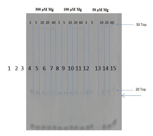
Figure 3 is the denaturing PAGE gel of three of the six MgCl2 Ion concentrations tested.
Denaturing PAGE gel that was got from this study is labelled as following lanes: 1-6 which is corresponded to
the following time points 3,5,10,20 and 60 minutes, at 500 µM of Mg+2 ion as an example of this gel, at the top of
gel are 50 mer (50 Top) and the bottom of gel are 20 mer (20 Top). The Bromophenol blue is the colour which is
used to check the bands on the denaturing gel.2154 Indian Journal of Forensic Medicine & Toxicology, January-March 2021, Vol. 15, No. 1
Discussion well 500 µM, which is indicated the similarity between
these ions activity on different enzymes (26). Moreover,
Because of the NAD+-dependent DNA ligase is
the results can be concluded that DNA ligases of E. coli
very essential enzyme for DNA replication and repair,
and Staphylococcus aureus are structurally similar. The
it had been reported as a prospective broad-spectrum
Km value (equilibrium association constant) in this study
antibacterial target, since there are a high conserved
was 5.2 µM, which is explained the binding affinity
phylogenetic and distinctly different from the Eukaryotic
between DNA ligase of Staphylococcus aureus SLE-A
DNA ligases (20-22). Thus, prokaryotic NAD+-dependent
and MgCl2 ion was slightly similar to the Km value for
ligases is present as a promising drug target for
NAD+ cofactor for S. aureus LigA (2.75 µM) that found
antimicrobial therapy comparing to ATP-dependent
by Gul et al, 2004 (27). However, different result by
in human (23-24). In 1973s, Modrich and Lehman were
Sriskanda & Shuman 2001 who obtained the Km value
referred that magnesium ion increase the activity of ligase
for E. coli DNA ligase enzyme LigA for singly-nicked
protein. Magnesium ion is very important component in
DNA substrate and was 12.2 nM, which is completely
the step1 and step2 of the ligation mechanism of ligases
different comparing with this study (7). Generally, it was
enzyme in general. Moreover, in the same study showed
mentioned above in the discussion that the main objective
that the magnesium ion is being involved to increase the
in the biology and medical sciences is to find a target in
rate of phosphodiester synthesis and DNA adenylation
the bacteria to be as novel drug target for antimicrobial
in the system of mechanism of ligases enzymes (25).
therapy, and DNA ligase is one of them since there are
The obtained results in this paper from the investigation
no similarity to the eukaryotic DNA ligases. However,
the effect of Mg+2 ion on Staphylococcus aureus-DNA
magnesium ions here did not inhibit the activity of
Ligases enzyme type A (SLE-A) activity showed that
Staphylococcus aureus-DNA Ligases enzyme type A
500 µM of Mg+2 ion had the best on ligases’s activity for
(SLE-A), but increase the rat of reaction. Therefore, Due
fixing the nick in the DNA between the 20 mer and 30
to the importance of the DNA ligase enzyme, further
mer, and transfer them to 50 mer. Comparing this result
study would be involved to identify more compounds
with different study by using ammonium sulphate ion to
that would be able to inhibit the activity of DNA ligase
determine how much this ion affected to the E. coli DNA
(SEL-A) but not inhibit the activity of eukaryotic DNA
Ligase activity (LigA), and the result was showed that
ligases by blocking the communication sites between the
the best concentration of ammonium sulphate was asIndian Journal of Forensic Medicine & Toxicology, January-March 2021, Vol. 15, No. 1 2155
magnesium ions (Mg+2) and the ligase of bacteria, and bound to nicked DNA-adenylate. Molecular cell.
eventually control the growth of them. 2007 Apr 27;26(2):257-71.
9- Shuman S. DNA ligases: progress and prospects.
Acknowledgment: The authors are very grateful
Journal of Biological Chemistry. 2009 Jun
to the University of Mosul for providing assistance and 26;284(26):17365-9.
facilities which allowed finishing this work properly, with
10- Poidevin L, MacNeill SA. Biochemical
many thanks to the College of Agriculture and Forestry,
characterisation of LigN, an NAD+-dependent
College of Environmental Science and Technology and
DNA ligase from the halophilic euryarchaeon
College of Education for Pure Science for kind support. Haloferax volcanii that displays maximal in vitro
Source of Funding: Self activity at high salt concentrations. BMC Molecular
Biology. 2006 Dec 1;7(1):44.
Conflict of Interest: Nil 11- Sriskanda V, Shuman S. Role of Nucleotidyl
Transferase Motif V in Strand Joining byChlorella
References Virus DNA Ligase. Journal of Biological
1- Olivera BM, Hall ZW, Anraku Y, Chien JR, Lehman Chemistry. 2002 Mar 22;277(12):9661-7.
IR. On the mechanism of the polynucleotide 12- Korycka-Machala M, Rychta E, Brzostek A,
joining reaction. InCold Spring Harbor Symposia Sayer HR, Rumijowska-Galewicz A, Bowater
on Quantitative Biology 1968 Jan 1 (Vol. 33, pp. RP, Dziadek J. Evaluation of NAD+-dependent
27-34). Cold Spring Harbor Laboratory Press. DNA ligase of mycobacteria as a potential
2- Gefter ML, Becker A, Hurwitz J. The enzymatic target for antibiotics. Antimicrobial agents and
repair of DNA. I. Formation of circular lambda- chemotherapy. 2007 Aug 1;51(8):2888-97.
DNA. Proceedings of the National Academy of 13- Lahiri SD, Gu RF, Gao N, Karantzeni I, Walkup
Sciences of the United States of America. 1967 GK, Mills SD. Structure guided understanding of
Jul;58(1):240. NAD+ recognition in bacterial DNA ligases. ACS
3- Gellert M. Formation of covalent circles of lambda Chemical Biology. 2012 Mar 16;7(3):571-80.
DNA by E. coli extracts. Proceedings of the 14- Lehnman IR. DNA ligase: structure, mechanism,
National Academy of Sciences of the United States and function. Science. 1974 Nov 29;186(4166):790-
of America. 1967 Jan;57(1):148. 7.
4- Zimmerman SB, Little JW, Oshinsky CK, Gellert M. 15- Wilkinson A, Day J, Bowater R. Bacterial
Enzymatic joining of DNA strands: a novel reaction DNA ligases. Molecular microbiology. 2001
of diphosphopyridine nucleotide. Proceedings of Jun;40(6):1241-8.
the National Academy of Sciences of the United 16- Sriskanda V, Shuman S. A second NAD+-
States of America. 1967 Jun;57(6):1841. dependent DNA ligase (LigB) in Escherichia coli.
5- Le D, Hua X, Huang L, Gao G, Lu H, Xu Z, Tian B, Nucleic acids research. 2001 Dec 15;29(24):4930-
Hua Y. Biochemical characterization of two DNA 4.
ligases from Deinococcus radiodurans. Protein and 17- Kaczmarek FS, Zaniewski RP, Gootz TD, Danley
peptide letters. 2008 Jul 1;15(6):600-5. DE, Mansour MN, Griffor M, Kamath AV, Cronan
6- Dwivedi N, Dube D, Pandey J, Singh B, Kukshal M, Mueller J, Sun D, Martin PK. Cloning and
V, Ramachandran R, Tripathi RP. NAD+‐ functional characterization of an NAD+-dependent
Dependent DNA Ligase: A novel target waiting DNA ligase from Staphylococcus aureus. Journal
for the right inhibitor. Medicinal research reviews. of Bacteriology. 2001 May 15;183(10):3016-24.
2008 Jul;28(4):545-68. 18- Lee JY, Chang C, Song HK, Moon J, Yang JK, Kim
7- Sriskanda V, Shuman S. A second NAD+- HK, Kwon ST, Suh SW. Crystal structure of NAD+‐
dependent DNA ligase (LigB) in Escherichia coli. dependent DNA ligase: modular architecture and
Nucleic acids research. 2001 Dec 15;29(24):4930- functional implications. The EMBO Journal. 2000
4. Mar 1;19(5):1119-29.
8- Nandakumar J, Nair PA, Shuman S. Last stop on 19- Georlette D, Blaise V, Bouillenne F, Damien B,
the road to repair: structure of E. coli DNA ligase Thorbjarnardóttir SH, Depiereux E, Gerday C,2156 Indian Journal of Forensic Medicine & Toxicology, January-March 2021, Vol. 15, No. 1
Uversky VN, Feller G. Adenylation-dependent 23- Brötz-Oesterhelt H, Knezevic I, Bartel S,
conformation and unfolding pathways of the Lampe T, Warnecke-Eberz U, Ziegelbauer K,
NAD+-dependent DNA ligase from the thermophile Häbich D, Labischinski H. Specific and potent
Thermus scotoductus. Biophysical journal. 2004 inhibition of NAD+-dependent DNA ligase
Feb 1;86(2):1089-104. by pyridochromanones. Journal of Biological
20- Benson EL, Tomich PK, Wolfe ML, Choi GH, Chemistry. 2003 Oct 10;278(41):39435-42.
Hagadorn JC, Mutchler VT, Garlick RL. A high- 24- Nandakumar J, Nair PA, Shuman S. Last stop on
throughput resonance energy transfer assay for the road to repair: structure of E. coli DNA ligase
Staphylococcus aureus DNA ligase. Analytical bound to nicked DNA-adenylate. Molecular cell.
biochemistry. 2004;2(324):298-300. 2007 Apr 27;26(2):257-71.
21- Gul S, Brown R, May E, Mazzulla M, Smyth MG, 25- Chauleau M, Shuman S. Kinetic mechanism and
Berry C, Morby A, Powell DJ. Staphylococcus fidelity of nick sealing by Escherichia coli NAD+-
aureus DNA ligase: characterization of its kinetics dependent DNA ligase (LigA). Nucleic acids
of catalysis and development of a high-throughput research. 2016 Mar 18;44(5):2298-309.
screening compatible chemiluminescent 26- Alomari A. Biophysical and Kinetic Analysis
hybridization protection assay. Biochemical of Escherichia coli DNA Ligase Activity and
Journal. 2004 Nov 1;383(3):551-9. Inhibition (Doctoral dissertation, University of
22- Chen XC, Hentz NG, Hubbard F, Meier TI, Portsmouth).
Sittampalam S, Zhao G. Development of a 27- Gul S, Brown R, May E, Mazzulla M, Smyth MG,
fluorescence resonance energy transfer assay Berry C, Morby A, Powell DJ. Staphylococcus
for measuring the activity of Streptococcus aureus DNA ligase: characterization of its kinetics
pneumoniae DNA ligase, an enzyme essential of catalysis and development of a high-throughput
for DNA replication, repair, and recombination. screening compatible chemiluminescent
Analytical biochemistry. 2002 Oct 15;309(2):232- hybridization protection assay. Biochemical
40. Journal. 2004 Nov 1;383(3):551-9.
Study of bacterial
Study of bacterialYou can also read