Structural characterization of human importin alpha 7 in its cargo free form at 2.5 Å resolution - Nature
←
→
Page content transcription
If your browser does not render page correctly, please read the page content below
www.nature.com/scientificreports
OPEN Structural characterization
of human importin alpha 7 in its
cargo‑free form at 2.5 Å resolution
S. Tsimbalyuk1, C. M. Donnelly1 & J. K. Forwood1,2*
Shuttling of macromolecules between nucleus and cytoplasm is a tightly regulated process mediated
through specific interactions between cargo and nuclear transport proteins. In the classical nuclear
import pathway, importin alpha recognizes cargo exhibiting a nuclear localization signal, and this
complex is transported through the nuclear pore complex by importin beta. Humans possess seven
importin alpha isoforms that can be grouped into three subfamilies, with many cargoes displaying
specificity towards these importin alpha isoforms. The cargo binding sites within importin alpha
isoforms are highly conserved in sequence, suggesting that specificity potentially relies on structural
differences. Structures of some importin alpha isoforms, both in cargo-bound and free states, have
been previously solved. However, there are currently no known structures of cargo free importin
alpha isoforms within subfamily 3 (importin alpha 5, 6, 7). Here, we present the first crystal structure
of human importin alpha 7 lacking the IBB domain solved at 2.5 Å resolution. The structure reveals
a typical importin alpha architecture comprised of ten armadillo repeats and is most structurally
conserved with importin alpha 5. Very little difference in structure was observed between the cargo-
bound and free states, implying that importin alpha 7 does not undergo conformational change when
binding cargo. These structural insights provide a strong platform for further evaluation of structure–
function relationships and understanding how isoform specificity within the importin alpha family
plays a role in nuclear transport in health and disease.
The shuttling of macromolecules such as RNA and proteins between the cytoplasm and nucleus is an important
and fundamental process for eukaryotic cells. The process is highly regulated, mediating a range of differentiation
and developmental pathways, but is also targeted during viral infections and implicated in cancer pathogenesis1–3.
Whilst molecules smaller than 40 kDa can diffuse passively through the nuclear pore complex (NPC), larger
molecules require active t ransport4. The classical nuclear import pathway is mediated by specific interactions
between proteins from the karyopherin family and cargo proteins harbouring a nuclear localization signal (NLS)5.
Members of the IMPα are responsible for binding NLSs displayed within cargo, and through interaction with
IMPβ, the heterotrimer is imported into the n ucleus6. Upon entry, the cargo is released by RanGTP binding, and
the importins are recycled back to the c ytoplasm7–9.
IMPα proteins consist of three functional domains, an N-terminal IMPβ-binding (IBB) domain that medi-
ates interaction with IMPβ, ten ARM domains that recognize and interact with cargo, and a C-terminal CAS
domain (involving ARM 10) that mediates nuclear export and r ecycling5,10,11. The ten tandem ARM repeats are
represented by three α-helices (H1, H2 and H3) spanning ~ 40 amino acids. Overall these ARM repeats form a
bean-shaped molecule with the H3 helices defining the inside of the concaved surface. The inside groove harbour
a number of Asn and Trp residues at the third and fourth turn of H3, and play roles in cargo binding12. The Trp
and Asn are absent in ARM repeats 5 and 6, resulting in the segregation of IMPα binding regions into major
(ARM 2–4) and minor (ARM 6–8) sites12. A monopartite NLS (consisting of a single basic region) can bind to
both the major and minor sites of IMPα, whereas a bipartite NLS (consisting of two basic regions separated by
a 10–12 amino acid linker) binds to both the major and minor s ites13.
There are seven isoforms of human IMPα, grouped into three subfamilies that exhibit specificity for specific
nuclear cargo14. The IMPα1 subfamily has the lowest sequence identity and conservation, and consists of IMPα1
and IMPα8 isoforms. IMPα1 isoform and mouse homologue IMPα2 have been extensively studied with multiple
structures available describing classical monopartite b inding15. The IMPα2 subfamily is comprised of two highly
similar IMPα isoforms, IMPα3 and α4. While there are several structures of IMPα3 available, the structure of
1
School of Dentistry and Medical Sciences, Charles Sturt University, Wagga Wagga, NSW 2678, Australia. 2School
of Dentistry and Medical Sciences, Charles Sturt University, Room 2, National Life Sciences Hub, Wagga Wagga,
NSW 2678, Australia. *email: jforwood@csu.edu.au
Scientific Reports | (2022) 12:315 | https://doi.org/10.1038/s41598-021-03729-3 1
Vol.:(0123456789)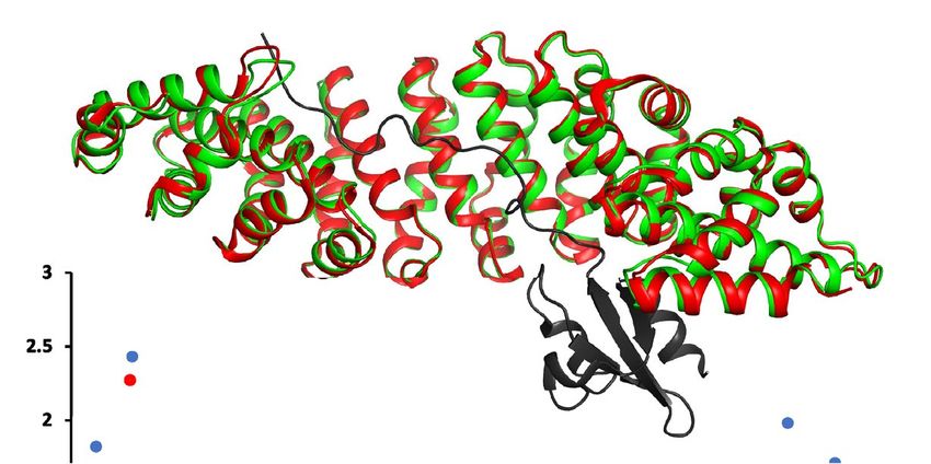
www.nature.com/scientificreports/
IMPα4 has not been solved to date15. Finally, the IMPα3 subfamily has the highest sequence homology and con-
servation among the subfamily members and includes IMPα5, α6, α7 i soforms15. Despite the high similarities,
different tissue expression profiles have been observed, including the limited expression of IMPα6 exclusively
in testis16. Furthermore, IMPα7 is critical for development in mice, with a knockout causing embryonic devel-
opment to halt at the two-cell stage17. More recently, IMPα7 has been shown to play a critical role in regulating
spermatogenesis and Sertoli cell f unction18.
Currently, there is only one structure of IMPα7 solved to date, and this is in its cargo bound state with the
influenza PB2 protein (PDB 4UAD)19. As have been previously reported, no significant variation in the core
ARM domains was observed upon binding of the cargo protein20. Here, we describe the first structure of IMPα7
NLS binding domain (ARMS 1–10) in the cargo-free state. We evaluate the similarities between IMPα isoforms
from other subfamilies and compare the structures of cargo-free and bound structures.
Materials and methods
Protein constructs, protein expression and purification. The gene encoding IMPα7 ARM domains
1–10 (lacking the importin-beta binding (IBB) domain) (KPNA6, Uniprot ID O60684, residues 74-536) was
codon optimized for Escherichia coli expression and cloned into the pET30a(+) vector at the BamHI site (Gene-
script, Picataway, NJ). The recombinant protein sequence incorporated the addition of the TEV protease amino
acid sequence and a cleavable N-terminal His-tag. Plasmids were transformed in BL21(DE3) pLysS E. coli cells
using the heat-shock method and were recombinantly expressed based on methods described previously for
other importin isoforms20. The protein was purified using a Ni–NTA affinity column pre-equilibrated with His
buffer A (50 mM phosphate buffer, 300 mM sodium chloride, 20 mM imidazole, pH 8.0) and eluted using a linear
gradient of imidazole over five column volumes using His buffer B (50 mM phosphate buffer, 300 mM sodium
chloride, 500 mM imidazole, pH 8.0). The protein was further purified using size exclusion chromatography on a
Superdex 200 pg 26/600 column (GE Healthcare) using Tris-Buffered saline (50 mM Tris–HCl, 125 mM sodium
chloride, pH 8.0). A single peak corresponding to a monomer was pooled together and analyzed on SDS-PAGE
and concentrated using a 10 kDa MW centrifugal filter and stored at − 80 °C.
Crystallization, data collection and processing. Crystallization trials were performed using 48 well
crystallization plates with 1.5 μl protein mixed with 1.5 μl of reservoir solution, equilibrated over 300 μl reservoir
solution using the hanging-drop vapour diffusion method. The IMPα7 protein crystallized at 15 mg/ml over a
reservoir solution containing 0.1 M MES pH 6.5 and 12% PEG 20,000 at 18 °C. Rod-shaped crystals appearing
in 30 days were cryoprotected in 25% glycerol and flash-cooled in liquid nitrogen. X-ray diffraction data from
a single crystal was collected over 3600 images at 0.1o oscillation at the Australian National Synchrotron MX2
beamline (Eiger X 16 M detector). The data were processed in iMosflm21, scaled in Aimless22 and phased using
molecular replacement in P haser23 with 4 UAD19 as the search model. The structure was modelled and refined in
Coot24 and P henix25,26, respectively.
PDB accession code. Coordinates and structure factors have been deposited in the PDB and released
under accession code 7RHT.
Results and discussion
Structure of IMPα7 in cargo‑free form. The cargo binding domain (ARMS 1–10) of IMPα7ΔIBB was
successfully cloned and expressed, and crystals diffracting at Australia National Synchrotron MX2 beamline
to 2.5 Å were indexed in the space group P21 21 21 with unit cell parameters of a = 64.85, b = 75.84, c = 88.97.
The structure was solved using molecular replacement in Phaser23 using chain A of PDB model 4UAD19. One
molecule of IMPα7 was present in the asymmetric unit, and following modelling and refinement in C OOT24
25,26
and Phenix , a model was produced with good stereochemistry and an Rwork and R free of 20.9% and 23.7%
respectively. Full data collection and refinement statistics are presented in Table 1.
The final model of IMPα7ΔIBB consists of 424 residues (80–503) and 34 waters, with 79 helix-helix intra-
molecular interactions (analyzed in PDBSum)27. Overall, the structure exhibited a conserved topology and
architecture similar to other IMPα isoforms. The structure is comprised of ten ARM domains (Fig. 1A,B), each
consisting of three α-helices H1, H2 and H3 in a triangular arrangement (Fig. 1C), and overall forming a concave
assembly28,29. Due to flexibility and lack of density at the N-terminus, the H1 of ARM1 could not be modelled.
The inner concave surface of IMPα7 contained highly conserved Trp and Asn residues within H3 α-helices on
ARMs 2–4 and 7–8, creating cargo NLS binding pockets at the major and minor sites, r espectively15 (Fig. 1B).
Minor cargo‑induced structural changes observed in IMPα7. Studies have reported that importins
may undergo conformational changes upon cargo binding, whilst other studies have reported little to no sig-
nificant variation in the core ARM domains upon cargo binding20. For example, IMPβ undergoes structural
changes upon binding to Ran and nuclear import cargo8,30–32. Similarly, IMPα1 has been shown to undergo con-
formational changes within the IBB domain to facilitate cargo b inding33 and the flexibility within hinge region
of IMPα3 has been reported to contribute to RCC1 specificity34. In contrast, structural comparisons between
both unbound IMPα1 and IMPα3, and their requisite cargo bound forms with Henipavirus W proteins, revealed
no major structural changes and similar positioning within the core ARM domains20. Since only one structure
of IMPα7 in a cargo bound form has been solved to date (IMPα7 in complex with Influenza PB2 protein (PDB
4UAD)19), and here we describe the first structure of IMPα7 in an unbound form, we performed structural com-
parisons between these two structures to examine how these observations extend to IMPα7. Structural align-
ment using Superpose in C CP435 revealed highly similar structures, with an r.m.s.d of 0.66 Å for IMPα7 main-
Scientific Reports | (2022) 12:315 | https://doi.org/10.1038/s41598-021-03729-3 2
Vol:.(1234567890)
www.nature.com/scientificreports/
Data collection and processing IMPα7 (PDB code: 7RHT)
Wavelength (Å) 0.9537
Resolution range (Å) 24.77–2.50 (2.60–2.50)
Space group P 21 21 21
Unit cell (Å, o) 64.85 75.84 88.97 90 90 90
Total reflections 77,300 (8824)
Unique reflections 15,096 (1703)
Multiplicity 5.1 (5.2)
Completeness (%) 97.2 (98.5)
Mean I/sigma(I) 8.1 (2.0)
Wilson B-factor Å2 38.58
R-merge 0.134 (0.958)
R-pim 0.09 (0.655)
Refinement
Number of reflections 15,070
Number of R-free reflections 751
R-work % 20.9
R-free % 23.7
RMS(bonds) Å 0.003
RMS(angles), o 0.50
Ramachandran plot
Favoured (%) 97.87
Allowed (%) 2.13
Outliers (%) 0
Validation
Clash score 1.66
PDB accession code 7RHT
Table 1. Data collection and refinement statistics.
chain residues of 80–503 (424 residues) (Fig. 2). We found that the positioning of ARM domains responsible
for binding cargo at the major and minor sites (ARMS 2–4 and 6–8) were highly similar across both structures,
whilst ARM domains at the extremities (ARMS 1, 9–10) exhibited minor structural changes that appeared to
coincide with cargo binding (Fig. 2).
IMPα7 structure comparisons with IMPα1, α3 and α5. Despite the relatively high sequence similari-
ties and conserved residues at the cargo binding site, IMPα isoforms exhibit structural differences that poten-
tially account for cargo specificity15. The structural basis for importin isoform specificity has been previously
investigated in a limited number of studies20,36, and so we compared IMPα7 with IMPα isoforms from other
subfamilies (Table 2). We found that IMPα7 was most structurally similar to IMPα5, ranging in r.m.s.d values
of 1.3–1.5 Å (Table 2), whilst IMPα1 structures ranged from r.m.s.d values of 1.4–2.2 Å, and IMPα3 structures
ranged from r.m.s.d values of 1.5–2.5 Å. We also found that comparing the unbound structure of IMPα1 with α7
revealed structural differences in the positioning at the major site (ARM 4) and ARMS 1, 9–10 (Fig. 3), whilst
comparisons between unbound IMPα3 and α7 revealed structural differences at both the major and minor
sites as well as ARM extremities. Finally, comparisons for IMPα5 (for which is there no unbound structure and
therefore PDB 6wx9 was used) exhibited far fewer structural changes, localized mainly within the C-terminal
ARM domains 9–10.
Previous research has highlighted the importance of the positioning of ARM domains 7 and 8 for isoform
specificity20,36. In addition, steric clashes between the cargo NLS and ARM7 and ARM8 domains of IMPα have
been reported when aligning cargo-bound structures of IMPα2 and IMPα5 with IMPα336. In this regard, to
Scientific Reports | (2022) 12:315 | https://doi.org/10.1038/s41598-021-03729-3 3
Vol.:(0123456789)
www.nature.com/scientificreports/
A B H1 H2 H3
ARM1 ARM1
80
..........VITREMVEMLFSD...DSDLQLATTQKFRKLLSKEPSP....... 115
ARM2
H2 ARM2 116.PIDEVINTPRVVDRFVEFLKRN....ENCTLQFEAAWALTNIASGTSQQ.....159
ARM3
Maj
H1
H3
ARM3 160
..TKIVIEA..GAVPIFIELLN...SDFEDVQEQAVWALGNIAGDSSVCRDY..204
or
ARM4
205
VLNCSILN....PLLTLLTKSTR..... LTMTRNAVWALSNLCRGKNP......243
ARM4 ARM5
244
PPEFAK.....VSPCLPVLSRLLFS .SDSDLLADACWALSYLSDGPNEKIQA.. 288
ARM6
289
VIDSG......VCRRLVELLMHND....YKVASPALRAVG NIVTGDDIQT....328
ARM5 ARM7
329
QVIL....NCSALPCLLHLLSSP....KESIRKEAC WTISNITAGNRAQ..... 367
ARM8
ARM6 371
.IQAVIDA..NIFPVLIEILQKAE....FRTRKEAAWAITNATSGGT.......410
ARM9
411
PEQIRYLVS..LGCIKPLCDLLTVM ..DSKIVQVALNGLENILRLGEQEG.... 456
ARM10
Mino
ARM7 457KRSGSGVNPYCGLIEEAYGLDKIEFLQSHENQEIYQKAFDLIEHYFG....... 503
r C
ARM8 H2
H1
ARM9 H3
N
ARM10 C
Figure 1. (A) Structure of IMPα7 ARM domains resolved to 2.5 Å resolution in cargo-free form shown
in ribbon-cartoon format containing 10 ARM-repeats. (B) IMPα7 ARM domain structure-based sequence
alignment with α-helices shown as colour-coded cylinders matching panel (A), with structural repeat H1, H2
and H3 indicated at the top. The residue numbers are shown in the beginning and at the end of each repeat.
Presence of highly conserved Trp and Asn residues highlighted in bold and underline. (C) Each ARM repeat
consists of three alpha helices: H1, H2 and H3, forming a stable triangular shape.
examine whether the IMPα7 structure described in this study could bind SOX2, we superimposed IMPα7 with
IMPα3:SOX2 (PDB 6wx8) and examined the structures for possible steric clashes. We observed 62 atomic clashes
(with a clash score of 0.8 or greater in Phenix validation) involving 14 residues with IMPα7 (Fig. 4). These clashes
were observed both within the major and minor sites, and the ARM extremities (Fig. 4). These clashes would
suggest that the binding between IMPα7 and SOX2 would be weaker than that observed between IMPα3 and
SOX2, which is consistent with both a previous report showing a lack of detectable binding between IMPα7 and
SOX236 and the notion that differential positioning of ARM domains in the IMPα isoforms can confer specificity
inding20,36. Moreover, the clashes observed at the major site within the superimposed model of IMPα7
of cargo b
and SOX2, together a previous report of IMPα5 binding with SOX2, is supported by the minor structural differ-
ences we observed between IMPα5 and IMPα7 at the major site (ARMS 2–4; Fig. 3).
Conclusion
Here, we describe the first structure of human IMPα7 in cargo-free form. Structural analysis revealed that the
IMPα7 protein exhibits the same structural architecture as other IMPα isoforms, and there were only minor
conformational changes upon cargo binding. IMPα7 was most structurally similar to IMPα5, supporting their
grouping within the same subfamily. The structural differences observed between IMPα3 and IMPα7 is consist-
ent with previous studies highlighting the role of ARM domains 7 and 8 in mediating cargo specificity between
these IMPα isoforms.
Scientific Reports | (2022) 12:315 | https://doi.org/10.1038/s41598-021-03729-3 4
Vol:.(1234567890)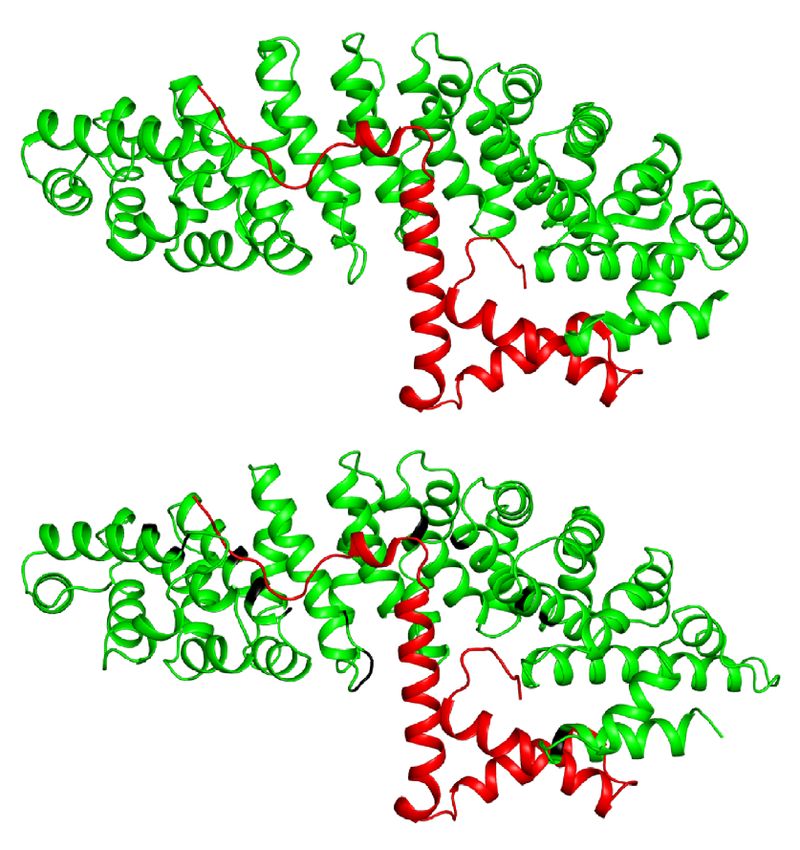
www.nature.com/scientificreports/
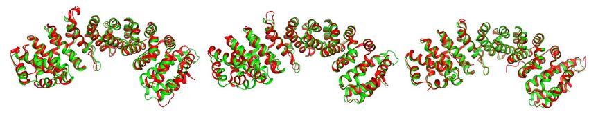
Figure 2. Structural alignment of IMPα7 in unbound (coloured green, PDB 7RHT) and bound (coloured red
and PB2 NLS in black, PDB 4 UAD19) forms. Graph inset represents distance differences between bound and
unbound structures. Red dots reflect the positions within IMPα7 mediating PB2 binding. Minor changes in the
positioning of some residues (listed in the graph inset) were observed in the cargo bound form, and these are
presented in the right panels.
PDB z-score r.m.s.d Aligned Res % Seq ID IMPA Subfamily
4uad 55.8 0.7 424 100 IMPα7 3
6wx9 53.7 1.3 423 86 IMPα5 3
4b18 53.6 1.4 424 86 IMPα5 3
2jdq 53.2 1.4 402 84 IMPα5 3
3tj3 52.6 1.5 424 86 IMPα5 3
7rg5 49.1 1.5 414 52 IMPα3 2
4uae 48.6 1.5 414 52 IMPα3 2
6bwb 48.6 1.5 414 52 IMPα3 2
6bwa 48.5 1.5 414 52 IMPα3 2
6bw9 48.4 1.5 414 52 IMPα3 2
6bvv 48.3 1.6 414 52 IMPα3 2
5xzx 47.5 1.6 413 52 IMPα3 2
6wx8 47.7 2.1 414 52 IMPα3 2
7jjl 47.9 2.2 411 52 IMPα3 2
6bvz 47.5 2.5 414 52 IMPα3 2
3fex 48.2 1.4 412 52 IMPα1 1
4wv6 49.4 1.5 415 52 IMPα1 1
3fey 49 1.5 415 52 IMPα1 1
7n8j 49 1.5 415 52 IMPα1 1
4e4v 46.7 2.2 415 52 IMPα1 1
Table 2. Structural comparison of IMPα7 structure with other human IMPα isoforms.
Scientific Reports | (2022) 12:315 | https://doi.org/10.1038/s41598-021-03729-3 5
Vol.:(0123456789)www.nature.com/scientificreports/
Figure 3. Structural comparisons of unbound IMPα7 with other IMPα subfamily members. IMPα1 (unbound,
PDB 4e4v), IMPα3 (unbound, PDB 6bvz), IMPα5 (unbound structure remains to be determined, PDB 6wx9)
isoforms were superimposed using CCP4 Superpose and the structural differences analyzed (see graph inset).
The positions of the ARM domains 2–4 and 6–8, mediating cargo binding at the major and minor sites,
respectively, are highlighted in bold.
Figure 4. Structural alignment of unbound IMPα7 with IMPα3 in complex with SOX2 (PDB 6WX8). Phenix
validation was used to analyze clash data. IMPα7 with merged SOX2 produced clashscore value of 20.7. The
positions of clashing residues of IMPα7 are highlighted in black. Clashes are also presented in the graph, with
clashes > 0.8 highlighted with black arrows.
Received: 21 September 2021; Accepted: 24 November 2021
References
1. Cook, A., Bono, F., Jinek, M. & Conti, E. Structural biology of nucleocytoplasmic transport. Annu. Rev. Biochem. 76, 647–671.
https://doi.org/10.1146/annurev.biochem.76.052705.161529 (2007).
2. Fang, X., Chen, T., Tran, K. & Parker, C. S. Developmental regulation of the heat shock response by nuclear transport factor
karyopherin-alpha3. Development 128, 3349–3358 (2001).
Scientific Reports | (2022) 12:315 | https://doi.org/10.1038/s41598-021-03729-3 6
Vol:.(1234567890)www.nature.com/scientificreports/
3. Wirthmueller, L., Roth, C., Banfield, M. J. & Wiermer, M. Hop-on hop-off: Importin-α-guided tours to the nucleus in innate
immune signaling. Front. Plant Sci. 4, 149. https://doi.org/10.3389/fpls.2013.00149 (2013).
4. Bednenko, J., Cingolani, G. & Gerace, L. Nucleocytoplasmic transport: Navigating the channel. Traffic 4, 127–135. https://doi.org/
10.1034/j.1600-0854.2003.00109.x (2003).
5. Marfori, M., Lonhienne, T. G., Forwood, J. K. & Kobe, B. Structural basis of high-affinity nuclear localization signal interactions
with importin-α. Traffic 13, 532–548. https://doi.org/10.1111/j.1600-0854.2012.01329.x (2012).
6. Yang, S. N. et al. Probing the specificity of binding to the major nuclear localization sequence-binding site of importin-alpha using
oriented peptide library screening. J. Biol. Chem. 285, 19935–19946. https://doi.org/10.1074/jbc.M109.079574 (2010).
7. Fontes, M. R., Teh, T. & Kobe, B. Structural basis of recognition of monopartite and bipartite nuclear localization sequences by
mammalian importin-alpha. J. Mol. Biol. 297, 1183–1194. https://doi.org/10.1006/jmbi.2000.3642 (2000).
8. Lee, S. J., Matsuura, Y., Liu, S. M. & Stewart, M. Structural basis for nuclear import complex dissociation by RanGTP. Nature 435,
693–696. https://doi.org/10.1038/nature03578 (2005).
9. Rexach, M. & Blobel, G. Protein import into nuclei: Association and dissociation reactions involving transport substrate, transport
factors, and nucleoporins. Cell 83, 683–692. https://doi.org/10.1016/0092-8674(95)90181-7 (1995).
10. Kobe, B. Autoinhibition by an internal nuclear localization signal revealed by the crystal structure of mammalian importin alpha.
Nat. Struct. Biol. 6, 388–397. https://doi.org/10.1038/7625 (1999).
11. Falces, J. et al. Recognition of nucleoplasmin by its nuclear transport receptor importin α/β: Insights into a complete import
complex. Biochemistry 49, 9756–9769. https://doi.org/10.1021/bi101179g (2010).
12. Conti, E., Uy, M., Leighton, L., Blobel, G. & Kuriyan, J. Crystallographic analysis of the recognition of a nuclear localization signal
by the nuclear import factor karyopherin alpha. Cell 94, 193–204. https://doi.org/10.1016/s0092-8674(00)81419-1 (1998).
13. Robbins, J., Dilworth, S. M., Laskey, R. A. & Dingwall, C. Two interdependent basic domains in nucleoplasmin nuclear targeting
sequence: Identification of a class of bipartite nuclear targeting sequence. Cell 64, 615–623. https://doi.org/10.1016/0092-8674(91)
90245-t (1991).
14. Kosugi, S. et al. Six classes of nuclear localization signals specific to different binding grooves of importin alpha. J. Biol. Chem. 284,
478–485. https://doi.org/10.1074/jbc.M807017200 (2009).
15. Pumroy, R. A. & Cingolani, G. Diversification of importin-α isoforms in cellular trafficking and disease states. Biochem. J. 466,
13–28. https://doi.org/10.1042/bj20141186 (2015).
16. Köhler, M. et al. Cloning of two novel human importin-alpha subunits and analysis of the expression pattern of the importin-alpha
protein family. FEBS Lett. 417, 104–108. https://doi.org/10.1016/s0014-5793(97)01265-9 (1997).
17. Rother, F. et al. Importin α7 is essential for zygotic genome activation and early mouse development. PLoS ONE 6, e18310. https://
doi.org/10.1371/journal.pone.0018310 (2011).
18. Liu, N. et al. Importin α7 deficiency causes infertility in male mice by disrupting spermatogenesis. Development https://doi.org/
10.1242/dev.198374 (2021).
19. Pumroy, R. A., Ke, S., Hart, D. J., Zachariae, U. & Cingolani, G. Molecular determinants for nuclear import of influenza A PB2 by
importin α isoforms 3 and 7. Structure 23, 374–384. https://doi.org/10.1016/j.str.2014.11.015 (2015).
20. Smith, K. M. et al. Structural basis for importin alpha 3 specificity of W proteins in Hendra and Nipah viruses. Nat. Commun. 9,
3703. https://doi.org/10.1038/s41467-018-05928-5 (2018).
21. Battye, T. G., Kontogiannis, L., Johnson, O., Powell, H. R. & Leslie, A. G. iMOSFLM: A new graphical interface for diffraction-image
processing with MOSFLM. Acta Crystallogr. D 67, 271–281. https://doi.org/10.1107/s0907444910048675 (2011).
22. Evans, P. Scaling and assessment of data quality. Acta Crystallogr D Biol Crystallogr 62, 72–82. https://doi.org/10.1107/s090744490
5036693 (2006).
23. McCoy, A. J. Solving structures of protein complexes by molecular replacement with Phaser. Acta Crystallogr. D 63, 32–41. https://
doi.org/10.1107/s0907444906045975 (2007).
24. Emsley, P. & Cowtan, K. Coot: Model-building tools for molecular graphics. Acta Crystallogr. D 60, 2126–2132. https://doi.org/
10.1107/s0907444904019158 (2004).
25. Adams, P. D. et al. PHENIX: A comprehensive Python-based system for macromolecular structure solution. Acta Crystallogr. D
66, 213–221. https://doi.org/10.1107/s0907444909052925 (2010).
26. Afonine, P. V. et al. Towards automated crystallographic structure refinement with phenix.refine. Acta Crystallogr. D 68, 352–367.
https://doi.org/10.1107/s0907444912001308 (2012).
27. Laskowski, R. A., Jabłońska, J., Pravda, L., Vařeková, R. S. & Thornton, J. M. PDBsum: Structural summaries of PDB entries. Protein
Sci. 27, 129–134. https://doi.org/10.1002/pro.3289 (2018).
28. Andrade, M. A., Petosa, C., O’Donoghue, S. I., Müller, C. W. & Bork, P. Comparison of ARM and HEAT protein repeats. J. Mol.
Biol. 309, 1–18. https://doi.org/10.1006/jmbi.2001.4624 (2001).
29. Conti, E., Müller, C. W. & Stewart, M. Karyopherin flexibility in nucleocytoplasmic transport. Curr. Opin. Struct. Biol. 16, 237–244.
https://doi.org/10.1016/j.sbi.2006.03.010 (2006).
30. Forwood, J. K. et al. Quantitative structural analysis of importin-β flexibility: Paradigm for solenoid protein structures. Structure
18, 1171–1183. https://doi.org/10.1016/j.str.2010.06.015 (2010).
31. Forwood, J. K. et al. Kap95p binding induces the switch loops of RanGDP to adopt the GTP-bound conformation: Implications
for nuclear import complex assembly dynamics. J. Mol. Biol. 383, 772–782. https://doi.org/10.1016/j.jmb.2008.07.090 (2008).
32. Lee, S. J. et al. The structure of importin-beta bound to SREBP-2: Nuclear import of a transcription factor. Science 302, 1571–1575.
https://doi.org/10.1126/science.1088372 (2003).
33. Cingolani, G., Petosa, C., Weis, K. & Müller, C. W. Structure of importin-beta bound to the IBB domain of importin-alpha. Nature
399, 221–229. https://doi.org/10.1038/20367 (1999).
34. Sankhala, R. S. et al. Three-dimensional context rather than NLS amino acid sequence determines importin α subtype specificity
for RCC1. Nat. Commun. 8, 979. https://doi.org/10.1038/s41467-017-01057-7 (2017).
35. Krissinel, E. & Henrick, K. Secondary-structure matching (SSM), a new tool for fast protein structure alignment in three dimen-
sions. Acta Crystallogr. D 60, 2256–2268. https://doi.org/10.1107/s0907444904026460 (2004).
36. Jagga, B. et al. Structural basis for nuclear import selectivity of pioneer transcription factor SOX2. Nat. Commun. 12, 28. https://
doi.org/10.1038/s41467-020-20194-0 (2021).
Author contributions
S.T. performed the experiments and assisted with manuscript writing. C.D. assisted with manuscript writing and
figure preparation. J.K.F. supervised the project and assisted with manuscript writing and figure preparation.
Competing interests
The authors declare no competing interests.
Additional information
Correspondence and requests for materials should be addressed to J.K.F.
Scientific Reports | (2022) 12:315 | https://doi.org/10.1038/s41598-021-03729-3 7
Vol.:(0123456789)www.nature.com/scientificreports/
Reprints and permissions information is available at www.nature.com/reprints.
Publisher’s note Springer Nature remains neutral with regard to jurisdictional claims in published maps and
institutional affiliations.
Open Access This article is licensed under a Creative Commons Attribution 4.0 International
License, which permits use, sharing, adaptation, distribution and reproduction in any medium or
format, as long as you give appropriate credit to the original author(s) and the source, provide a link to the
Creative Commons licence, and indicate if changes were made. The images or other third party material in this
article are included in the article’s Creative Commons licence, unless indicated otherwise in a credit line to the
material. If material is not included in the article’s Creative Commons licence and your intended use is not
permitted by statutory regulation or exceeds the permitted use, you will need to obtain permission directly from
the copyright holder. To view a copy of this licence, visit http://creativecommons.org/licenses/by/4.0/.
© The Author(s) 2022
Scientific Reports | (2022) 12:315 | https://doi.org/10.1038/s41598-021-03729-3 8
Vol:.(1234567890)You can also read