Study of Local Fields of Dendrite Nanostructures in Hot Spots Formed on SERS-Active Substrates Produced via Template-Assisted Synthesis
←
→
Page content transcription
If your browser does not render page correctly, please read the page content below
ISSN 1062-8738, Bulletin of the Russian Academy of Sciences: Physics, 2020, Vol. 84, No. 12, pp. 1465–1468. © Allerton Press, Inc., 2020.
Russian Text © The Author(s), 2020, published in Izvestiya Rossiiskoi Akademii Nauk, Seriya Fizicheskaya, 2020, Vol. 84, No. 12, pp. 1725–1728.

                Study of Local Fields of Dendrite Nanostructures
           in Hot Spots Formed on SERS-Active Substrates Produced
                         via Template-Assisted Synthesis
                            E. P. Kozhinaa, *, S. N. Andreeva, b, V. P. Tarakanovc, S. A. Bedina, d,
                                            I. M. Doludenkod, and A. V. Naumova
                                          aMoscow
                                           Pedagogical State University, Moscow, 119435 Russia
                                              b
                                      Moscow Polytechnic University, Moscow, 107023 Russia
                 c
                  Joint Institute for High Temperatures, Russian Academy of Sciences, Moscow, 125412 Russia
       dFederal Research Center “Crystallography and Photonics,” Russian Academy of Sciences, Moscow, 119333 Russia

                                               *e-mail: Liza.kozhina.99@mail.ru
                                      Received July 15, 2020; revised August 10, 2020; accepted August 26, 2020

         Abstract—A procedure is proposed for template-assisted synthesis on track membranes using iodide electro-
         lyte to produce substrates with dendritic nanostructures formed on the tips of silver nanowires. The distribu-
         tion of the electromagnetic field near a silver nanorhombus irradiated with visible laser radiation is modeled
         because the dendrite branches are rhombus-shaped nanoparticles. Calculations indicate considerable
         enhancement of the local electric fields near the sharp tops of the nanorhombus.

         DOI: 10.3103/S1062873820120205

                  INTRODUCTION                                                         sized peaks or irregularities. Examples of such struc-
    Nanostructures made of precious metals are of                                      tures include substrates with arrays of nanowires
special interest in giant Raman scattering (SERS, or                                   (NWs) on their surfaces [10, 11].
surface-enhanced Raman scattering), due to their res-                                      Since the optical properties of metallic nanostruc-
onant behavior in the optical range [1]. The extremely                                 tures depend not only on their size but largely on the
high electromagnetic field amplification associated                                    shape as well, nanoparticles with irregular shapes (e.g.,
with plasmon resonance plays an important role in the                                  polyhedrons) display more unique physical properties
amplification of the Raman signal of molecules                                         than nanostructures of simple shape (spheres or rods)
adsorbed in domains with high scattering capacity                                      [12]. Triangle-shaped nanoparticles thus exhibit not
(also known as hot spots) [2, 3]. There are currently a                                only the effect of multiple plasmon resonances, but
number of synthesis procedures and ways of manufac-                                    the strong amplification of local fields on sharp tips of
turing metallic nanoparticles with different shapes.                                   a triangle as well (in contrast to an ellipsoid) [13].
The modeling of local electric fields near the surfaces                                Strong amplification can also be achieved in a gap of
of such structures allows us to study the nature of                                    less than 3 nm between two nanotriangles. This is
SERS signal amplification in the domain of hot spots.                                  explained by the more complicated spectrum of reso-
When studying the influence of local fields, it is                                     nance for interacting structures, in which the number
important to consider their fluctuations in dielectric                                 and magnitude of different resonances depend on the
solutions [4, 5] and the results from concentration                                    direction of irradiation and the distance between par-
effects [6, 7]. The parameters of amplification can                                    ticles [14, 15].
therefore vary from molecule to molecule.                                                  Strong amplification of SERS signals can also be
    Hot spots can arise between adjacent plasmon                                       obtained on a more developed surface of a substrate,
nanostructures if they are less than 4 nm from one                                     since the area of the contact surface does not increase
another and form near single nanosized particles with                                  (e.g., on substrates with highly-branched dendritic
high surface curvature (e.g., on the poles of a nano-                                  nanostructures that amplify electric fields in a wide
sphere, or on the tips of nanorods and nanotriangles                                   spectral range). It has been shown that more branched
because of the glowing peak effect) [3, 8, 9]. A sub-                                  surfaces provide the strongest amplification of SERS
stantial increase in the intensity of the electric field is                            signals, and electric fields reach their maximum values
then observed, due to the electric component of an                                     at the vertices of dendrite branches (sharp tips), and at
electromagnetic field being concentrated on nano-                                      points where multilayer dendritic branches are in con-

                                                                                 1465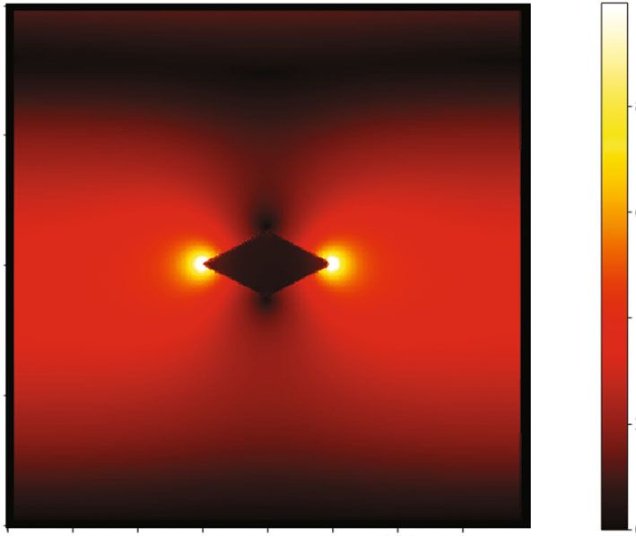
1466                                                             KOZHINA et al.

                                                                                                                            Amplitude of electric field, conventional units
                                                                          z, nm
                                                                          400

                                                                                                                        8
                                                                          300

                                                                                                                        6
                                                                          200
                                                                                                                        4

                                                                          100
                                                                                                                        2
                                         2 µm
         High-vac. SED PC-std.   15 kV    ×10 000   18.03.2019   000012                                                 0
                                                                            0       100     200      300     400
                                                                                                           x, nm

   Fig. 1. (a) Image of a dendrite nanostructure with branches, obtained on a raster electronic microscope. (b) Distribution of the
   coefficient of amplification (E/E0) of an electric field near a nanorhombus.

tact with one another [16, 17]. Compared to simple                        at room temperature with a constant current density of
irregular silver substrates, dendritic nanostructures                     80 to 250 mA/cm2 through the pores of the TM. By
made of silver have a great many hot spots, ensuring                      varying the period of deposition from 10 to 100 s, we
strong amplification of SERS signals. Such structures                     achieved complete filling of pores in the polymer tem-
can be produced via galvanic displacement [18], along                     plate with metal emerging on its surface in the form of
with the simpler and cheaper technique of electro-                        dendritic structures (Fig. 1a). When electrochemical
chemical deposition in power of a porous aluminum                         deposition was complete, the polymer matrix was
oxide [19–21].                                                            removed by dissolving it in a concentrated alkali solu-
    In this work, we propose manufacturing substrates                     tion (25% NaOH).
with dendritic nanostructures formed on the vertices
of silver NWs via template synthesis with iodide-con-                        The amplification of local electromagnetic fields
taining electrolyte on tracking membranes (TMs).                          near nanorhombus was modeled mathematically using
The advantage of using TM is that we can vary the                         the fully electromagnetic KARAT code [25] in planar
diameter of pores and, in contrast to porous aluminum                     geometry (Х, Z). The domain of calculation was a
oxide, TMs do not have a high density of pores; as a                      square 400 by 400 nm in size. In the center of the
result, dendritic nanostructures do not form layers on                    square, there was a silver nanoparticle in the form of a
one another, preserving the unique pattern of hot                         rhombus with large diagonal of 100 nm along X axis,
spots on a surface. This allows optimization of the                       and a small diagonal of variable length (10 to 100 nm)
density of pores in order to form more hot spots. Since                   along the Z axis.
branches of dendrites are rhombus-shaped nanoparti-                           We used a Drude model with parameters for silver
cles, we modeled the distribution of an electromag-                       [26] to describe the electromagnetic properties of a
netic field near a silver nanorhombus irradiated with a                   nanoparticle: plasma frequency, 8.78 eV; constant of
laser in the visible range. Calculations indicate local                   attenuation, 0.02 eV. Laser irradiation with flat polar-
amplification of electric fields near the vertices of the                 ization (Ex, By) and a wavelength of 785 nm propa-
nanorhombus.                                                              gated in the positive direction of the Z axis and inter-
                                                                          acted with the nanorhombus, resulting in a local
                  EXPERIMENTAL                                            change in the electromagnetic field of incident radia-
                                                                          tion in the domains near its vertices. As an example,
    Substrates with dendritic nanostructures formed                       Fig. 1b shows the distribution of the coefficient of
on the vertices of silver NWs were prepared via pattern                   electric field amplification (E/E0) near a nanorhom-
synthesis. A polymer TM 12 μm thick with a pore                           bus of 100 by 50 nm, where E0 is the amplitude of the
diameter of 100 nm and a surface pore density of                          electric field of incident radiation. It can be seen in
108 cm−2 was used as a synthesis template. Pores in the                   Fig. 1b that there was considerable amplification on
TM were filled with silver [22].                                          the acute vertices of the rhombus, while it attenuated
    Iodide-containing silvering electrolyte (metallic                     on obtuse vertices. Figure 2a shows the dependence of
silver, 15–20 g/L; potassium iodide, 230–300 g/L                          the coefficient of electric field amplification (E/E0) at
[23, 24]) was used in this work. Deposition was done                      points on the continuation of the rhombus’s diagonals

                       BULLETIN OF THE RUSSIAN ACADEMY OF SCIENCES: PHYSICS                            Vol. 84     No. 12                                                    2020
STUDY OF LOCAL FIELDS OF DENDRITE NANOSTRUCTURES 1467
(a) (b)
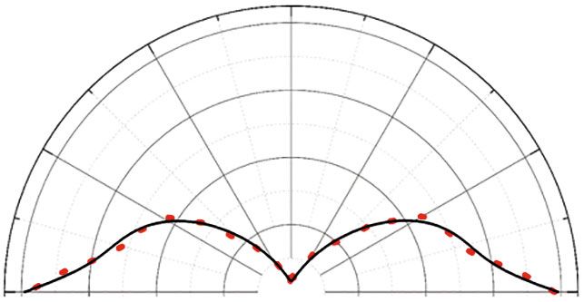
Acute angle of the triangle
16 Obtuse angle of the triangle
14
12 90
8 120 60
10
E/E0
8 6
150 30
E/E0
6 4
4
2
2
0 180 0
0 2 4 6 8 10
Aspect ratio
Fig. 2. (a) Dependence of the coefficient of the amplification of an electric field on the aspect ratio of the diagonals of a nanor-
hombus in spots near a vertex with an acute angle and one with an obtuse angle. (b) Dependence E/E0 on the angle of rotation of
the large diagonal of a nanorhombus with respect to the vector of the electric field of the incident radiation.
at a distance of 2 nm from its vertices on the aspect ones. We obtained the dependence of the coefficient
ratio of its diagonals (d). of electric field amplification on aspect ratio d of the
When d > 3, we observe a general tendency of both diagonals of a rhombus, and showed in particular that
vertices: the higher the aspect ratio, the stronger the when d > 3, the coefficient of field amplification grows
field amplification. There is a local maximum (E/E0) along with the aspect ratio. The coefficient of field
when 1 < d < 3, but it is much lower when d = 2 than amplification also depends on the orientation of the
the coefficient of amplification when d > 4. Near the nanorhombus with respect to the direction of the
vertex of the rhombus with an acute angle, E/E0 thus intensity of the electric field of the incident radiation:
if the large diagonal of the rhombus is parallel to the
reaches a value of 14 when d = 10. As noted above, vector of the electric field’s intensity, the amplifica-
there is virtually no amplification of the field near the tion of local electric field will be maximal. The maxi-
vertex of the rhombus with an obtuse angle, and E/E0 mum efficiency of the local amplification of the field
is greater than 1 only when d > 8. will thus be displayed by the rhombus-shaped edges of
Both the aspect ratio of the rhombus’s diagonals dendrites with acute angles oriented in parallel to the
and its dislocation with respect to incident radiation electric component of the incident laser radiation.
are important parameters for the local amplification of
the electric field. Silver nanoparticles were modeled in
the form of a rhombus with large diagonal of 100 nm ACKNOWLEDGMENTS
and small diagonal of 20 nm. The coefficient of ampli- The measurements were made using equipment at the
fication was determined for a point at a distance of shared resource center of the Russian Academy of Sciences’
2 nm from a vertex with an acute angle on the contin- Center “Crystallography and Photonics.”
uation of the rhombus’s large diagonal. Figure 2b
shows the dependence of E/E0 on the angle of rotation
of the rhombus’s large diagonal with respect to the FUNDING
vector of the electric field of the incident radiation. It This work was performed as part of a State Task for Mos-
can be seen that the field amplification is maximal if cow State Pedagogical University on the topic “Physics of
the large diagonal is parallel to the vector of electric Nanostructured Materials: Fundamental Research and
field E, but the coefficient of amplification tends Applications in Materials Science, Nanotechnology, and
toward zero if the large diagonal is perpendicular to Photonics.”
field vector E.
We obtained metallic substrates made of silver with
dendrites on their surfaces in the form of nanorhombi. REFERENCES
Interaction between a nanorhombus and laser radia- 1. Radziuk, D. and Moehwalda, H., Phys. Chem. Chem.
tion of the visible spectrum in planar geometry was Phys., 2010, vol. 17, p. 21072.
modeled, and the local distribution of electric fields 2. Etchegoina, P.G. and Le Ru, E.C., Phys. Chem. Chem.
near the vertices of the nanorhombus was studied Phys., 2008, vol. 10, p. 6079.
using the KARAT fully electromagnetic 3D code. We 3. Kuttner, C., Plasmonics in sensing: From colorimetry
found there was strong amplification of electric field to SERS analytics, in Plasmonics, Gric, T., Ed., Inte-
on acute vertices, while it was attenuated on obtuse chOpen, 2018.
BULLETIN OF THE RUSSIAN ACADEMY OF SCIENCES: PHYSICS Vol. 84 No. 12 20201468 KOZHINA et al.
4. Naumov, A.V., Gorshelev, A.A., Gladush, M.G., 16. 16.Orságová Králová, Z., Oriňak, A., Oriňaková, R.,
et al., Nano Lett., 2018, vol. 18, p. 6129. et al., J. Biomed. Opt., 2018, vol. 23, no. 7, 075002.
5. Gladush, M.G., Anikushina, T.A., Gorshelev, A.A., 17. Ye, Y., Chen, C., Hua, H., et al., Cell Rep. Phys. Sci.,
et al., J. Exp. Theor. Phys., 2019, vol. 128, no. 5, p. 655. 2020, vol. 1, no. 3, 100031.
6. Eskova, A.E., Arzhanov, A.I., Magaryan, K.A., et al., 18. Yakimchuk, D.V., Kaniukov, E.Yu., Lepeshov, S.,
Bull. Russ. Acad. Sci.: Phys., 2020. vol. 84, no. 1, p. 40. et al., J. Appl. Phys., 2019, vol. 126, no. 23, 233105.
7. Eskova, A.E., Arzhanov, A.I., Magaryan, K.A., et al., 19. Rafailović, L.D., Gammer, C., Srajer, J., et al.,
EPJ Web Conf., 2019, vol. 220, 03014. RCS Adv., 2016, vol. 6, no. 40, p. 33348.
8. Ostroukhov, N., Sleptsov, V., Tyanginskii, A., et al., 20. Gutés, A., Carraro, C., and Maboudian, R., J. Am.
Fotonika, 2011, no. 5, p. 38. Chem. Soc., 2010, vol. 132, no. 5, p. 1476.
21. Cheng, Z.-Q., Li, Z.-W., Xu, J.-H., et al., Nanoscale
9. Karpov, S., Fotonika, 2012, no. 3, p. 52.
Res. Lett., 2019, vol. 14, 89.
10. Kozhina, E.P., Bedin, S.A., Razumovskaya, I.V., et al., 22. Bedin, S.A., Rybalko, O.G., Polyakov, N.B., et al., Inorg.
J. Phys.: Conf. Ser., 2019, vol. 1283, 012009. Mater.: Appl. Res., 2010, vol. 1, p. 359.
11. Ermushev, A.V., Mchedlishvili, B.V., Oleinikov, V.A., 23. Burkat, G.K., Elektroosazhdenie dragotsennykh metallov
et al., Quantum Electron., 1993, vol. 23, p. 435. (Electrodeposition of Precious Metals), St. Petersburg:
12. Hu, J., Wang, Z., and Li, J., Sensors, 2007, no. 7, Politekhnika, 2009.
p. 3299. 24. Fourcade, F. and Tzedakis, T., J. Electroanal. Chem.
13. Kottmann, J.P., Martin, O.J.F., Smith, D.R., et al., Interfacial Electrochem., 2000, vol. 493, p. 20.
Opt. Express, 2000, vol. 6, no. 11, p. 213. 25. Tarakanov, V.P., EPJ Web Conf., 2017, vol. 149, 04024.
14. Simeone, D., Esposito, M., Scuderi, M., et al., 26. Johnson, P.B. and Christy, R.W., Phys. Rev. B: Solid
ACS Photonics, 2018, vol. 5, no. 8, p. 3399. State, 1972, vol. 6, p. 4370.
15. Merlen, A. and Lagugné-Labarthet, F., Appl. Spectrosc.,
2014, vol. 68, no. 12, p. 1307. Translated by K. Gumerov
BULLETIN OF THE RUSSIAN ACADEMY OF SCIENCES: PHYSICS Vol. 84 No. 12 2020You can also read