Systematic Review of Thigh Symptoms after Lateral Transpsoas Interbody Fusion for Adult Patients with Degenerative Lumbar Spine Disease
←
→
Page content transcription
If your browser does not render page correctly, please read the page content below
Systematic Review of Thigh Symptoms after Lateral Transpsoas
Interbody Fusion for Adult Patients with Degenerative Lumbar
Spine Disease
Isaac D. Gammal, Jeffrey M. Spivak and John A. Bendo

Int J Spine Surg 2015, 9 ()
doi: https://doi.org/10.14444/2062
http://ijssurgery.com/content/9/62
This information is current as of May 5, 2021.

   Email Alerts       Receive free email-alerts when new articles cite this article. Sign up at:
                      http://ijssurgery.com/alerts

The International Journal of Spine Surgery
                             Downloaded from http://ijssurgery.com/ by guest on May 5, 2021
2397 Waterbury Circle, Suite 1,
Aurora, IL 60504, Phone: +1-630-375-1432
© 2015 ISASS. All Rights Reserved.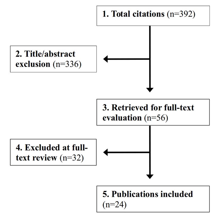
Systematic Review of Thigh Symptoms after Lateral
Transpsoas Interbody Fusion for Adult Patients with
Degenerative Lumbar Spine Disease
Isaac D. Gammal, BA, Jeffrey M. Spivak, MD, John A. Bendo, MD
Division of Spine Surgery, Hospital for Joint Diseases, NYU Langone Medical Center, New York, NY

Abstract
Background
Lateral transpsoas interbody fusion (LTIF) is a minimally invasive technique for achieving lumbar spinal fusion.
While it has many advantages over open techniques it carries with it a distinct set of risks, most commonly post-
operative ipsilateral thigh pain, weakness and sensory disturbances. It is vital for both the surgeon and patient to
understand the risks for and outcomes of injury associated with this procedure. We conducted a systematic review
of the literature to evaluate the incidence, risks, and long-term clinical outcomes of post-operative thigh symptoms
in patients treated with LTIF.

Methods
We conducted a search of MEDLINE, EMBASE, CINAHL, Scopus, Web of Science and the Cochrane Collabora-
tion Library, using keywords and MeSH terms, for English-language literature published through September 2014,
as well as reference lists from key articles. Studies were then manually filtered to retrieve articles that met inclusion
criteria. We were interested in studies that reported postoperative lower extremity symptoms after LTIF, such as
pain, weakness and changes in sensation. The strength of evidence was determined based on precepts outlined by
the Grades of Recommendation Assessment, Development and Evaluation Working Group (GRADE).

Results
A total of 392 articles were initially retrieved, with 24 ultimately meeting criteria for inclusion. The incidence of
any post-operative thigh symptom varied, ranging as high as 60.7%, with 9.3% of patients experiencing a motor
deficit related to direct nerve injury. Several studies reported cases of persistent symptoms at 6 months follow up.
Additionally, inclusion of the L4-5 disc space and a longer duration of surgery were both identified as risks for de-
veloping postoperative thigh symptoms.

Conclusion
The risk of postoperative thigh symptoms after LTIF is high. Thigh pain, paresthesias and weakness were the most
commonly reported symptoms. While most patients' symptoms resolved by 6 months follow up, several studies re-
ported patients with symptoms persistent as far as 12 months removed from surgery. Surgery at the L4-5 disc
space and longer surgical duration place the patient at greater risk for developing postoperative and long-term
thigh symptoms.

keywords: minimally invasive spine surgery, lateral lumbar interbody fusion, transpsoas interbody fusion
volume 9 article 62 doi: 10.14444/2062

                                                                               bilization of abdominal contents and great vessels,
Introduction                                                                   avoided extensive muscle stripping and denervation,
McAfee et al.1 first described a minimally invasive                            and left normal spinal stabilizing elements intact (e.g.
retroperitoneal approach for lumbar spinal fusion in                           anterior and posterior longitudinal ligaments), thus
1998, which involved psoas muscle retraction poste-                            avoiding many of the complications associated with
riorly to expose the disc space. Unlike traditional                            anterior and posterior approaches to spinal fusion.
open approaches, this approach did not require mo-                             Following advances in this technique, Bergey et al.2
                                       Downloaded from http://ijssurgery.com/ by guest on May 5, 2021
doi: 10.14444/2062
described the first endoscopic lateral transpsoas ap- through September 2014. Our results were limited to
proach with the use of intra-operative electromyelog- human studies published in the English language.
raphy (EMG) to assess nerve root proximity. In Search terms were identified and combined with ap-
2006, Ozgur et al.3 first described the use of the now propriate Boolean connectors and included “lumbar
popular Extreme Lateral Interbody Fusion system spine AND lateral interbody fusion,” “lateral inter-
(XLIF, NuVasive, Inc, San Diego, CA), which also body fusion AND complications,” and “transpsoas
utilizes intra-operative EMG and permits direct visu- interbody fusion AND complications.” Reference
alization of the disc space. This system utilizes se- lists from relevant articles were hand searched for ar-
quential dilators of increasing diameter to traverse ticles not identified during our electronic search.
the psoas muscle and expose the disc space.
Inclusion and Exclusion Criteria
In light of the growing popularity of this technique, Titles, abstracts and full-text articles were screened
studies were undertaken to define the intrapsoas by two independent reviewers at each stage by apply-
nerve anatomy and establish safe corridors for tra- ing the inclusion and exclusion criteria, with dis-
versing the psoas muscle.4-7 Several surgically- agreement settled by a third reviewer. We included
relevant findings were reported in the literature: articles evaluating adult patients with degenerative
nerve elements course along the anterior, middle, lumbar spine disease including, but not limited to,
and posterior thirds of the psoas muscle; the L2-4 degenerative disc disease, acquired spondylolisthe-
nerve roots migrate anteriorly at lower levels; the sis, scoliosis, and spinal stenosis that were treated us-
sympathetic chain overlies the L1-L5 disc spaces; ing a lateral retroperitoneal transpsoas approach. We
and the genitofemoral nerve exits at L3 and overlies only included prognostic studies (Table 1).
the anterior third of the psoas muscle. Because of its
intimate relationship with neural structures, it is not To answer our first study question we included arti-
surprising that the most commonly reported postop- cles reporting postoperative complications of this
erative complications of this approach include lower procedure, specifically lower extremity pain, sensory
extremity sensory disturbances, motor deficits, and disturbances and weakness. To answer our second
hip flexion weakness. With this in mind, the purpose study question we included articles that delineated
of this review is to attempt to answer the following risk factors for developing complications, specifically
three clinical questions: patient factors, disease factors, and surgical factors
Table 1. Inclusion and exclusion criteria.
1. What is the incidence of lower extremity symp- Adults (>18 years old) treated for:
toms (e.g. thigh pain, hip flexion weakness, sensory Degenerative disc disease Pediatric and
adolescent
and motor deficits, etc.) following lateral transpsoas Disc herniation
Patients Spondylolisthesis Tumor
interbody fusion? Infection
Scoliosis
2. What are the patient, disease and surgical risk Spinal stenosis Trauma
factors for the development of lower extremity symp- Adjacent segment disease
toms?
Lateral lumbar interbody fusion
3. What are the long-term clinical consequences Intervention
Transpsoas interbody fusion
and outcomes for patients with postoperative lower
Non-English
extremity symptoms?
English language Review articles
RCT Case reports
Study
Material and Methods Observational studies
Level I/II/III evidence
Level IV evi-
dence
ndoi: 10.14444/2062
from multivariate statistical analyses. To answer our estimate of effect and is likely to change the esti-
third study question we included articles reporting mate.12
clinical consequences of these complications, specifi-
cally time to resolution of symptoms. Data Analysis
The rate of lower extremity symptoms was reported
Excluded from our study were articles that did not as the proportion of patients who experienced thigh
report complications, studies that included pediatric or groin pain, hip flexion weakness, sensory distur-
patients and studies that included patients with dis- bances, and motor deficits. These symptoms were ei-
ease etiology of infection, trauma or tumor. We also ther subjectively reported by patients using question-
excluded review articles, case reports and case se- naires or noted on detailed neurological exam. We re-
ries’. In addition, animal and cadaveric studies were port the cumulative incidence of postoperative lower
excluded. The PRISMA checklist8 was used to guide extremity symptoms using the number of events in
reporting of this systematic review. the numerator and the total number of patients in the
denominator. The cumulative incidence of postoper-
Data Extraction ative lower extremity symptoms was defined as the
From the included articles the following data were proportion of patients who had been free of similar
extracted using a standardized form: study design symptoms at the time of index fusion and had subse-
and purpose, patient demographics, primary diag- quent development of these symptoms in the postop-
noses, surgical treatment, follow-up time period, erative period. To evaluate the incidence of persis-
follow-up rate, duration of surgery, complications, tent lower extremity symptoms, we used the number
and risk factors for and long-term consequences of of patients experiencing symptoms at 6 months
postoperative complications. follow-up in the numerator and total number of pa-
tients in the denominator. Six months was chosen as
Study Quality and Overall Strength of Body of the cut-off point since transient postoperative symp-
Literature toms would be expected to resolve by this point and
Levels of evidence ratings were assigned to each arti- this was the most commonly reported follow up visit
cle independently by two reviewers using criteria set among the studies reviewed.
by The Journal of Bone and Joint Surgery, America
Volume 9for prognostic studies and modified to delin-
eate criteria associated with methodological quality Results
described elsewhere.10 Study Selection
We identified 392 total citations from the literature
The overall body of evidence with respect to each search. Of these, 336 were excluded by title/abstract
clinical question was determined based on the pre- review, leaving 56 articles for full-text review. From
cepts outlined by Grades of Recommendation As- these 56 articles, 32 were excluded at full-text review.
sessment, Development and Evaluation Working Of these 32 excluded articles, 25 reported other com-
Group (GRADE) 11 and recommendations made by plications including vertebral body fracture, cage
the Agency for Healthcare Research and Quality subsidence and medical complications and did not
(AHRQ ).10 This system derives strength of evidence report lower extremity symptoms as a complication.
grades for each clinical question of “high,” “moder- Five articles were excluded as review articles and
ate,” or “low.” An overall strength of “high” means case reports. One article by Bergey at al.2 described
that further research is unlikely to change our confi- an older endoscopic technique and was also exclud-
dence in the estimate of effect and will not change ed. The selection process is summarized in Figure 1.
the estimate. Overall strength of “moderate” means
that further research is likely to change our confi- All of the remaining 24 articles provide information
dence in the estimate of effect and may change the on the frequency of postoperative lower extremity
estimate. A grade of “low” means that further re- symptoms. Of the 24 included articles, 17 are retro-
search is very likely to impact our confidence in the spective cohort studies and 7 are prospective nonran-
Downloaded from http://ijssurgery.com/ by guest on May 5, 2021
International Journal of Spine Surgery 3 / 11doi: 10.14444/2062
domized clinical studies. Of the 17 retrospective thesias, dysesthesias and hypoesthesias was most fre-
studies, 5 were graded as level of evidence II13-17 and quent among all complications and ranged from 3.1%
12 graded as level of evidence III.18-29 Of the 7 to 60.7%. This figure includes 6 cases of meralgia
prospective studies, 3 were graded as level of evi- paraesthetica due to lateral femoral cutaneous nerve
dence I,30-32 3 graded as level of evidence II,23,33,34 and injury.36 Finally, the incidence of neurological deficit
one graded as level of evidence III.35 Intraoperative ranged from 0% to 23.9%, including two cases of
neuromonitoring was used in all studies with the ex- femoral nerve injury.21 These motor deficits resulted
ception of two,14,36 where its use was at the surgeon’s from direct lumbar nerve root injury, which was visu-
discretion. In addition, the use of anterior plate fixa- alized intraoperatively and resulted in quadriceps
tion, whether via minimally invasive or open ap- weakness, anterior tibialis weakness and ankle dorsi-
proaches, was observed in 4 studies.18,19,28,37 Further flexion weakness.
details on the level of evidence ratings and study
characteristics for each included article can be found What are the factors associated with increased risk of
in the supplementary materials. Table 2. Studies reporting incidence of postoperative lower extremity
symptoms.
Anterior Hip
What is the incidence of lower extremity symptoms Author Population thigh or flexion Sensory
changes Neurological
size groin pain weakness (%) deficit (%)
after lateral transpsoas interbody fusion? (%) (%)
The incidence of any postoperative lower extremity Anand et
al n=12 - - 3 (25.0) 1 (8.3)
symptom ranged from 0% to 60.7% (Table 2). The in- Anand et n=28 - - 17 2 (7.1)
al (60.7)
cidence of anterior thigh or groin pain ranged from Berjano et n=97 9 (9.3) - 3 (3.1) 4 (4.1)
9.3% to 43.0%. The incidence of hip flexion weakness al
Cahill et al n=118 - - - 2 (1.7)*
ranged from 13.6% to 30.8%. It bears mentioning that
Cummock n=59 23 (39.0) 14 (23.7) 32 0
many authors attributed postoperative thigh pain and et al (54.3)
hip flexion weakness to psoas manipulation during Dakwar et
al n=25 - - 3 (8.0) 0
the procedure which causes an edematous reac- Isaacs et al n=107 - 33 (30.8) - 3 (2.8)
tion.13,21,26,31 Many viewed these complications as a Kepler et
al n=13 - 3 (23.1) 1 (7.7) 0
side-effect of the procedure, rather than a true com- Knight et n=58 - - 6 2 (3.4)
al (10.3)†
plication, and therefore did not report the incidence.
Lykissas et n=451 174 (38.5) - 171 108 (23.9)
The incidence of sensory changes including pares- al (38.0)
Malham et n=30 - - 5 (16.7) 1 (3.3)
al
Moller et n=53 12 (22.6) - 13 0
al (24.5)
Oliveira et n=21 - 3 (14.3) - 0
al
Pimenta et n=36 - 5 (13.9) 3 (8.3) 1 (2.8)
al
Pumberger n=235 101 (43.0) 32 (13.6) 70 12 (5.1)
et al (29.8)
Rodgers et n=600 - - - 4 (0.7)
al
Rodgers et n=100 - - - 1 (1.0)
al
Rodgers et n=313 - - - 4 (1.3)
al
Sharma et n=43 15 (34.9) 11 (25.6) - 4 (9.3)
al
Sofianos et n=45 - 10 (22.2) 8 (17.8) 6 (13.3)
al
Tohmeh et n=102 - 28 (27.5) 18 3 (1.0)
al (17.6)
Uribe et al n=323 - 91 (28.2) 38 13 (4.0)
(11.8)
Waddell et n=21 3 (14.3) - - 3 (14.3)
al
Wang et al n=23 5 (21.7) - 6 (26.1) 0
Fig. 1. Summary of study selection. *Two femoral nerve injuries; †Meralgia paraesthetica due to lateral femoral
cutaneous nerve irritation.
Downloaded from http://ijssurgery.com/ by guest on May 5, 2021
International Journal of Spine Surgery 4 / 11doi: 10.14444/2062 postoperative lower extremity symptoms? independent risk factor for the development of psoas Six studies reported significant risk factors associated mechanical flexion deficit (OR=1.00; 95% CI: with lower extremity symptoms. 14-17,30,32 Three of 1.00-1.01, p=0.03). Duration of surgery was also the these studies reported patient risk factors associated only independent risk factor associated with lumbar with postoperative lower extremity symptoms. 14,17,30 plexus related motor deficits (OR=1.01, 95% CI: The Pumberger study reported patient risk factors 1.01-1.01, p=0.01) over a 12-month period. The au- from a multivariate regression analysis for psoas me- thors also mention an increased odds ratio for both chanical flexion deficit and lumbar plexus related mo- psoas mechanical flexion deficits and lumbar plexus tor deficits after lateral interbody fusion. Results motor deficits when L4-5 was included, however this from this analysis revealed gender to be associated was not statistically significant (p=0.17 and p=0.15, with psoas mechanical flexion deficits, with female respectively). The Rodgers study (2011) found inclu- patients nearly 4 times as likely (OR=3.86; 95% CI: sion of the L4-5 disc space to be an independent risk 1.10-13.5, p=0.03) to develop psoas mechanical flex- factor for developing any complication postopera- ion deficits than males. The Isaacs study showed that tively (p
doi: 10.14444/2062
operatively, however, these could not be detected on search is very likely to impact our confidence in the
physical exam. Cahill et al reported 0.8% (1/118) of estimate of effect, and is likely to change the esti-
patients with persistent symptoms at 9 months fol- mates.
low up. This patient was noted to have new grade 3/5
weakness in ipsilateral iliopsoas and grade 1/5 weak- With respect to patient risk factors for lower extremi-
ness in ipsilateral quadriceps, which was persistent at ty symptoms after lateral interbody fusion, female
9 months follow-up and due to femoral nerve injury. patients are nearly 4 times as likely to develop psoas
Malham et al reported 6.7% (2/30) of patients with mechanical flexion deficits as males. Also, the pres-
persistent symptoms at 6 months follow up. One of ence of at least one comorbid condition was found to
the cases was a new motor deficit due to a posteriorly affect the incidence of developing major postopera-
placed cage resulting in an L2 radiculopathy and tive complications. In addition, age is associated with
grade 4/5 weakness in ipsilateral quadriceps. The an increased risk of sensory and motor deficits in the
symptoms only partially resolved by 12 months postoperative period and over long-term follow up.
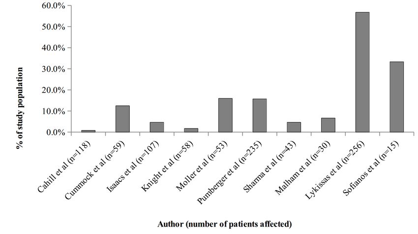
follow-up. Lykissas et al reported 56.8% (256/451) of However, since each of these associations is based on
patients with persistent symptoms at 6 months fol- only one study, the strength of evidence for each
low up, including 43 patients with anterior thigh finding is “low”.
pain, 127 with sensory deficits, and 86 with motor
deficits. Of these patients, 5.9% (15/256) had symp- With respect to disease factors, primary diagnosis
toms at least 18 months out from surgery. Sofianos et was found to significantly affect whether a complica-
al reported 33.3% of patients (15/45) with symptoms tion occurred postoperatively; however, since this is
persistent at least 9 months removed from surgery, based on only one study, the strength of evidence is
including 5 patients with persistent hip flexion weak- “low”.
ness at 11 months follow up, 7 with sensory deficits at
9 months, and 3 with neurologic deficits at 11 With respect to surgical factors, duration of surgery
months. Isaacs et al (4.7% of patients at 6 months), was found to be an independent risk factor for devel-
Knight et al (1.7% at 12 months), and Sharma et al opment of psoas mechanical flexion deficit, lumbar
(4.7% at 12 months) also reported the incidence of plexus related motor deficits, and sensory deficits
persistent lower extremity symptoms. postoperatively. In addition, inclusion of the L4-5
disc space was found to be an independent risk factor
Evidence Summary for development of any postoperative complication,
With respect to the incidence of lower-extremity including sensory deficits. The strength of evidence
symptoms after lateral interbody fusion, the risk is 0% is "high." That is, we have high confidence that our
to 60.7%. However, due to low levels of evidence and estimates represent the true effect and further re-
inconsistency of results the overall strength of evi- search is unlikely to change our estimates of the ef-
dence is “low”, that is we have low confidence that fect. Psoas retraction time was also found to be asso-
the evidence reflects the true effect and further re- ciated with the development of postoperative neuro-
logic deficit, however, the strength of evidence sup-
porting this association is low since it is based on on-
ly one study’s findings.
With respect to long-term consequences of postoper-
ative lower extremity symptoms, the estimates are
broad, ranging from 1.7% to 56.8% of patients with
symptoms at 6 months follow up. Due to the incon-
sistency of results and low levels of evidence, the
strength of evidence is “low.”
Fig. 2. Persistent lower extremity symptoms at 6 months follow-up.
Downloaded from http://ijssurgery.com/ by guest on May 5, 2021
International Journal of Spine Surgery 6 / 11doi: 10.14444/2062
dure.4-6 Finally, this technique relies on indirect
Discussion foraminal decompression via distraction of the disc
With the aging of the U.S. population degenerative space rather than direct decompression with
lumbar spine disease has been increasing in preva- laminectomy, which is often performed as part of
lence and is now one of the most commonly treated posterior spinal fusion. The technique also has its
orthopedic conditions. Since 2001 there has been a own set of distinct complications related to travers-
70% rise in spinal fusion procedure volume in the ing the psoas muscle, which is the focus of this re-
United States.38 Patients with this condition have tra- view.
ditionally been treated with lumbar spinal fusion via
either an anterior, posterior, or combined approach. Our review revealed several important findings relat-
While this procedure is associated with excellent im- ing to postoperative complications. The most ubiqui-
provement in patient pain and disability, there are a tous complication reported in the literature is post-
host of potential complications and limitations, such operative thigh pain, which was present in up to
as bowel perforation, major vessel injury and inciden- 60.7% of patients. Many authors attribute this com-
tal durotomy, leading to a complication rate between plication to an edematous reaction related to manipu-
11% and 24% and additional unplanned surgeries in lation of the psoas muscle and is viewed as a minor
up to 12% of cases.39 complication.31 Indeed, many authors fail to report
the incidence of this complication since most cases
Lateral transpsoas interbody fusion was introduced are transient in nature.16 However, it must be empha-
amidst the growing popularity of minimally invasive sized that these complications may not be transient
surgical techniques over the last two decades.40 Sev- in all cases, as our review showed 1.7% to 56.8% of pa-
eral popular systems are now offered, many of which tients may present with persistent symptoms at least
utilize intraoperative EMG technology to assess 6 months out from surgery and more than 12 months
proximity to neural structures. This approach offers removed in some cases. In addition, while it may be
numerous benefits over the traditional open ap- convenient to educate patients to expect thigh pain
proach to lumbar interbody fusion, owing in large following surgery, this explanation may be limited in
part to avoidance of peritoneal violation, lack of ex- scope and further elaboration on what symptoms to
tensive spinal muscle stripping, sparing of normal expect is necessary for proper patient education. Our
stabilizing elements of the spine (anterior and poste- review revealed that the incidence of any complica-
rior longitudinal ligaments) and the minimally inva- tion occurring ranged up to 60.7%, most commonly
sive nature of the technique. Furthermore, fusion sensory disturbances, followed by thigh pain in up to
rates and improvement in patient satisfaction have 43%, and hip flexion weakness in up to 30.8%. While
been on par with open techniques, ranging from these complications are transient in most cases, and
91-97% and 90%, respectively.41,42 the vast majority will improve by 6 months follow up,
it is important to emphasize the risk for more serious
Despite the many benefits of this technique, it is far long-term complications due to femoral and lumbar
from a panacea for patients with lumbar degenerative nerve root injury, resulting in neurological deficits in
pathology and is limited in several ways. Access to up to 23.9% of patients. We were also able to eluci-
the L5-S1 disc space is limited given its location be- date risk factors associated with this procedure with a
low the aortic bifurcation, which would require ex- high level of confidence. We have shown that dura-
tensive vessel dissection. Also the presence of the tion of surgery and inclusion of the L4-5 disc space
sacrum and pelvis at this level renders it inaccessible are independent risk factors for the development of
without the creation of an iliac “window.” In addi- postoperative complications. This has multiple im-
tion, many studies have mapped the intrapsoas neur- plications. First, given the steep learning curve for
al anatomy and a confluence of nerves exists lateral this procedure, inexperienced surgeons may require
to the L4-5 disc space including the femoral, gen- longer operative times until they are more comfort-
itofemoral, ilioinguinal and iliohypogastric nerves, able with this technique. It may be appropriate for in-
which are all at risk for injury during this proce- experienced surgeons to familiarize themselves with
Downloaded from http://ijssurgery.com/ by guest on May 5, 2021
International Journal of Spine Surgery 7 / 11doi: 10.14444/2062
the approach beforehand and assist in multiple pro- the significant heterogeneity in the selected studies
cedures in order to reduce their operative times and with respect to sample size and diagnosis. A meta-
rate of complications. While no studies defined spe- analysis was considered to address the apparent het-
cific cut-off time points at which the risk of symp- erogeneity, however, due to the significant variability
toms increases, our review has found that operative in study quality, and paucity of high quality prospec-
time ranged from 165 minutes to 178 minutes in stud- tive studies (only three level I studies) on this topic
ies reporting duration of surgery as a significant risk reported in the literature, we decided against this
factor associated with complications. To the extent methodology. It is important for the reader to note
that this represents a reliable cut-off point above that variability in surgical technique between studies
which the risk of symptoms increases, it may be ben- incorporates an additional confounding variable and
eficial to use this cut-off point as a guide in preopera- should be considered in the context of this review’s
tive surgical planning. Furthermore, while not sup- findings. Three high quality studies varied from the
ported by data, it is reasonable to suspect that in- rest in terms of their inconsistent use of intraopera-
creasing surgical duration is associated with longer tive neuromonitoring (Pumberger et al.14 and Knight
psoas retraction times, a risk factor for postoperative et al.36) and anterior plate fixation (Isaacs et al.37),
neuropraxia. Uribe et al found a significant positive which may diminish the accuracy of this review’s
relationship between the presence of a neurologic findings. In addition, other potential confounding
deficit and total retraction time. The authors recom- factors such as retraction time, retractor opening
mend monitoring EMG for increasing thresholds size, bed flexion, and adherence to non-muscular
during prolonged retraction times in an effort to re- pharmaco-blockade were not reported by the studies
duce the incidence of complications.32 Inclusion of included in this review but should also be considered
the L4-5 disc space has been implicated in many cas- when interpreting these findings. Regarding the nov-
es of postoperative complications and it is easy to un- elty of this study’s findings, there have been few re-
derstand why, given the confluence of major neural view articles and meta-analyses published on related
structures at this location. Prudent use of neural topics, including direct nerve injury after LTIF; how-
monitoring is all the more important with inclusion ever, a systematic review of nerve injury and tran-
of this disc space, and the psoas should be parted be- sient thigh pain after LTIF is lacking, and we believe
tween the middle and anterior third of the muscle, the comprehensive overview and recommendations
ensuring that nerves of the lumbar plexus are located contained herein will bolster surgeons’ knowledge
posteriorly and out of the surgical corridor. and understanding of complications associated with
this popular technique.
While our study focuses on lower extremity symp-
toms associated with this procedure, it is important
to consider other complications reported in the liter- Conclusion
ature. Vertebral body fracture occurred in 0.96%-15% The lateral transpsoas approach is a popular mini-
of reported cases, cage subsidence in 1.2%-18.2%, and mally invasive technique for achieving spinal fusion
abdominal wall paresis in 1.8%-5.1%.15,16,20-23,25,34,43-50 in patients with degenerative lumbar spine disease.
Concerning abdominal dissection, many authors rec- The literature is replete with studies reporting post-
ommend the use of blunt dissection over electro- operative complications of this procedure including
cautery, which has been associated with increased transient lower extremity sensory changes and lum-
risk of paresis.43 In addition, Tohmeh et al reported bar nerve root injury. Numerous risk factors were al-
on 2 cases of peritoneal perforation, which has the so identified including lengthier duration of surgery
potential to go unnoticed given the limited visualiza- and inclusion of L4-5 intervertebral disc space.
tion with this procedure, so care should be taken While most patients experienced transient symp-
when sweeping the peritoneum anteriorly and in toms, the occurrence of long term postoperative
placing the dilators. complications is not uncommon. Future high-quality
research focused on risk reduction strategies may im-
There are several limitations of this review including prove complication rates associated with this proce-
Downloaded from http://ijssurgery.com/ by guest on May 5, 2021
International Journal of Spine Surgery 8 / 11doi: 10.14444/2062
dure. 11. Atkins D, Best D, Briss PA et al. Education and
debate. BMJ. 2004;328:1-8.
12. Dettori JR, Norvell DC, Dekutoski M, Fisher C,
References Chapman JR. Methods for the systematic reviews on
1. McAfee PC, Regan JJ, Geis WP, Fedder IL. Mini- patient safety during spine surgery. Spine (Phila Pa
mally invasive anterior retroperitoneal approach to 1976). 2010;35(9 Suppl):S22-S27.
the lumbar spine. Emphasis on the lateral BAK. 13. Cummock MD, Vanni S, Levi AD, Yu Y, Wang
Spine (Phila Pa 1976). 1998;23(13):1476-1484. MY, Cummock MD. Vanni S. Levi AD. Yu Y. Wang
2. Bergey DL, Villavicencio AT, Goldstein T, Regan M. An analysis of postoperative thigh symptoms af-
JJ. Endoscopic lateral transpsoas approach to the ter minimally invasive transpsoas interbody fusion. J
lumbar spine. Spine (Phila Pa 1976). Neurosurg Spine. 2011;15(1):11-18.
2004;29(15):1681-1688. 14. Pumberger M, Hughes AP, Huang RR, Sama
3. Ozgur BM, Aryan HE, Pimenta L, Taylor WR. AA, Cammisa FP, Girardi FP. Neurologic deficit fol-
Extreme Lateral Interbody Fusion (XLIF): a novel lowing lateral lumbar interbody fusion. Eur Spine J.
surgical technique for anterior lumbar interbody fu- 2012;21(6):1192-1199.
sion. Spine J. 2006;6(4):435-443. 15. Rodgers WB, Cox CS, Gerber EJ. Early compli-
4. Banagan K, Gelb D, Poelstra K, Ludwig S. cations of extreme lateral interbody fusion in the
Anatomic mapping of lumbar nerve roots during a di- obese. J Spinal Disord Tech. 2010;23(6):393-397.
rect lateral transpsoas approach to the spine: a cadav- 16. Rodgers WB, Gerber EJ, Patterson J. Intraopera-
eric study. Spine (Phila Pa 1976). tive and early postoperative complications in extreme
2011;36(11):E687-E691. lateral interbody fusion: an analysis of 600 cases.
5. Benglis DM. Vanni S. Levi A, Benglis DM, Vanni Spine (Phila Pa 1976). 2011;36(1):26-32.
S, Levi AD. An anatomical study of the lumbosacral 17. Lykissas MG, Aichmair A, Hughes AP, et al.
plexus as related to the minimally invasive transpsoas Nerve injury after lateral lumbar interbody fusion: a
approach to the lumbar spine. J Neurosurg Spine. review of 919 treated levels with identification of risk
2009;10(2):139-144. factors. Spine J. 2014;14(5):749-758.
6. Guérin P, Obeid I, Bourghli A, et al. The lum- 18. Anand N, Baron EM, Thaiyananthan G, Khalsa
bosacral plexus: anatomic considerations for mini- K, Goldstein TB. Minimally invasive multilevel per-
mally invasive retroperitoneal transpsoas approach. cutaneous correction and fusion for adult lumbar de-
Surg Radiol Anat. 2012;34(2):151-157. generative scoliosis: a technique and feasibility study.
7. Park DK, Lee MJ, Lin EL, Singh K, An HS, J Spinal Disord Tech. 2008;21(7):459-467.
Phillips FM. The relationship of intrapsoas nerves 19. Anand N, Rosemann R, Khalsa B, Baron EM.
during a transpsoas approach to the lumbar spine: Mid-term to long-term clinical and functional out-
anatomic study. J Spinal Disord Tech. comes of minimally invasive correction and fusion
2010;23(4):223-228. for adults with scoliosis. Neurosurg Focus.
8. Moher D, Liberati A, Tetzlaff J, Altman DG. Pre- 2010;28(3):E6.
ferred reporting items for systematic reviews and 20. Berjano P, Balsano M, Buric J, Petruzzi M,
meta-analyses: the PRISMA statement. PLoS Med. Lamartina C. Direct lateral access lumbar and thora-
2009;6(7):e1000097. columbar fusion: preliminary results. Eur Spine J.
9. Jour- T, Journal T, Sur- J. Introducing Levels of 2012;21 Suppl 1:S37-S42.
Evidence to The Journal Deputy Editor for Outcome 21. Cahill KS, Martinez JL, Wang MY, Vanni S,
Studies. 2003. Levi AD. Motor nerve injuries following the mini-
10. Norvell DC, Dettori JR, Fehlings MG, Fourney mally invasive lateral transpsoas approach. J Neuro-
DR, Chapman JR. Methodology for the systematic surg Spine. 2012.
reviews on an evidence-based approach for the man- 22. Dakwar E, Cardona RF, Smith DA, Uribe JS.
agement of chronic low back pain. Spine (Phila Pa Early outcomes and safety of the minimally invasive,
1976). 2011;36(21 Suppl):S10-S18. lateral retroperitoneal transpsoas approach for adult
Downloaded from http://ijssurgery.com/ by guest on May 5, 2021
International Journal of Spine Surgery 9 / 11doi: 10.14444/2062
degenerative scoliosis. Neurosurg Focus. praxia after XLIF? Results from a prospective multi-
2010;28(3):E8. center trial. Eur Spine J. 2015;24 Suppl 3:378-385.
23. Kepler CK, Sharma AK, Huang RC. Lateral 33. Pimenta L, Oliveira L, Schaffa T, Coutinho E,
transpsoas interbody fusion (LTIF) with plate fixa- Marchi L. Lumbar total disc replacement from an ex-
tion and unilateral pedicle screws: a preliminary re- treme lateral approach: clinical experience with a
port. J Spinal Disord Tech. 2011;24(6):363-367. minimum of 2 years’ follow-up. J Neurosurg Spine.
24. Malham GM, Ellis NJ, Parker RM, Seex KA. 2011;14(1):38-45.
Clinical outcome and fusion rates after the first 30 34. Tohmeh AG, Rodgers WB, Peterson MD. Dy-
extreme lateral interbody fusions. Scientific World namically evoked, discrete-threshold electromyogra-
Journal. 2012;2012:246989. phy in the extreme lateral interbody fusion approach.
25. Rodgers WB, Cox CS, Gerber EJ. Minimally In- J Neurosurg Spine. 2011;14(1):31-37.
vasive Treatment (XLIF) of Adjacent Segment Dis- 35. Oliveira L, Marchi L, Coutinho E, Pimenta L. A
ease after prior Lumbar Fusions. Internet J Minim In- radiographic assessment of the ability of the extreme
vasive Spinal Technol. 2009;3(4). lateral interbody fusion procedure to indirectly de-
26. Sharma AK, Kepler CK, Girardi FP, Cammisa compress the neural elements. Spine (Phila Pa 1976).
FP, Huang RC, Sama AA. Lateral lumbar interbody 2010;35(26 Suppl):S331-S337.
fusion: clinical and radiographic outcomes at 1 year: a 36. Knight RQ, Schwaegler P, Hanscom D, Roh J.
preliminary report. J Spinal Disord Tech. Direct lateral lumbar interbody fusion for degenera-
2011;24(4):242-250. tive conditions: early complication profile. J Spinal
27. Wang MY, Mummaneni P V, Wang MY. Mum- Disord Tech. 2009;22(1):34-37.
maneni P. Minimally invasive surgery for thora- 37. Isaacs RE, Hyde J, Goodrich JA, Rodgers WB,
columbar spinal deformity: initial clinical experience Phillips FM. A prospective, nonrandomized, multi-
with clinical and radiographic outcomes. Neurosurg center evaluation of extreme lateral interbody fusion
Focus. 2010;28(3):E9. for the treatment of adult degenerative scoliosis: pe-
28. Sofianos DA, Briseño MR, Abrams J, Patel AA. rioperative outcomes and complications. Spine (Phila
Complications of the lateral transpsoas approach for Pa 1976). 2010;35(26 Suppl):S322-S330.
lumbar interbody arthrodesis: a case series and litera- 38. Weiss AJ, Elixhauser A. Trends in Operating
ture review. Clin Orthop Relat Res. Room Procedures in U.S. Hospitals, 2001—2011 -
2012;470(6):1621-1632. Statistical Brief #171. HCUP. 2014.
29. Waddell B, Briski D, Qadir R, et al. Lateral lum- 39. Phillips FM, Slosar PJ, Youssef JA, Andersson
bar interbody fusion for the correction of spondy- G, Papatheofanis F. Lumbar spine fusion for chronic
lolisthesis and adult degenerative scoliosis in high- low back pain due to degenerative disc disease: a sys-
risk patients: early radiographic results and complica- tematic review. Spine (Phila Pa 1976).
tions. Ochsner J. 2014;14(1):23-31. 2013;38(7):E409-E422.
30. Isaacs RE, Hyde J, Goodrich JA, Rodgers WB, 40. Ozgur BM, Aryan HE, Pimenta L, Taylor WR.
Phillips FM. A prospective, nonrandomized, multi- Extreme Lateral Interbody Fusion (XLIF): a novel
center evaluation of extreme lateral interbody fusion surgical technique for anterior lumbar interbody fu-
for the treatment of adult degenerative scoliosis: pe- sion. Spine J. 2006;6(4):435-443.
rioperative outcomes and complications. Spine (Phila 41. Rodgers WB, Gerber EJ, Patterson JR. Fusion
Pa 1976). 2010;35(26 Suppl):S322-S330. after minimally disruptive anterior lumbar interbody
31. Moller DJ, Slimack NP, Acosta FL, Koski TR, fusion: Analysis of extreme lateral interbody fusion
Fessler RG, Liu JC. Minimally invasive lateral lum- by computed tomography. SAS J. 2010;4(2):63-66.
bar interbody fusion and transpsoas approach-related 42. Ozgur BM, Agarwal V, Nail E, Pimenta L. Two-
morbidity. Neurosurg Focus. 2011;31(4):E4. year clinical and radiographic success of minimally
32. Uribe JS, Isaacs RE, Youssef JA, et al. Can trig- invasive lateral transpsoas approach for the treat-
gered electromyography monitoring throughout re- ment of degenerative lumbar conditions. SAS J.
traction predict postoperative symptomatic neuro- 2010;4(2):41-46.
Downloaded from http://ijssurgery.com/ by guest on May 5, 2021
International Journal of Spine Surgery 10 / 11doi: 10.14444/2062
43. Dakwar E, Le T V, Baaj AA, et al. Abdominal Coutinho E, Pimenta L. Stand-alone lateral inter-
wall paresis as a complication of minimally invasive body fusion for the treatment of low-grade degenera-
lateral transpsoas interbody fusion. Neurosurg Focus. tive spondylolisthesis. Scientific World Journal.
2011;31(4):E18. 2012;2012:456346.
44. Karikari IO, Grossi PM, Nimjee SM, et al. Min- 50. Rodgers WB, Lehmen JA, Gerber EJ, Rodgers
imally invasive lumbar interbody fusion in patients JA. Grade 2 spondylolisthesis at L4-5 treated by
older than 70 years of age: analysis of peri- and post- XLIF: safety and midterm results in the “worst case
operative complications. Neurosurgery. scenario”. Scientific World Journal.
2011;68(4):897-902; discussion 902. 2012;2012:356712.
45. Brier-Jones JE, Palmer DK, Ĭnceoğlu S, Cheng
WK. Vertebral body fractures after transpsoas inter-
body fusion procedures. Spine J. Disclosures
2011;11(11):1068-1072. The authors declare no relevant disclosures.
46. Le T V, Baaj AA, Dakwar E, et al. Subsidence of
polyetheretherketone intervertebral cages in mini-
mally invasive lateral retroperitoneal transpsoas lum- Corresponding Author
bar interbody fusion. Spine (Phila Pa 1976). Jeffrey M. Spivak, MD, Director, Spine Cen-
2012;37(14):1268-1273. ter, NYU Hospital for Joint Diseases, Depart-
47. Le T V, Smith DA, Greenberg MS, Dakwar E, ment of Orthopaedic Surgery, 301 East 17th
Baaj AA, Uribe JS. Complications of lateral plating in Street, New York NY 10003. Jef-
the minimally invasive lateral transpsoas approach. J frey.Spivak@nyumc.org.
Neurosurg Spine. 2012;16(3):302-307. Published 12 November 2015.
48. Marchi L, Oliveira L, Amaral R, et al. Lateral This manuscript is generously published free of
interbody fusion for treatment of discogenic low back charge by ISASS, the International Society for the
pain: minimally invasive surgical techniques. Adv Or- Advancement of Spine Surgery. Copyright © 2015
thop. 2012;2012:282068. ISASS. To see more or order reprints or permissions,
49. Marchi L; A, Abdala N, Oliveira L, Amaral R, see http://ijssurgery.com.
Downloaded from http://ijssurgery.com/ by guest on May 5, 2021
International Journal of Spine Surgery 11 / 11You can also read