Deep Orthogonal Fusion: Multimodal Prognostic Biomarker Discovery Integrating Radiology, Pathology, Genomic, and Clinical Data
←
→
Page content transcription
If your browser does not render page correctly, please read the page content below
Deep Orthogonal Fusion: Multimodal Prognostic
                                          Biomarker Discovery Integrating Radiology,
                                            Pathology, Genomic, and Clinical Data

                                            Nathaniel Braman, Jacob W. H. Gordon, Emery T. Goossens, Caleb Willis,
                                                          Martin C. Stumpe, Jagadish Venkataraman
arXiv:2107.00648v1 [cs.CV] 1 Jul 2021

                                                               Tempus Labs, Inc., Chicago, IL, USA
                                                     {nathaniel.braman, jagadish.venkataraman}@tempus.com
                                                                   https://www.tempus.com/

                                                Abstract. Clinical decision-making in oncology involves multimodal data
                                                such as radiology scans, molecular profiling, histopathology slides, and
                                                clinical factors. Despite the importance of these modalities individually,
                                                no deep learning framework to date has combined them all to predict
                                                patient prognosis. Here, we predict the overall survival (OS) of glioma
                                                patients from diverse multimodal data with a Deep Orthogonal Fusion
                                                (DOF) model. The model learns to combine information from multipara-
                                                metric MRI exams, biopsy-based modalities (such as H&E slide images
                                                and/or DNA sequencing), and clinical variables into a comprehensive
                                                multimodal risk score. The model learns to combine embeddings from
                                                each modality via attention-gated tensor fusion. To maximize the infor-
                                                mation gleaned from each modality, we introduce a multimodal orthogo-
                                                nalization (MMO) loss term that increases model performance by incen-
                                                tivizing constituent embeddings to be more complementary. DOF pre-
                                                dicts OS in glioma patients with a median C-index of 0.788 ± 0.067, sig-
                                                nificantly outperforming (p=0.023) the best performing unimodal model
                                                with a median C-index of 0.718 ± 0.064. The prognostic model signif-
                                                icantly stratifies glioma patients by OS within clinical subsets, adding
                                                further granularity to prognostic clinical grading and molecular subtyp-
                                                ing.

                                        1     Introduction

                                        Cancer diagnosis and treatment plans are guided by multiple streams of data
                                        acquired from several modalities, such as radiology scans, molecular profiling,
                                        histology slides, and clinical variables. Each characterizes unique aspects of tu-
                                        mor biology and, collectively, they help clinicians understand patient prognosis
                                        and assess therapeutic options. Advances in molecular profiling techniques have
                                        enabled the discovery of prognostic gene signatures, bringing precision medicine
                                        to the forefront of clinical practice [1]. More recently, computational techniques
                                        in the field of radiology have identified potential imaging-based phenotypes of
                                            Accepted for presentation at MICCAI 2021.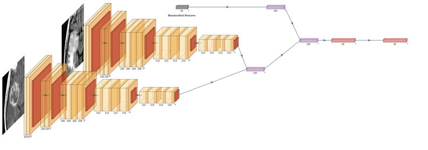
2      N. Braman et al.

treatment response and patient survival. Such approaches leverage large sets of
explicitly designed image features (commonly known as radiomics [2]) or entail
the novel discovery of image patterns by optimizing highly parameterized deep
learning models such as convolutional neural networks (CNN) [3] for prediction.
Along similar lines, the digitization of histopathology slides has opened new av-
enues for tissue-based assays that can stratify patients by risk from H&E slide
images alone [4]. Given the complementary nature of these various modalities
in comprehensive clinical assessment, we hypothesize that their combination in
a rigorous machine learning framework may predict patient outcomes more ro-
bustly than qualitative clinical assessment or unimodal strategies.
    Glioma is an intuitive candidate for deep learning-based multimodal biomark-
ers owing to the presence of well-characterized prognostic information across
modalities [5], as well as its severity [6]. Gliomas can be subdivided by their
malignancy into histological grades II-IV [5]. Grades differ in their morphologic
and molecular heterogeneity [7], which correspond to treatment resistance and
short-term recurrence [8, 9]. Quantitative analysis of glioma [10] and its tumor
habitat [11] on MRI has demonstrated strong prognostic potential, as well as
complex interactions with genotype [12] and clinical variables [13].
    Most deep multimodal prediction strategies to date have focused on the fusion
of biopsy-based modalities [14, 15, 16]. For instance, previous work integrating
molecular data with pathology analysis via CNN or graph convolutional neural
networks (GCN) has shown that a deep, multimodal approach improves prog-
nosis prediction in glioma patients [14, 15]. Likewise, Cheerla et al. integrated
histology, clinical, and sequencing data across cancer types by condensing each to
a correlated prognostic feature representation [16]. Multimodal research involv-
ing radiology has been predominantly correlative in nature [13, 12]. Some have
explored late-stage fusion approaches combining feature-based representations
from radiology with similar pathology [17] or genomic features [18] to predict
recurrence. While promising, these strategies rely on hand-crafted feature sets
and simple multimodal classifiers that likely limit their ability to learn complex
prognostic interactions between modalities and realize the full additive benefit
of integrating diverse clinical modalities.
    To our knowledge, no study to date has combined radiology, pathology, and
genomic data within a single deep learning framework for outcome prediction
or patient stratification. Doing so requires overcoming several challenges. First,
owing to the difficulty of assembling multimodal datasets with corresponding
outcome data in large quantities, fusion schemes must be highly data efficient
in learning complex multimodal interactions. Second, the presence of strongly
correlated prognostic signals between modalities [16] can create redundancy and
hinder model performance.
    In this paper, we introduce a deep learning framework that combines radi-
ologic, histologic, genomic, and clinical data into a fused prognostic risk score.
Using a novel technique referred to as Deep Orthogonal Fusion (DOF), we train
models using a Multimodal Orthogonalization (MMO) loss function to maxi-
mize the independent contribution of each data modality, effectively improving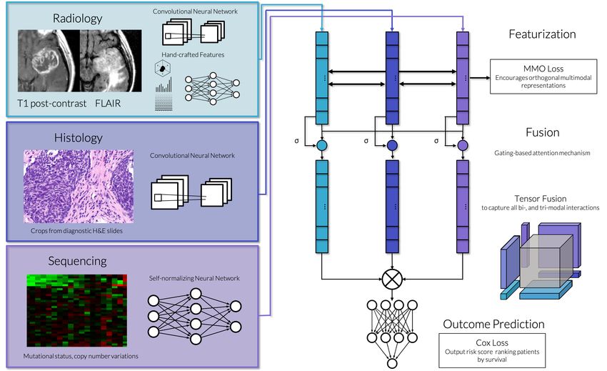
Deep Orthogonal Fusion for Multimodal Prognostic Biomarker Discovery         3

predictive performance. Our approach, depicted in Fig. 1, first trains unimodal
embeddings for overall survival (OS) prediction through a Cox partial likelihood
loss function. Next, these embeddings are combined through an attention-gated
tensor fusion to capture all possible interactions between each data modality.
Fusion models are trained simultaneously to predict OS and minimize the corre-
lation between unimodal embeddings. We emphasize the following contributions:
    Deep Fusion of Radiology, Pathology, and Omics Data: We present a
powerful, data-efficient framework for combining oncologic data across modali-
ties. Our approach enabled a previously unexplored deep integration of radiology
with tissue-based modalities and clinical variables for patient risk stratification.
This fusion model significantly improved upon unimodal deep learning mod-
els. In particular, we found that integrating radiology into deep multimodal
models, which is under-explored in previous prognostic studies, conferred the
single greatest performance increase. This finding suggests the presence of inde-
pendent, complementary prognostic information between radiology and biopsy-
based modalities and warrants their combination in future prognostic studies.
    MMO: To mitigate the effect of inherent correlations between data modal-
ities, we present an MMO loss function that penalizes correlation between uni-
modal embeddings and encourages each to provide independent prognostic in-
formation. We find that this training scheme, which we call DOF, improves
prediction by learning and fusing disentangled, prognostic representations from
each modality. DOF was also found to outperform a fusion scheme that en-
forces correlated representations between modalities [16], emphasizing that the
dissimilarity of these clinical data streams is crucial to their collective strength.
    Multi-parametric Radiology FeatureNet: A neural network architec-
ture that can fuse CNN-extracted deep features from local tumor regions on
multiple image sequences (e.g., Gd-T1w and T2w-FLAIR scans) with global
hand-crafted radiomics features extracted across the full 3D region-of-interest.
    Independent prognostic biomarker of OS in glioma patients: Using
15-fold Monte Carlo cross-validation with a 20% holdout test set, we evaluate
deep fusion models to predict glioma prognosis. We compare this multimodal risk
score with existing prognostic clinical subsets and biomarkers (grade, IDH sta-
tus) and investigate its prognostic value within these outcome-associated groups.

2     Methodology

Let X be a training minibatch of data for N patients, each containing M modal-
ities such that X = [x1 , x2 , ..., xM ]. For each modality m, xm includes data from
for N patients. Φm denotes a trainable unimodal network, which accepts xm and
generates a deep embedding hm = Φm (xm ) ∈ Rl1 xN .

2.1    Multimodal Fusion

When M > 1, we combine embeddings from each modality in a multimodal
fusion network. For each hm , an attention mechanism is applied to control its
4 N. Braman et al.
Fig. 1. DOF model architecture and training.
expressiveness based on information from the other modalities. An additional
fully connected layer results in hSm of length l2 . Attention weights of length l2
are obtained through a bilinear transformation of hm with all other embeddings
S
(denoted as Hm ), then applied to hm to yield the attention-gated embedding:
h∗m = am ∗ hSm = σ(hTm ∗ WA ∗ H S
m ) ∗ hm . (1)
To capture all possible interactions between modalities, we combine attention-
weighted embeddings through an outer product between modalities, known as
tensor fusion [19]. A value of 1 is also included in each vector, allowing for partial
interactions between modalities and for the constituent unimodal embeddings to
be retained. The output matrix
1 1 1
F = ∗ ⊗ ∗ ⊗ ... ⊗ ∗ (2)
h1 h2 hM
is an M -dimensional hypercube of all multimodal interactions with sides of
length l2 + 1. Fig. 1 depicts F for the fusion of radiology, pathology, and genomic
data. It contains subregions corresponding to unaltered unimodal embeddings,
pairwise fusions between 2 modalities, and trilinear fusion between all three of
the modalities. A final set of fully connected layers, denoted by ΦF , is applied
to tensor fusion features for a final fused embedding hF = ΦF (F ).
2.2 MMO Loss
To address the shortcoming of multimodal models converging to correlated pre-
dictors, we introduce MMO loss. Inspired by Orthogonal Low-rank EmbeddingDeep Orthogonal Fusion for Multimodal Prognostic Biomarker Discovery 5
[20], we stipulate that unimodal embeddings preceding fusion should be orthog-
onal. This criterion enforces that each modality introduced contributes unique
information to outcome prediction, rather than relying on signal redundancy
between modalities. Each Φm is updated through MMO loss to yield embed-
dings that better complement other modalities. Let H ∈ Rl1 xM ∗N be the set of
embeddings from all modalities. MMO loss is computed as
M
1 X
LM M O = max(1, ||hm ||∗ ) − ||H||∗ (3)
M ∗ N m=1
where || · ||∗ denotes the matrix nuclear norm (i.e., the sum of the matrix sin-
gular values). This loss is the difference between the sum of nuclear norms per
embedding and the nuclear norm of all embeddings combined. It penalizes the
scenario where the variance of two modalities separately is decreased when com-
bined and minimized when all unimodal embeddings are fully orthogonal. The
per-modality norm is bounded to a minimum of 1 to prevent the collapse of
embedding features to zero.
2.3 Cox Partial Likelihood Loss
The final layer of each network, parameterized by β, is a fully connected layer
with a single unit. This output functions as a Cox proportional hazards model
using the deep embedding from the previous layer, h, as its covariates. This final
layer’s output, θ, is the log hazard ratio, which is used as a risk score. The log
hazard ratio for patient i is denoted as θi = hTi ∗ β.
We define the negative log likelihood Lpl as our cost function
X X
Lpl = − θi − log eθj (4)
i:Ei =1 j:ti ≥tj
where t ∈ RN x1 indicates the time to date of last follow up. The event vector,
E ∈ {0, 1}N x1 , equals 1 if an event was observed (death) or 0 if a patient was
censored (still alive) at time of last follow up. Each patient i with an observed
event is compared against all patients whose observation time was greater than
or equal to ti . Networks are trained using the final loss L which is a linear
combination of the two loss functions specified above
L = Lpl + γLM M O (5)
where γ is a scalar weighting the contribution of MMO loss relative to Cox partial
likelihood loss. When training unimodal networks, γ is always zero. Performance
for various values of γ are included in the Table S4.
2.4 Modality-specific Networks for Outcome Prediction
Radiology: A multiple-input CNN was designed to incorporate multiparametric
MRI data and global lesion measurements, shown in Fig. S1. The backbone of6 N. Braman et al.
the network is a VGG-19 CNN [21] with batch normalization, substituting the
final max pooling layer with a 4x4 adaptive average pooling. Two pre-trained
[22] CNN branches separately extract features from Gd-T1w and T2w-FLAIR
images, which are then concatenated and passed through a fully connected layer.
A third branch passes hand-crafted features (described in section 3) through a
similar fully connected layer. Concatenated embeddings from all branches are
fed to 2 additional fully connected layers. All fully connected layers preceding
the final embedding layer have 128 units.
Histology, genomic, and clinical data: We reused the models proposed
in [14] - a pre-trained VGG-19 CNN with pretrained convolutional layers for
Histology and a Self-Normalizing Neural Network (SNN) for genomic data. We
also use this SNN for analysis of clinical data, which was not explored in [14].
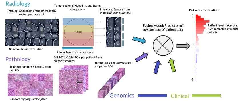
Fig. 2. Sampling multiple radiology & pathology images for patient level risk scores.
3 Experimental Details
Radiology: 176 patients (see patient selection in Fig. S2) with Gd-T1w and
T2w-FLAIR scans from the TCGA-GBM [23] and TCGA-LGG [24] studies
were obtained from TCIA [25] and annotated by 7 radiologists to delineate
the enhancing lesion and edema region. Volumes were registered to the MNI-
ICBM standardized brain atlas with 1 mm isotropic resolution, processed with
N4 bias correction, and intensity normalized. 96x96x3 patches were generated
from matching regions of Gd-T1w and T2w-FLAIR images within the enhancing
lesion. For each patient, 4 samples were generated from four even quadrants of
the tumor along the z-axis. Patch slice position was randomized in unimodal
training and fixed to the middles of quadrants during inference and fusion net-
work training. Nine features including size, shape, and intensity measures were
extracted separately from Gd-T1w and T2w-FLAIR images, and summarized in
three fashions for a total of 56 handcrafted features, listed in Table S1.Deep Orthogonal Fusion for Multimodal Prognostic Biomarker Discovery 7
Pathology and Genomics: We obtained 1024×1024 normalized regions-of-
interest (ROIs) and DNA sequencing data curated by [15]. Each patient had 1-3
ROIs from diagnostic H&E slides, totaling 372 images. DNA data consisted of
80 features including mutational status and copy number variation (Table S2).
Clinical information: 14 clinical features were included into an SNN for
the prediction of prognosis. The feature set included demographic and treatment
details, as well as subjective histological subtype (see Table S3).
Implementation Details: The embedding size for unimodal networks, l1 ,
was set to 32. Pre-fusion scaled embedding size, l2 , was 32 for M =2, 16 for M =3,
and 8 for M =4. Post-fusion fully connected layers consisted of 128 units each.
The final layer of each network had a single unit with sigmoid activation, but its
outputs were rescaled between -3 and 3 to function as a prognostic risk score.
Unimodal networks were trained for 50 epochs with linear learning rate decay,
while multimodal networks were trained for 30 epochs with learning rate decay
beginning at the 10th epoch. When training multimodal networks, the unimodal
embedding layers were frozen for 5 epochs to train the fusion layers only, then
unfrozen for joint training of embeddings and fusion layers.
Statistical Analysis: All models were trained via 15-fold Monte Carlo cross-
validation with 20% holdout using the patient-level splits provided in [15]. The
primary performance metric was the median observed concordance index (C-
index) across folds, a global metric of prognostic model discriminant power. We
evaluated all possible combinations of a patient’s data (see sampling strategy in
Fig. 2) and used the 75th percentile of predicted risk score as their overall predic-
tion. C-indexes of the best-performing unimodal model and the DOF multimodal
model were compared with a Mann-Whitney U test [26]. Binary low/high-risk
groups were derived from the risk scores, where a risk score >0 corresponded to
high risk. For Kaplan-Meier (KM) curves and calculation of hazard ratio (HR),
patient-level risk scores were pooled across validation folds.
4 Results and Discussion
Genomic- and pathology-only model performance metrics are practically similar
(Table 1). However, the CNN-only (C-index=0.687 ± 0.067) and feature-only
(C-index=0.653 ± 0.057) configurations of the radiology model underperform
relative to the aforementioned unimodal models. Combining the radiology CNN
features with the handcrafted features results in the strongest unimodal model.
In contrast, clinical features are the least prognostic unimodal model.
Deep fusion models integrating radiology outperform individual unimodal
models, naive ensembles of unimodal models, as well as fusions of only clini-
cal and/or biopsy-derived modalities. The full fusion model (C-index=0.775 ±
0.061) achieves the best performance when trained with Cox loss [27] alone, sec-
ond only to the Rad+Path+Gen model trained with MMO loss. Naive late fusion
ensembles (i.e., averaging unimodal risk scores) exhibit inferior performance for
Rad+Path+Gen with (C-index=0.735 ± 0.063) and without (C-index=0.739 ±
0.062) clinical features, confirming the benefits of deep fusion.8 N. Braman et al.
Table 1. Median C-index of unimodal and fusion models with and without MMO loss.
Group Model Cox Loss Only With MMO Loss
Unimodal Rad 0.718 ± 0.064 –
Path 0.715 ± 0.054 –
Gen 0.716 ± 0.063 –
Clin 0.702 ± 0.049 –
Pairwise Fusion Path+Gen 0.711 ± 0.055 0.752 ± 0.072
Gen+Clin 0.702 ± 0.053 0.703 ± 0.052
Rad+Gen 0.761 ± 0.071 0.766 ± 0.067
Rad+Path 0.742 ± 0.067 0.752 ± 0.072
Rad+Clin 0.746 ± 0.068 0.736 ± 0.067
Path+Clin 0.696 ± 0.051 0.690 ± 0.043
Triple Fusion Path+Gen+Clin 0.704 ± 0.059 0.720 ± 0.056
Rad+Path+Clin 0.748 ± 0.067 0.741 ± 0.067
Rad+Gen+Clin 0.754 ± 0.066 0.755 ± 0.067
Rad+Path+Gen 0.764 ± 0.062 0.788 ± 0.067
Full Fusion Rad+Path+Gen+Clin 0.775 ± 0.061 0.785 ± 0.077
Fig. 3. Stratification by (a) grade, (b) IDH mutation status, and (c) DOF risk groups.
Fig. 4. DOF risk groups stratify patients by OS within (a,b) grade & (c,d) IDH subsets.Deep Orthogonal Fusion for Multimodal Prognostic Biomarker Discovery 9
The addition of MMO loss when training these deep fusion models consis-
tently improves their performance at five different weightings (Table S4), with
best performance for both at γ = .5. When all fusion models are trained at this
weighting, 8 of 11 improve in performance. DOF combining radiology, pathology,
and genomic data predicts glioma survival best overall with a median C-index
of 0.788 ± 0.067, a significant increase over the best unimodal model (p=0.023).
An ablation study was conducted to investigate the contributions of compo-
nents of the fusion module (modality attention-gating and tensor fusion). We
found that a configuration including both yields the best performance, but that
strong results can also be achieved with a simplified fusion module (Table S5).
In Fig. 3, KM plots show that the stratification of patients by OS in risk
groups derived from this model perform comparably to established clinical mark-
ers. In Fig. 4, risk groups further stratify OS within grade and IDH status groups.
In sum, these results suggest that the DOF model provides useful prognostic
value beyond existing clinical subsets and/or individual biomarkers.
To further benchmark our approach, we implemented the fusion scheme of
[16], who combined pathology images, DNA, miRNA, and clinical data, which
we further modified to also include radiology data. The network and learning
approach is described in-depth in Table S6. In contrast to DOF, [16] instead
seeks to maximize the correlation between modality embeddings prior to pre-
diction. A model combining radiology, pathology, and genomic data achieved
C-index=0.730 ± 0.05, while a model excluding the added radiology arm strat-
ified patients by OS with C-index=0.715 ± 0.05.
5 Conclusions
We present DOF, a data efficient scheme for the novel fusion of radiology, histol-
ogy, genomic, and clinical data for multimodal prognostic biomarker discovery.
The integration of multi-dimensional data from biopsy-based modalities and
radiology strongly boosts the ability to stratify glioma patients by OS. The
addition of a novel MMO loss component, which forces unimodal embeddings
to provide independent and complementary information to the fused predic-
tion, further improves prognostic performance. Our DOF model incorporating
radiology, histology, and genomic data significantly stratifies glioma patients by
OS within outcome-associated subsets, offering additional granularity to routine
clinical markers. DOF can be applied to any number of cancer domains, modality
combinations, or new clinical endpoints including treatment response.
References
[1] Wafik S. El-Deiry et al. “The current state of molecular testing in the
treatment of patients with solid tumors, 2019”. In: CA: a cancer journal
for clinicians 69.4 (July 2019), pp. 305–343.10 N. Braman et al.
[2] Robert J. Gillies, Paul E. Kinahan, and Hedvig Hricak. “Radiomics: Im-
ages Are More than Pictures, They Are Data”. In: Radiology 278.2 (Nov. 18,
2015), pp. 563–577.
[3] Luca Saba et al. “The present and future of deep learning in radiology”.
In: European Journal of Radiology 114 (May 1, 2019), pp. 14–24.
[4] Ole-Johan Skrede et al. “Deep learning for prediction of colorectal cancer
outcome: a discovery and validation study”. In: Lancet (London, England)
395.10221 (Feb. 1, 2020), pp. 350–360.
[5] David N. Louis et al. “The 2016 World Health Organization Classification
of Tumors of the Central Nervous System: a summary”. In: Acta Neu-
ropathologica 131.6 (June 2016), pp. 803–820.
[6] Rebecca L. Siegel, Kimberly D. Miller, and Ahmedin Jemal. “Cancer Statis-
tics, 2017”. In: CA: a cancer journal for clinicians 67.1 (Jan. 2017), pp. 7–
30.
[7] Adriana Olar and Kenneth D. Aldape. “Using the molecular classifica-
tion of glioblastoma to inform personalized treatment”. In: The Journal of
Pathology 232.2 (Jan. 2014), pp. 165–177.
[8] Roger Stupp et al. “Radiotherapy plus concomitant and adjuvant temo-
zolomide for glioblastoma”. In: The New England Journal of Medicine
352.10 (Mar. 10, 2005), pp. 987–996.
[9] Nicole R. Parker et al. “Intratumoral heterogeneity identified at the epi-
genetic, genetic and transcriptional level in glioblastoma”. In: Scientific
Reports 6 (Mar. 4, 2016), p. 22477.
[10] Sohi Bae et al. “Radiomic MRI Phenotyping of Glioblastoma: Improving
Survival Prediction”. In: Radiology 289.3 (Oct. 2, 2018). Publisher: Radi-
ological Society of North America, pp. 797–806.
[11] Prateek Prasanna et al. “Radiomic features from the peritumoral brain
parenchyma on treatment-naı̈ve multi-parametric MR imaging predict long
versus short-term survival in glioblastoma multiforme: Preliminary find-
ings”. In: European Radiology 27.10 (Oct. 2017), pp. 4188–4197.
[12] Niha Beig et al. “Radiogenomic-Based Survival Risk Stratification of Tu-
mor Habitat on Gd-T1w MRI Is Associated with Biological Processes in
Glioblastoma”. In: Clinical Cancer Research 26.8 (Apr. 15, 2020). Pub-
lisher: American Association for Cancer Research Section: Precision Medicine
and Imaging, pp. 1866–1876.
[13] Niha Beig et al. “Sexually dimorphic radiogenomic models identify distinct
imaging and biological pathways that are prognostic of overall survival in
glioblastoma”. In: Neuro-Oncology 23.2 (Feb. 1, 2021), pp. 251–263.
[14] Richard J. Chen et al. “Pathomic Fusion: An Integrated Framework for
Fusing Histopathology and Genomic Features for Cancer Diagnosis and
Prognosis”. In: arXiv:1912.08937 [cs, q-bio] (Dec. 18, 2019). version: 1.
[15] Pooya Mobadersany et al. “Predicting cancer outcomes from histology and
genomics using convolutional networks”. In: Proceedings of the National
Academy of Sciences 115.13 (Mar. 27, 2018), E2970–E2979.Deep Orthogonal Fusion for Multimodal Prognostic Biomarker Discovery 11
[16] Anika Cheerla and Olivier Gevaert. “Deep learning with multimodal rep-
resentation for pancancer prognosis prediction”. In: Bioinformatics 35.14
(July 15, 2019), pp. i446–i454.
[17] Pranjal Vaidya et al. “RaPtomics: integrating radiomic and pathomic fea-
tures for predicting recurrence in early stage lung cancer”. In: Medical
Imaging 2018: Digital Pathology. Medical Imaging 2018: Digital Pathol-
ogy. Vol. 10581. International Society for Optics and Photonics, Mar. 6,
2018, p. 105810M.
[18] Vaishnavi Subramanian, Minh N. Do, and Tanveer Syeda-Mahmood. “Mul-
timodal fusion of imaging and genomics for lung cancer recurrence predic-
tion”. In: arXiv:2002.01982 [cs, eess, q-bio] (Feb. 5, 2020).
[19] Amir Zadeh et al. “Tensor Fusion Network for Multimodal Sentiment Anal-
ysis”. In: arXiv:1707.07250 [cs] (July 23, 2017).
[20] José Lezama et al. “OL\’E: Orthogonal Low-rank Embedding, A Plug and
Play Geometric Loss for Deep Learning”. In: arXiv:1712.01727 [cs, stat]
(Dec. 5, 2017).
[21] Karen Simonyan and Andrew Zisserman. “Very Deep Convolutional Net-
works for Large-Scale Image Recognition”. In: arXiv:1409.1556 [cs] (Apr. 10,
2015).
[22] Jia Deng et al. “ImageNet: A large-scale hierarchical image database”. In:
2009 IEEE Conference on Computer Vision and Pattern Recognition. 2009
IEEE Conference on Computer Vision and Pattern Recognition. ISSN:
1063-6919. June 2009, pp. 248–255.
[23] Lisa Scarpace et al. Radiology Data from The Cancer Genome Atlas Glioblas-
toma Multiforme [TCGA-GBM] collection. In collab. with TCIA Team.
type: dataset. 2016.
[24] Nancy Pedano et al. Radiology Data from The Cancer Genome Atlas Low
Grade Glioma [TCGA-LGG] collection. In collab. with TCIA Team. type:
dataset. 2016.
[25] Kenneth Clark et al. “The Cancer Imaging Archive (TCIA): Maintaining
and Operating a Public Information Repository”. In: Journal of Digital
Imaging 26.6 (Dec. 2013), pp. 1045–1057.
[26] H. B. Mann and D. R. Whitney. “On a Test of Whether one of Two Ran-
dom Variables is Stochastically Larger than the Other”. In: The Annals
of Mathematical Statistics 18.1 (Mar. 1947). Publisher: Institute of Math-
ematical Statistics, pp. 50–60.
[27] Travers Ching, Xun Zhu, and Lana X. Garmire. “Cox-nnet: An artificial
neural network method for prognosis prediction of high-throughput omics
data”. In: PLOS Computational Biology 14.4 (Apr. 10, 2018). Publisher:
Public Library of Science, e1006076.12 N. Braman et al.
6 Supplemental Information
Fig. S1. Radiology FeatureNet combining images and features from Gd-T1w and T2w-
FLAIR scans.
Fig. S2. Patient selection flowchart.
Table S1. List of handcrafted radiology features.
Feature name/number Feature Description Summarization of annotated regions
No. regions (f1, f2) # annotated lesions on Gd-T1w, edema on T2w-FLAIR N/A
Volume (f3-f8) Volume of 3D ROI, measured in mm3 sum, largest, & avg on Gd-T1w and T2w-FLAIR
Longest axis (f9-f14) Longest distance between a contour’s vertices sum, largest, & avg on Gd-T1w and T2w-FLAIR
SA/V Ratio (f15-f20) Ratio of the surface area to volume. sum, largest, & avg on Gd-T1w and T2w-FLAIR
Sphericity (f21-f26) How closely a region’s shape resembles a sphere sum, largest, & avg on Gd-T1w and T2w-FLAIR
Mean I (f27-f32) Mean intensity in contoured region sum, largest, & avg on Gd-T1w and T2w-FLAIR
10th percentile (f33-f38) 10th % of intensities in contoured region sum, largest, & avg on Gd-T1w and T2w-FLAIR
90th percentile (f39-f44) 90th % of intensities in contoured region sum, largest, & avg on Gd-T1w and T2w-FLAIR
Skewness (f45-f50) Skewness of intensities in contoured region sum, largest, & avg on Gd-T1w and T2w-FLAIR
Variance (f51-f56) Variance of intensities in contoured region sum, largest, & avg on Gd-T1w and T2w-FLAIRDeep Orthogonal Fusion for Multimodal Prognostic Biomarker Discovery 13
Table S2. List of molecular features
Category Variable Value Type
Gene-level CNV (f1-f41) EGFR, MDM4, MYC, BRAF, EZH2, MET, SMO, KIAA1549, CREB3L2, Categorical
NTRK1, PRCC, BLM, NTRK3, CRTC3, CDKN2A, CDKN2B, FGFR2,
TSHR, TCL1A, TRIP11, GOLGA5, GPHN, DICER1, TCL6, EBF1, ITK,
RPL22, CDKN2C, LCP1, RB1, IRF4, FGFR1OP, MLLT4, MYB, ROS1, TN-
FAIP3, GOPC, CARD11, JAK2, STK11, PTEN
Arm-level CNV (f42-f78) 1q, 2p, 2q, 3p, 3q, 4p, 4q, 5p, 5q, 6p, 6q, 7p, 7q, 8p, 8q, 9p, 9q, 10p, 10q, Continuous
11p, 11q, 12p, 12q, 13q, 14q, 15q, 16p, 16q, 17p, 17q, 18p, 18q, 19p, 20p,
20q, 21q, 22q
Biomarkers (f79, f80) IDH Mutation, 1p/19q Codeletion Binary
Table S3. List of clinical features.
Variable Value Type
Age (f1) Continuous
Karnofsky Performance Score (f2) Continuous
Grade (f3) Categorical
Sex: Male vs. Female (f4) Binary
Treatment: any (f5), radiation (f6), chemotherapy (f7) Binary
Histological diagnosis: LGG (f8), Astrocytoma (f9), Glioblastoma (f10), Oligoastrocytoma (f11), Oligodendroglioma (f12) Binary
Race/ethnicity: White vs. Non-white (f13), Hispanic vs. Non-hispanic (f14) Binary
Table S4. Median C-index for fusion models at various MMO loss weightings.
γ Rad + Path + Gen Rad + Path + Gen + Clin
0 0.764 ± 0.062 0.775 ± 0.061
.1 0.768 ± 0.064 0.745 ± 0.068
.25 0.777 ± 0.066 0.782 ± 0.066
.5 0.788 ± 0.067 0.785 ± 0.077
1 0.779 ± 0.070 0.776 ± 0.075
2.5 0.781 ± 0.073 0.760 ± 0.072
Table S5. Ablation study investigating impact of components of fusion module for
best-performing modality combination (Rad + Path + Gen)
Attention Gating Combination Strategy Median C-index
Yes Tensor Fusion 0.79 ± 0.07
No Tensor Fusion 0.77 ± 0.08
Yes Concatenation 0.78 ± 0.07
No Concatenation 0.76 ± 0.07
Table S6. Correlation-based deep fusion framework [16], adapted to include radiology.
Module Description
DNA Fully connected (FC) layer. Unmodified from [16].
Pathology Squeezenet applied to histology ROIs, followed by a FC layer. While [16] trained from scratch, we found
better results when using pretrained ImageNet weights and freezing the convolutional layers
Radiology Not included in [16]. We applied pre-trained, frozen squeezenet to Gd-T1w and T2w-FLAIR ROIs. FC
layers were applied to CNN-extracted features and radiomics features, yielding 128 features from each.
These were concatenated and processed by another FC layer
Fusion The output of each unimodal arm is a feature vector of length 256, which were averaged together. The
averaged feature representation is then processed by a 10-layer highway network. Unmodified from [16]
Loss Combination of Cox proportional hazard-based loss for prognosis prediction and similarity loss that
enforces correlated representations between modalities. Unmodified from [16].
*All changes from [16] made by us are noted specifically above. Any further differences from the description of [16]
are due to discrepancies between the paper and its codebase.You can also read