TDCS effects on pointing task learning in young and old adults
←
→
Page content transcription
If your browser does not render page correctly, please read the page content below
www.nature.com/scientificreports
OPEN TDCS effects on pointing task
learning in young and old adults
E. Kaminski1,2*, M. Engelhardt3,4, M. Hoff2, C. Steele2,5, A.Villringer2,6,7 & P. Ragert1,2
Skill increase in motor performance can be defined as explicitly measuring task success but also via
more implicit measures of movement kinematics. Even though these measures are often related,
there is evidence that they represent distinct concepts of learning. In the present study, the effect
of multiple tDCS-sessions on both explicit and implicit measures of learning are investigated in a
pointing task in 30 young adults (YA) between 27.07 ± 3.8 years and 30 old adults (OA) between
67.97 years ± 5.3 years. We hypothesized, that OA would show slower explicit skill learning indicated
by higher movement times/lower accuracy and slower implicit learning indicated by higher spatial
variability but profit more from anodal tDCS compared with YA. We found age-related differences in
movement time but not in accuracy or spatial variability. TDCS did not facilitate learning neither in
explicit nor implicit parameters. However, contrary to our hypotheses, we found tDCS-associated
higher accuracy only in YA but not in spatial variability. Taken together, our data shows limited
overlapping of tDCS effects in explicit and implicit skill parameters. Furthermore, it supports the
assumption that tDCS is capable of producing a performance-enhancing brain state at least for explicit
skill acquisition.
Motor skill learning is an essential part of our everyday life, ranging from experts performance in s ports1 or
music2 to recovery of motor functions after pathological brain lesions3. Skilled motor performance can be meas-
ured as task success, an often explicit categorical measure, but also via more implicit continuous measures of
movement kinematics4. Task success in motor learning studies is mainly characterized by performance param-
eters such as speed, accuracy or efficiency5–8 measured by accuracy rates, reaction times or the trade-off between
both parameters7–10, which are usually reported as a single value per trial. Measures of movement kinemat-
ics allow the characterization of a variety of parameters such as position, velocity, acceleration or movement
direction11 but also temporal and spatial characteristics of motor output v ariability12–16. Variability in motor
movement performance stems from a variety of different adaptational processes, including integration of sen-
sory feedback17. In line with the approach that unstable sensory conditions need to be integrated into internal
models17,18, motor variability can be considered as noise in the neuro-motor system causing variability in motor
output performance16,19. Learning a new motor movement includes variability reduction in motor p erformance10
but also for example implicit learning of spatial accuracy or learning the best task-specific strategy21. Even
20
though parameters of task success such as accuracy and movement kinematics are often related, there is evidence
that they represent distinct concepts of learning with regard to its explicitness4,22. Explicit and implicit compo-
nents of learning are likely to be maintained in separate but interacting s ystems20,23,24. Therefore, investigating
whether learning a novel motor skill task is associated with both increases in explicit task success and more
implicit decreases in motor output variability might help understanding the relationship between both explicit
and implicit learning components.
Aging is one major factor modulating motor skill learning25–30. Presumably, one reason is that aging pro-
foundly impacts the sensorimotor network31–34, resulting in a progressive decline in motor functions32,35–38.
Learning a novel motor task in older age involves a much more distributed neural network compared with
younger adults (YA)39,40. This phenomenon, however, cannot merely be attributed to increased neural noise but
presumably serves as a necessary compensatory mechanism for successful task performance in older age39–41.
Even though there is evidence showing intact explicit skill learning in older adults (OA)25,27, rates of motor learn-
ing are usually smaller26,28. Therefore, extended practice periods and different strategies are necessary to obtain
1
Institute for General Kinesiology and Exercise Science, Faculty of Sport Science, University of Leipzig, Jahnallee
59, 04109 Leipzig, Germany. 2Department of Neurology, Max Planck Institute for Human Cognitive and Brain
Sciences, Leipzig, Germany. 3Einstein Center for Neurosciences, Charite-Universitätsmedizin Berlin, Berlin,
Germany. 4Department of Neurosurgery, Charite-Universitätsmedizin Berlin, Berlin, Germany. 5Department of
Psychology, Concordia University, Montreal, QC, Canada. 6Berlin School of Mind and Brain, Humboldt-Universität
zu Berlin, Berlin, Germany. 7Charité-Universitätsmedizin, Berlin, Germany. *email: Elisabeth.kaminski@
uni-leipzig.de
Scientific Reports | (2021) 11:3421 | https://doi.org/10.1038/s41598-021-82275-4 1
Vol.:(0123456789)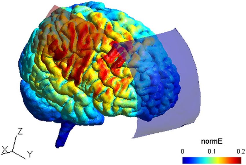
www.nature.com/scientificreports/
Group Age (years) Gender (f/m) LQ Regular video gaming Joystick experience
YA (n = 30) 27.7 ± 3.8 17/13 83.3 ± 13.98 11/30 9/30
OA (n = 30) 67.97 ± 5.32 15/15 82.7 ± 25.3 9/30 8/30
Table 1. Sample characteristics. Age groups did not differ regarding their laterality quotient (LQ),
independent-samples t-test: t(45.21) = 0.12, p = 0.91), the amount of regular video gamers (Chi-square test:
χ2(1) = 0.07, p = 0.79) and the amount of people with joystick experience (Chi-square test: χ2(1) = 0.08,
p = 0.77).
similar explicit skill levels as younger cohorts27,29,42. Furthermore, there is some evidence suggesting that OA
perform motor tasks with higher motor variability38,43,44. Age-related increase in variability may be caused by
age-related changes in motor unit morphology and properties38 but may furthermore also represent a decreased
ability to flexibly adapt to varying task constraints43. In this regard, adding kinematic measures in motor learning
investigations in OA may help unraveling more implicit mechanisms of motor control in the learning process.
In recent years, non-invasive brain stimulation techniques such as transcranial direct current stimulation
(tDCS) became popular with the potential to improve motor performance and learning in a variety of different
motor tasks (for review s ee45). Mechanistically, tDCS is capable of enhancing motor e xcitability46 at postsynaptic
cortical neurons47–49. Since long-lasting synaptic potentiation50 is considered a mechanism of plasticity, tDCS
combined with motor practice seems to be capable of systematically augmenting brain function. The primary
motor cortex (M1) is one of the key regions involved in the motor learning p rocess7 showing reduced activity
with extended p ractice51, which is why anodal tDCS over M1 has been predominantly used to facilitate early
skill acquisition52,53. The facilitatory effect of M1 tDCS was shown in sequential motor tasks52,54 and continuous
cursor navigation tasks, for example during continuous force t ransduction55, joint flexion and e xtention56,57 or
reaching movements58 but mainly for explicit learning parameters such as accuracy or movement time. Interest-
ingly, it seems that enhancing cortical excitability via M1 tDCS does not influence the motor act per se but only
those synaptic connections, previously selected by training, indicating specificity in learning with regards to the
sequence or the movement59. However, since there is some evidence suggesting that tDCS effects also transfer
to unlearned aspects of motor t asks60, one can assume that the amount of transfer depends on the underlying
function of the stimulated brain structure and therefore on the motor task that is performed. Furthermore, M1
plays a major role in consolidating a learned motor movement. In fact, skill acquisition over multiple days is
mainly enhanced via an effect of tDCS on consolidation9,54,61, indicated by major increases in offline gain scores.
Interestingly, over multiple tDCS sessions, offline gain enhancement accumulates, leading to observable effects on
long-term retention up to months after the experimental s ession9,54,61. In OA, tDCS over M1 has also been used
to improve motor learning rates62–64 and as one study indicates, tDCS may even produce greater improvement
than in YA65. Authors mainly attribute this effect to a larger room for improvement, which is also supported by
a positive association between age and skill improvement64. Since M1 stimulation is also capable of facilitating
motor memory consolidation in O A66, one may speculate that multiple tDCS sessions in OA have the potential
to produce similar effects to those seen in Y A 9.
In the present study, we aimed to investigate the effects of multiple tDCS-sessions on an arc pointing task
(APT) in YA and OA. APT can be considered a complex motor skill4 that requires highly precise pointing move-
ments which allow the analysis of specific kinematics on a single movement basis. Furthermore, since APT
learning is associated with increased activation in M167, it provides a suitable target for modification with tDCS.
Anodal tDCS was applied over M1 during APT training on 3 consecutive days to investigate tDCS-induced
improvements in both explicit and implicit measures of motor performance53. We hypothesized that (i) OA would
perform APT with lower accuracy and higher movement times and also show smaller learning r ates25,68. Fur-
thermore, we also expect them to show higher spatial variability compared to YA, indicating also lower implicit
learning. Since lower performance values at baseline provides greater room for improvement, we hypothesized
that (ii) OA would show greater gain than YA in both explicit and implicit measures of learning as a result of
tDCS. Furthermore, we hypothesized that (iii) multiple tDCS-sessions will mainly enhance offline gain9,54,61 in
a movement-specific way, suggesting that only learning in trained but not transfer movements are facilitated.
Results
Demographics. Age groups did not differ regarding demographic variables besides age (see Table 1 for
details). All participants tolerated the stimulation well and none reported any unexpected side effects from tDCS
stimulation. Across both age groups, a chi-squared test revealed no difference between members of the a-tDCS
and s-tDCS group in the ability to judge their group belonging (χ2(1) = 0.29, p = 0.59), indicating that the blind-
ing of conditions was effective. Additionally, differences between pre and post experimental ratings of attention,
fatigue and discomfort did not differ between age groups nor between stimulation groups.
Aging effects on APT learning. Movement time (3 × 20 × 2 Repeated Measures-Analysis of variance
(RM-ANOVA)) decrease in training trials (TT) over time was not different across age groups (interaction (IA)
trial*day*group, F (5.93, 160.1) = 1.38, p = 0.23, ηp2 = 0.05). Movement time also decreased within each training
day (main effect (ME) trial, F (4, 160.1) = 1.35, p = 0.26, ηp2 = 0.05) but interestingly not across training days (ME
day, F (1.64, 160.1 = 0.003, p = 0.99, ηp2 = 0). Generally, OA exhibited higher movement time values than YA (ME
group, F (1,27) = 38.7, p < 0.001, ηp2 = 0.59), see also Fig. 1. In transfer trials (TrT), movement time (3 × 2 × 2 RM-
Scientific Reports | (2021) 11:3421 | https://doi.org/10.1038/s41598-021-82275-4 2
Vol:.(1234567890)www.nature.com/scientificreports/
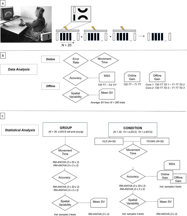
Figure 1. (a) Study design. Participants sat facing a computer screen with their left wrist on a joystick while
they received 20 min of a-tDCS or s-tDCS over the contralateral primary motor cortex. Their main task was
to move the cursor from the start to the target box through the arc channel without exceeding the channels
boundaries as fast and as accurately as possible. The study consisted of three consecutive training days with
tDCS (indicated by yellow flash) seperated by 24 h and a retention day without tDCS 1 week later. Each training
day consisted of 20 learning (black rectangle) and 2 transfer trials (white rectangle). Learning trials included
upward movements in clockwise direction, transfer trials downward movements in counter-clockwise direction.
(b) Data analysis. MSA: movement specific accuracy, TT: training trial, TrT: transfer trial, TD1: training day 1,
TD2: training day 2, TD3: training day 3, SV: Spatial Variability. Online calculations included movement time
and error rate calculation. Offline calculations included accuracy calculation (1-error rate) and calculation of SV
(variability of time-normalized radial position data). Furthermore, subparameters of accuracy were calculated:
MSA corresponds to the absolute difference between last TT (T20) and second TrT (Tr2). Online gain scores
were calculated as difference between T20 TT and T1 TT. Offline gain scores were calculated as differences
between last and first TT of consecutive TDs, values > 0 represent skill consolidation and values < 0 represent
skill loss. (c) Statistical analysis. RM-ANOVA: repeated-measures analysis of variances, ind-samples t-tests:
independent samples t-tests.
Scientific Reports | (2021) 11:3421 | https://doi.org/10.1038/s41598-021-82275-4 3
Vol.:(0123456789)www.nature.com/scientificreports/
15
Movement Time
10
5
0
1
Accuracy
0.8
old
0.6 young
Variability e+3
2
1
0
1
2
3
TD
TD
TD
Figure 2. Line Graph shows median values ± 95% Confidence Interval. Green line represents OA, orange line
represents YA.
ANOVA) did not decrease within training days (TDs) (ME trial, F (1, 54) = 1.26, p = 0.27, ηp2 = 0.05) but between
training days (ME day, F (1.48, 49.7) = 3.99, p = 0.04, ηp2 = 0.13). Furthermore, movement time was higher in
OA than in YA also in TrT (ME group, F (1,27) = 13.46, p = 0.001, ηp2 = 0.33) but the decrease over time was not
modulated by age (IA trial*day*group, F (2,54) = 0.25, p = 0.78, ηp2 = 0.01).
Accuracy (3 × 20 × 2 RM-ANOVA) increase in TT over time was not modulated by age (IA trial*day*group,
F (13.77, 358.18) = 0.65, p = 0.95, ηp2 = 0.02), however across both age groups accuracy significantly increased
within (ME trial, F (8.03, 358.18) = 7.97, p < 0.001, ηp2 = 0.24) and also across all training days (ME day, F (1.36,
358.18) = 13.28, p < 0.001, ηp2 = 0.34). Furthermore, accuracy in TT did not differ across age groups (ME group,
F (1,26) = 0.81, p = 0.38, ηp2 = 0.03), see also Fig. 1. Also in TrT, accuracy (3 × 2 × 2 RM-ANOVA) changes over
time were not modulated by age (IA trial*day*group, F (2,54) = 1.18, p = 0.32, ηp2 = 0.04). TrT accuracy increased
within (ME trial, F (1,49.65) = 6.2, p = 0.02, ηp2 = 0.19) and across training days (ME day, F (1.44, 49.65) = 4.27, p
= 0.03, ηp2 = 0.14) and did not differ between age groups (ME group, F (1, 27) = 0, p = 1, ηp2 = 0).
Spatial variability did not differ between age groups in none of the time-normalized data points (all p-val-
ues > 0.05/200, bonferoni threshold: 0.00025). Mean spatial variability (3 × 2 RM-ANOVA) decreased over time
(ME day, F (1.54, 43.25) = 3.65, p = 0.04, ηp2 = 0.12), but the variability decrease was not modulated by group (IA
day*group, F (1.54, 43.25) = 0.69, p = 0.47, ηp2 = 0.02). We found no effect of age group on mean spatial variability
(ME group, F (1,28) = 3.03, p = 0.09, ηp2 = 0.1), even though descriptively, OA showed higher higher spatial vari-
ability (525.43 ± 99) than YA (336.45 ± 198), see also Fig. 2.
TDCS effects on APT learning. OA. Movement time (3 × 20 × 2 RM-ANOVA) decrease in TT was not
affected by tDCS (IA trial*day*condition, (F (8.52, 213.08) = 1.13, p = 0.35, ηp2 = 0.04, BF10 = 0.15). Generally,
average movement times did not differ across stimulation conditions (ME condition, F (1,25) = 0.1, p = 0.76,
ηp2 = 0.004, BF10 = 0.48) indicating no effect of tDCS on movement times. Also in TrT (3 × 2 × 2 RM-ANOVA),
tDCS did not affect movement times (ME condition, F (1, 26) = 0.01, p = 0.91, ηp2 = 0 .001, BF10 = 0.43) and also
movement time decrease over time did not differ across stimulation conditions (IA trial*day*condition, (F (2,
52) = 0.04, p = 0.96, ηp2 = 0.002, BF10 = 0.16).
Accuracy in TT (3 × 20 × 2 RM-ANOVA) was not affected by tDCS (IA trial*day*condition, (F (38, 950) = 1.25,
p = 0.14, ηp2 = 0.05, BF10 = 0.008, ME condition, F (1,25) = 0.69, p = 0.42, ηp2 = 0.03, BF10 = 0.377), see also Fig. 3a.
Also TrT accuracy (3 × 2 × 2 RM-ANOVA) was not modulated by tDCS (IA trial*day*condition, F (1,26) = 0.06,
p = 0.81, ηp2 = 0.002, BF10 = 0.24, ME condition, F (1,26) = 0.06, p = 0.81, ηp2 = 0.002, BF10 = 0.39). Movement spe-
cific accuracy (MSA, see Fig. 2 for details on calculation) was not affected by tDCS (MWU, TD1: U = 138, p = 0.3,
BF10 = 0.56; TD2: U = 121, p = 0.74, BF10 = 0.35; TD3: U = 120, p = 0.52, BF10 = 0.41) and also online gain scores did
not differ between stimulation conditions (TD 1: MWU, U = 121.5, p = 0.47, B F10 = 0.42, TD 2: MWU, U = 121,
p = 0.72, BF10 = 0.37 TD 3: MWU, U = 121, p = 0.72, BF10 = 0.36). Furthermore, offline gain scores did not differ
neither between TD 1 and 2 (consolidation 1: MWU, U = 138.5, p = 0.28, B F10 = 0.53) nor between TD 2 and 3
(consolidation 2: MWU, U = 99, p = 0.58, B F10 = 0.42), see also Fig. 3a.
Spatial variability did not differ between stimulation conditions in none of the time-normalized data
points (all p-values > 0.05/200, bonferoni threshold: 0.00025), see Fig. 4a. Also regarding mean spatial vari-
ability (3 × 2 RM-ANOVA), we found no effect of the stimulation condition (ME condition, F (1, 28) = 1.09,
p = 0.31, ηp2 = 0.038, BF10 = 0.47) and also the amount of variability reduction did not differ across conditions
(IA day*condition, F (1.63, 45.78) = 1.27, p = 0.29, ηp2 = 0.04, BF10 = 0.41).
Scientific Reports | (2021) 11:3421 | https://doi.org/10.1038/s41598-021-82275-4 4
Vol:.(1234567890)www.nature.com/scientificreports/
a
Accuracy OA
1
0.9
0.8
a-tDCS
0.7
s-tDCS
0.6
1
2
3
TD
TD
TD
offline gain OA
online gain OA
0.6 0.4
MSA OA 0.4 0
0.2
0.2 -0.2
0 0
-0.4
-0.2 -0.2
s1
s2
1
2
3
1
2
3
on
on
TD
TD
TD
TD
TD
TD
C
C
b
Accuracy YA
1
0.9
0.8
a-tDCS
0.7
s-tDCS
0.6
1
2
3
TD
TD
TD
offline gain YA
online gain YA
0.6
0.2
0.4 0.4
MSA YA
0
0.2 0.2
0 -0.2
0
-0.2 -0.4
-0.2
s1
s2
1
2
3
on
on
1
2
3
TD
TD
TD
TD
TD
TD
C
C
Figure 3. (a) OA. (b) YA. Line Graph shows median values ± 95% Confidence Interval. Red line represents
a-tDCS group, blue line represents s-tDCS group. Separated values represent transfer trials, connected lines
represent training trials. Dotted lines indicate separation of training days. Boxplots show median with 25th and
75th percentiles. Outliers are depicted as dots. Red boxes represent the a-tDCS group, blue boxes represent the
s-tDCS group.
YA. Movement time (3 × 20 × 2 RM-ANOVA) decrease in TT was not affected by tDCS (IA trial*day*condition,
(F (38, 1064) = 1.35, p = 0.08, ηp2 = 0.05, BF10 = 0.004). Generally, average movement times did not differ across
stimulation conditions (ME condition, F (1, 28) = 4.05, p = 0.05, ηp2 = 0.13, B F10 = 1.55). Also in TrT (3 × 2 × 2 RM-
ANOVA), tDCS did not affect movement times (ME condition, F (1, 28) = 0.65, p = 0.43, ηp2 = 0.02, BF10 = 0.53)
and also movement time decrease over time did not differ across stimulation conditions (IA trial*day*condition,
(F (1.36, 38.18) = 0.32, p = 0.64, ηp2 = 0.01, B
F10 = 0.22).
TDCS did not affect accuracy (3 × 20 × 2 RM-ANOVA) learning in TT (IA trial*day*condition, F (38,
1064) = 1.006, p = 0.46, ηp2 = 0.04, BF10 = 0.004). However, accuracy was higher in the a-tDCS compared with
s-tDCS group in TT (ME condition, F (1, 28) = 15.65, p < 0.001, ηp2 = 0.36, BF10 = 39.45), already starting from
the first learning trial (t (25.57) = 2.68, p = 0.013, BF10 = 4.39), see also Fig. 3b. To evaluate potential baseline dif-
ferences between conditions in more detail, the RM-ANOVA was rerun using the accuracy value from the first
transfer trial (TrT 1) as a covariate. Here, we still found higher accuracy values in a-tDCS compared with s-tDCS
conditions (ME condition, F (1, 27) = 10.91, p = 0.003, ηp2 = 0.29, BF10 = 18.47) but still no difference in accuracy
increase over time (IA trial*day*condition, F (38, 1026) = 0.94, p = 0.58, ηp2 = 0.03, BF10 = 0.004). Accuracy dif-
ferences between conditions were still present at the retention session 1 week after the last training session (ME
condition, F (1,28) = 5.19, p = 0.03, ηp2 = 0.16, BF10 = 1.62). Interestingly, TrT accuracy (3 × 2 × 2 RM-ANOVA)
did not differ between conditions (ME condition, F (1, 28) = 0.9, p = 0.35, ηp2 = 0.03, BF10 = 0.44), also not over
time (IA trial*day*condition, F (2, 56) = 0.64, p = 0.53, ηp2 = 0.02, BF10 = 0.26). MSA was not affected by tDCS
(MWU, TD1: U = 85, p = 0.26, BF10 = 0.45; TD2: U = 131.5, p = 0.44, BF10 = 0.47; U = 155, p = 0.08, BF10 = 1.03)
and also regarding online gain, no tDCS effects were found (TD 1: MWU, U = 152, p = 0.1, B F10 = 0.82, TD 2:
MWU, U = 123, p = 0.66, BF10 = 0.35, TD 3: MWU, U = 116, p = 0.86, BF10 = 0.35). Furthermore, consolidation
was not affected by tDCS (consolidation 1: MWU, U = 70, p = 0.08, B F10 = 1.05, consolidation 2: MWU, U = 103,
p = 0.7, BF10 = 0.37).
Spatial variability of each time-normalized data point did not differ between stimulation groups (all p-val-
ues > 0.05/200, bonferoni threshold: 0.00025, see also Fig. 4b), indicating no group difference in spatial variabil-
ity. Mean spatial Variability (3 × 2 RM-ANOVA) was comparable across tDCS conditions (ME condition, F (1,
28) = 0.47, p = 0.5, ηp2 = 0.02, BF10 = 0.37) and we found no difference in variability reduction depending on the
stimulation condition (IA day*condition, F (1.12, 31.39) = 0.13, p = 0.75, ηp2 = 0.01, BF10 = 0.38), see also Fig. 4b.
Scientific Reports | (2021) 11:3421 | https://doi.org/10.1038/s41598-021-82275-4 5
Vol.:(0123456789)www.nature.com/scientificreports/
a
6000 6000
a-tDCS
Mean SV OA
Mean SV OA
4000 s-tDCS 4000
2000 2000
0 0
-2000 -2000
0 50 100 150 200 0 50 100 150 200
TD 1 Normalized Time TD 2 Normalized Time
6000 TD1 TD2 TD3
Mean SV OA
4000 1
diff p-values
2000
0.5
0
-2000 0
0 50 100 150 200 0 50 100 150 200
TD 3 Normalized Time Normalized Time
b 6000 6000
a-tDCS
Mean SV YA
Mean SV YA
4000 s-tDCS 4000
2000 2000
0 0
-2000 -2000
0 50 100 150 200 0 50 100 150 200
TD 1 Normalized Time TD 2 Normalized Time
6000 TD1 TD2 TD3
Mean SV YA
4000 1
diff p-values
2000
0.5
0
-2000 0
0 50 100 150 200 0 50 100 150 200
TD 3 Normalized Time Normalized Time
Figure 4. (a) OA. (b) YA. Plots 1–3 show spatial variability across all training days, lines represent mean
values ± standard deviation. Plot 4 shows p-values for each of the 200 t-tests across all three training days
investigating group differences in spatial variability of the cursors’ radial position (solid lines: blue: day 1,
orange: day 2, yellow: day 3) as well as the p-value threshold corrected for multiple comparisons (dashed line,
bonferoni-corrected p-value = 0.00025).
Discussion
The present study investigated the effect of multiple tDCS sessions over the M1 hand area on explicit and implicit
measures of motor skill learning comparing YA and OA. Our results revealed that both age groups were able to
learn the APT. While movement time reduced and accuracy increased over time, spatial variability decreased over
time, indicating both explicit and implicit APT learning. As hypothesized, OA showed higher movement times
than YA, while the amount of reduction over time did not differ, indicating similar learning rates. Contrary to
our hypothesis (i), accuracy and spatial variability did not differ between YA and OA. Furthermore, against our
hypothesis (ii), tDCS did not affect offline nor online APT learning in either group. However, in YA, accuracy was
higher in the a-tDCS group compared with s-tDCS. This enhancement in accuracy, however, was not restricted
to the learned movement since MSA was not affected by the stimulation and was also not accompanied by a
related reduction in spatial variability. We also did not find a cumulative effect of multiple stimulation sessions,
as hypothesized in (iii), which has previously been described in similar multi-session tDCS learning s tudies9,54.
Our results are in line with previous findings showing that APT-learning is associated with improvements
in accuracy4. However, participants showed much higher accuracy than those of the original study, and longer
movement times. We found mean movement times between three to six seconds, while in the previous study
participants trained at a medium movement time range of 620 ms4. Since Shmuelof et al.4 showed a generaliza-
tion of skill learning across all included movement time categories, we assumed that it is not necessary to restrict
Scientific Reports | (2021) 11:3421 | https://doi.org/10.1038/s41598-021-82275-4 6
Vol:.(1234567890)www.nature.com/scientificreports/
movement times to a certain range but instruct participants to perform as fast as possible. However, we suspect
participants were not aware of how fast they could perform the task and therefore performed at a convenient
speed level. Participants’ strategy may have been to prioritize error avoidance over speed, which is potentially
why high accuracy values were observed. This behavior may have altered the relative duration of learning stages7
but also the relative recruitment of brain regions69–71. Furthermore, in young participants, accuracy values were
close to 100 percent at the end of the training sessions. The missing effect of tDCS-induced APT skill learning
could be attributed to the fact, that participants performed at their peak performance (ceiling) level. However,
since we observed a main effect of stimulation and no initial ceiling was observed, we argue, that further improve-
ment was still possible.
Furthermore, our findings underline the importance of investigating motor skill acquisition in different
cohorts since learning strategies may differ tremendously between age groups. It is well known that aging is asso-
ciated with a progressive decline in motor functioning with evidence for reaction or response time slowing27,36,72,
diminished accuracy in movement execution30 and increased motor output variability38,43,44,73. Different age
groups have been shown to use different strategies to learn novel tasks especially during the fast initial phase of
learning74,75 and compensatory strategies in aged individuals such as slowing of movement time are especially
relevant for perceptual and higher-order cognitive p rocessing29,42,76. Our data supports these findings showing
that OA perform the APT with slower reaction times and potentially thereby increase accuracy up to a level of
YA29. Thus, one could speculate that high levels of accuracy in OA are achieved with a different behavioral strategy
than YA. In line with this argument are the different trajectory variability courses visible in YA and OA, even
though mean spatial variability was not affected by age. While OA show similar spatial variability throughout the
movement, YA show greater variability at the end of the movement. Shmuelof and colleagues interpret decreases
in trajectory variability around the average path as improvements in feedforward control, while trajectories with
large deviations at the end of the movement represent feedback improvements4. Following this idea, OA APT
performance may improve mostly from information during the motor task (knowledge of performance) for
example via immediate feedback of the cursor position. YA, instead, may rather profit from information after
task performance for example via feedback on movement time and error rates (knowledge of result). However,
this needs to be explicitly tested in future studies examining the effect of different feedback conditions on
APT learning in different age cohorts. Age-related alterations on motor behavior are related to functional and
structural alterations on a brain level including grey and white matter loss but also differential neural activation
patterns30,77–83. It cannot be ruled out that APT learning in OA is not primarily associated with M1 activation
but rather relies on higher-order brain regions such as (pre)-frontal cortices. This question needs to be addressed
by future studies, which should aim at identifying brain regions associated with APT learning in OA to unravel
potential targets for the application of non-invasive brain stimulation. Furthermore, aging also changes the pro-
pensity for plasticity modulation84,85, which may limit the potential of tDCS to induce a facilitatory learning effect
in OA. However, tDCS over M1 did not facilitate APT learning in YA either. Therefore, methodological issues
such as duration or time point of the stimulation but also insufficient current intensity could be held responsible
for the limited tDCS effect on learning86. Our current density (0.028 mA/cm2) was a little bit lower as the one
previously used9,54,61 which may have reduced tDCS e fficacy87. In contrast, stimulation duration was set to 20 min
which falls within the range, commonly u sed9,54. Furthermore, also subject factors such as biological variation
but also the current neuronal state of the target region influence the responsiveness to tDCS88, making direkt
monocausal inferences from missing tDCS effects on learning impossible. Similar multi-session tDCS learning
studies9,54 mainly found a cumulative M1 tDCS effect on offline learning gains, highlighting the importance
of M1 in early consolidation. In our study design, no tDCS effect on offline learning was found, contradicting
previous findings. Additional analyses using BF indicate inconclusive evidence to interpret this finding as a null
effect. One potential explanation for the divergent results between previous and our study may originate from
differences in the motor task since the exact role of M1 differs depending on the type of motor learning task
being learned45,54. APT learning is mainly associated with the reduction of variability of a motor action. In this
regard, acquisition of task-specific synergies, which mainly update during task performance, could have mainly
happened in M167, explaining at least the missing tDCS effect on offline learning performance. Furthermore,
a previous study interpreted the APT learning-related increase in M1 activation as a recruitment of additional
neurons67. However, tDCS effects are mainly based on mechanisms enhancing synaptic p lasticity50, thus may
not be suitable for enhancing APT learning-associated recruitment processes within M1. Furthermore, the
calculation of BFs in addition to conventional statistical analyses allow judgements for or against H0. However,
even though BFs provide important information89, one has to keep in mind their negative aspects such that BFs
are very sensitive to prior distributions, which can be too difficult to choose or depend on the belief that one
true model exists, which may not always be the c ase90. Therefore, our assumption that our data provides only
inconclusive evidence should be handled with caution. However, future studies in this field should nevertheless
aim to use bigger samples to gain clear evidence for or against tDCS effects on offline APT accuracy learning.
We did see a difference in accuracy between both tDCS groups in YA, potentially indicating an involvement
of M1 in APT performance at least in young age. BFs support this suggestion for a main stimulation effect on
accuracy. However, even though we used appropriate randomization to select group membership, we cannot
completely rule out that group differences already existed prior to tDCS since no baseline measurement was
included to maintain task naivety. In favour of an existing effect, we hypothesize that M1-stimulation induced
a performance-enhancing state which facilitated APT performance from the very beginning. This finding is
consistent with Shmuelof et al., showing M1-activation from the beginning of APT learning67 and highlights
the important role of M1 in initial skill acquisition in Y A7,91. Brain activity prior to learning is undoubtedly an
important predictor of subsequent motor p erformance92 and previous studies also found tDCS applied before
learning facilitated subsequent skill acquisition rates93–95. By contrast, no tDCS-induced effect was found on
spatial variability in either group. This finding contradicts our primary hypothesis that tDCS affects both explicit
Scientific Reports | (2021) 11:3421 | https://doi.org/10.1038/s41598-021-82275-4 7
Vol.:(0123456789)www.nature.com/scientificreports/
and implicit APT learning. We do see within-session and between-session reductions of spatial variability, indi-
cating APT learning. In a taxonomy proposed by Krakauer and Mazzoni, M1 is mainly involved in variability
reduction during skill learning10—which is in line with other research67. However, we found no evidence for
M1 tDCS-induced reductions in spatial variability. One reason for this mismatch may be the the low statistical
power induced by the relatively low number of participants but also the multiple testing. Looking at the p-value
figure, it is observable that group-differences in spatial variability are closest to significance at position points
95–110, which indicate the turning point from an upwards to a downwards movement in the arc channel during
the training trials. Thus, it could be speculated that lower spatial variability values at these critical turning points
may have resulted in higher accuracy values in the a-tDCS group. However, since our data does not support this
hypothesis, so far our results do not show that enhancing explicit parameters of task success such as accuracy
necessarily correlate with differences in implicit parameters of movement execution such as spatial variability.
In summary, we provide novel evidence that APT learning occurs both in YA and OA but also reveal age-
related differences in learning strategies related to higher movement times in OA. While no tDCS-induced
differences in APT learning were observable over time, we did see a group effect of M1 tDCS on accuracy in YA
from the very beginning, potentially indicating a tDCS-induced performance-enhancing brain state. However,
tDCS-induced differences in accuracy values did not translate into tDCS-induced differences in spatial vari-
ability, indicating no necessary interaction between explicit and implicit APT learning. Mechanisms of action
of tDCS-suppported motor training should be oberseved more carefully, while also considering age-related
differences in motor learning abilities.
Methods
Sample characteristics. In total, 60 healthy individuals aged between 21 and 78 years (32 females) were
enrolled in this double‐blinded, sham‐controlled study. 30 individuals were aged between 18 and 35 years
(YA, mean age: 27.07 ± 3.8 years, 17 females) and 30 individuals were older than 55 years (OA, mean age:
67.97 years ± 5.3 years, 15 females). Participants were right-handed, indicated by a score > 40 in the Edinburgh
Handedness Inventory96 and a similar number of people within each age group reported regular computer gam-
ing and joystick eperience (see also Table 1). Highly-skilled participants such as professional musicians and ath-
letes were excluded from participation. All participants gave written informed consent and the study procedures
were approved by the local ethics committee of the University of Leipzig and conducted in accordance with the
declaration of Helsinki. To exclude the presence of any neurological disease and/or contraindications relevant
for the study procedures outlined below, all participants underwent a detailed neurological examination prior to
the testing phase. Additionally, all participants were free of any medication affecting the central nervous system
and were task naïve.
Study design. The study comprised of four training days with the first three conducted on consecutive days
separated by a 24-h break. Training day 4 was conducted 1 week after training day 3 to investigate potential
effects on long-term retention. Before training day 1, participants were randomly allocated by a second experi-
mentor to either receive 20 min of daily anodal tDCS (a-tDCS) or sham tDCS (s-tDCS), where the stimulator
linding52. TDCS was put on five minutes prior to the start of the motor task.
was put on for 30 s only to ensure b
During the APT, participants performed 20 tt and two trt to investigate short-term transfer effects of each TD.
On TD 4, participants performed another 20 learning and 2 transfer trials but no tDCS was applied (see also
Fig. 2 for details).
APT. Motor task. The APT4 required participants to move a cursor on the computer screen by moving a
joystick with their left wrist. Similar to the original task, we also chose the left wrist in our right-handed study
cohort to maximize the dynamic learning range. In all four training sessions, participants sat facing a computer
screen where a semicircular channel was presented using the Presentation software (Neurobehavioral Systems,
Inc., version 14.7). They were instructed to guide a cursor through the channel from one end to the other without
exceeding the channels boundaries as fast and as accurately as possible (see also Fig. 4 for details, please also note
that informed consent was obtained to publish the participants’ image in an online open access publication).
Before the training started, participants familiarized themselves with the joystick by moving the cursor freely
within a white square field on the screen. At the beginning of each trial, the semicircular channel was presented
and participants were instructed to start the trial only when ready. As a result, the experiment was not consid-
ered to be a speeded reaction time task protocol. For starting the trial, participants had to move the cursor into
the start box positioned on the left side of the channel. Entering this start zone initiated time-keeping and was in-
dicated to the participant by a change in the start box colour to yellow. Time-keeping continued until the cursor
reached the stop box on the right side of the channel. During TT, participants performed an upwards movement
in clockwise direction followed by a downwards movement to enter the stop box. During TrT, a mirrored version
of the channel was presented, where a downwards movement in a counter-clockwise direction was followed by
an upwards movement to enter the stop box (see also Fig. 1). Maximum trial duration was set to 30 s. After this
time interval, the trial stopped and was marked as incomplete.
Information on performance. Cursor position was visible throughout the whole movement. Thereby, partici-
pants received feedback about their current position in the channel. Additionally, after each trial, the trajectory
of the cursor was projected on the screen. To maximize information gain, portions of the participant’s move-
ments inside the channel were colored in white and portions outside the channel in red. Furthermore, total time
(in seconds) and error (in percent of the fraction of movements outside the channel), were calculated online and
Scientific Reports | (2021) 11:3421 | https://doi.org/10.1038/s41598-021-82275-4 8
Vol:.(1234567890)www.nature.com/scientificreports/
Figure 5. TDCS current flow simulation. Anode is depicted as red rectangle and cathode as blue rectangle
projected on a standard head model. Normalized electrical field strength (V/m) is indicated through colormaps
with blue representing lowest and red representing highest field strengths, respectively. The current flow image
was created using the SIMNIBS software version 3.1.2100.
displayed on the screen after each trial. Thereby, participants received knowledge of performance in the form of
trajectory projections as well as knowledge of result by the information about time and error rates.
Transcranial direct current stimulation (tDCS). For tDCS, a weak direct current of 1 mA generated
from a battery driven stimulator (neuroConn, Ilmenau, Germany) was delivered for 20 min via saline-soaked
sponge electrodes. Stimulation was switched on five minutes prior to the start of training since a previous study
suggested that this time period is sufficient to induce enduring excitability increases within M197. Either a-tDCS
or s-tDCS was applied to the right M1 contralateral to the left wrist. Stimulation was performed double-blinded
to ensure unbiased results. Specifically, a second experimenter was responsible for controlling the stimulation
but was otherwise not in contact with participants or the primary experimenter. To evaluate, whether blinding of
conditions was effective, participants were asked to judge their group belonging after the third day of experimen-
tal testing by choosing between the options: “real stimulation” or “no stimulation”. The anatomical landmark for
the right M1 hand area was identified with neuronavigation (Brainsight Version 2; Rogue Research, Montreal,
QC, Canada) using the MNI coordinates (x, y, and z) 40, 20, and 5498,99. After localization with the neuronavi-
gation system, the skin was prepared using alcohol pads to ensure good contact of the stimulation electrodes
applied to the head. The impedance of stimulation electrodes was always kept below 10 kΩ for each participant.
The anode (7 × 5 cm) was positioned over the right M1, the cathode (10 × 10 cm) was placed over the frontal
orbit. Flexible elastic straps were used to additionally fixate the electrodes on the head. Current was ramped up
for 30 s in the beginning of tDCS eliciting a transient tingling sensation on the scalp that faded over seconds
and also ramped down for 30 s as described p reviously52. During s-tDCS, the current was increased, maintained
and decreased for 30 s each. Before and after tDCS, participants rated their level of attention (1 = not attentive,
10 = very attentive), fatigue (1 = very fatiqued, 10 = no fatigue) and discomfort (1 = no discomfort, 10 = strong
discomfort) on a visual analogue scale (VAS) to ensure potential differences between groups did not originate
from differences in these global parameters.
tDCS current flow simulation. We simulated electric field distributions based on a finite element model of a
representative head inside the open-source SimNIBS software100 to approximate current flow. The anode was
defined according to our anatomical landmark (40, 20, 54 as x, y and z) with a size of 7 × 5 cm, the cathodes
center position was defined at Fpz with a size of 10 × 10 cm. A current of 1 mA was selected. Maximum electrical
field strength (0.2 V/m) was determined below the anode, corresponding to the hand area of right M1 but also
in premotor areas between both electrodes (see Fig. 5).
Data analysis. Data was analyzed both online and offline. Online analysis for movement time and error
rates was performed by Presentation that controlled the experiment. Calculation of movement times was done by
counting the number of data points captured from start to stop signal multiplied with 13.333 ms (sampling rate:
75 Hz, 1 data point every 13.333 ms) divided by 1000. For error rate calculation, each captured data point was
screened for being inside or outside the channel. The number of data points outside the channel divided by the
total number of data points was then multiplied with 100 to compute percentage error rates. For offline analysis,
we used custom routines written in MATrix LABoratory R2018B (MATLAB, The MathWorks, Natick, MA). For
offline calculations, movement times and error rates were extracted from the logfiles created by Presentation.
Accuracy rates were calculated as 1-error rate which corresponds to the percentage of fraction of movements
in channel. For investigating potential stimulation effects on accuracy in more detail, we additionally calculated
MSA as well as online and offline gain scores. MSA was calculated for each training day as the difference of the
accuracy value of the last learning trial per training day and the second transfer trial accuracy (t20–tr 2), see also
Scientific Reports | (2021) 11:3421 | https://doi.org/10.1038/s41598-021-82275-4 9
Vol.:(0123456789)www.nature.com/scientificreports/
Fig. 2. Online gain scores were calculated for each training day representing within-day learning as the difference
between first and last learning trial accuracy (t20–t1), while offline gain scores were calculated between training
days analog to9 to investigate between-day consolidation effects.
Cursor position data was low-pass filtered (zero-lag, third-order Butterworth filter, cutoff frequency 14 Hz)
analog to Shmuelof et al.4 and filtering was applied at the trial level to remove any artifacts of returning the
joystick to the home position. Averaging was performed only for data values outside the start box, data points
within the start box were discarded from the analysis. Trial-by-trial variability was calculated to investigate the
effect of tDCS-combined practice on trajectory on the cursors time-normalized radial position data. Data was
resampled to 200 evenly spaced data points and then variance and average radial position was computed for each
subject and time point. To additionally investigate mean spatial variability, average radial position was averaged
across all 200 data points, see also Fig. 2.
Statistical analysis. Statistical analyses were performed using the Statistical Software Package for Social
Sciences (SPSS Version 27, IBM, Armonk, NY, USA). Difference scores of pre-post ratings for the VAS score
ratings were compared across all subgroups using the between-subject factors group (old, young) and condition
(a-tDCS, s-tDCS) in a repeated-measures analyses of variance (RM-ANOVA).
Aging effects on APT learning were calculated using only the data of the s-tDCS groups (N = 15 young and
N = 15 old). Effects of age on movement time and accuracy were calculated for tt using RM-ANOVAs with
between-subject factor group (young, old) and within-subject factors day (TD1–3) and trial (t1–t20). Likewise,
effects on transfer movement time accuracy were calculated using RM-ANOVAs, see also Fig. 2. To investigate
whether spatial variability of radial position differed between groups, we performed independent-samples t-tests
or the non-parametric equivalent for every normalized time point, resulting in n = 200 tests for each training
day. For investigating aging effects on mean spatial variability, we additionally performed a RM-ANOVA with
factor group (old, young) and factor day (TD 1–3).
TDCS effects were calculated for each age group, separately to be able to parcellate aging effects from stimula-
tion effects and decrease the number of factors in the analysis. TDCS effects on movement time and accuracy
were calculated for training trials using RM-ANOVAs with between-subject factor condition (a-tDCS, s-tDCS)
and within-subject factors day (TD 1–3) and trial (t1–t20) for each age cohort. Likewise, effects on trt move-
ment time and accuracy were calculated using RM-ANOVAs. Furthermore, MSA, online and offline gain scores
were compared across conditions using independent-samples t-tests or the non-parametric equivalent in case of
non-normal distribution. TDCS effects on spatial variability were also investigated using independent-samples
t-tests or the non-parametric equivalent for every normalized time point. Mean spatial variability was compared
across conditions using an RM-ANOVA with factor condition (a-tDCS, s-tDCS) and factor day (TD 1–3), see
also Fig. 2c (Supplementary Information).
Partial eta-squared (ηp2) for ANOVA’s are provided as measures of effect size and used to aid in the inter-
pretation of inferential statistics. To control for multiple comparisions, p-values were adjusted according to the
false-discovery-rate101. Conventional inferential statistics analyses are used to quantify our research hypotheses
(H1) against the null hypothesis (H0). However, since conventional significance testing does not allow to state
evidence for H 0102, non-significant outcomes provide no information whether they represent real null findings
or inconclusive evidence for example due to low sample sizes. To add this information, Bayes Factors (BF10) were
calculated and provided in addition to the inferential data, quantifying how well H1 predicts the empirical data
relative to H 0103,104. Following recent recommendations, we considered B F10 > 1 as evidence for H1 over H0 with
values > 3 suggesting noteworthy evidence, while BF10 < 1 indicated evidence for H0 over H1 with values < 0.33
suggesting noteworthy evidence105. Furthermore, BF10s between 0.33 and 3 are considered inconclusive evidence
for either hypothesis105. BF10 were calculated using the statistical software package JASP (Jeffrey’s Amazing
Statistics Program106).
Data availability
The data that support the findings of this study are available on request from the corresponding author, E.K.
The data are not publicly available due to data protection policies practiced at our institute, e.g. their containing
information that could compromise the privacy of research participants.
Received: 14 August 2020; Accepted: 14 January 2021
References
1. Nielsen, J. B. & Cohen, L. G. The Olympic brain. Does corticospinal plasticity play a role in acquisition of skills required for
high-performance sports?. J. Physiol. 586, 65–70 (2008).
2. Schlaug, G. Musicians and music making as a model for the study of brain plasticity. Prog. Brain Res. 217, 37–55 (2015).
3. Kitago, T. & Krakauer, J. W. Motor learning principles for neurorehabilitation. Handb. Clin. Neurol. 110, 93–103 (2013).
4. Shmuelof, L., Krakauer, J. W. & Mazzoni, P. How is a motor skill learned? Change and invariance at the levels of task success and
trajectory control. J. Neurophysiol. 108, 578–594 (2012).
5. Willingham, D. B. A neuropsychological theory of motor skill learning. Psychol. Rev. 105, 558–584 (1998).
6. Kantak, S. S., Mummidisetty, C. K. & Stinear, J. W. Primary motor and premotor cortex in implicit sequence learning–evidence
for competition between implicit and explicit human motor memory systems. Eur. J. Neurosci. 36, 2710–2715 (2012).
7. Dayan, E. & Cohen, L. G. Neuroplasticity subserving motor skill learning. Neuron 72, 443–454 (2011).
8. Wolpert, D. M., Diedrichsen, J. & Flanagan, J. R. Principles of sensorimotor learning. Nat. Rev. Neurosci. 12, 739–751 (2011).
9. Reis, J. et al. Noninvasive cortical stimulation enhances motor skill acquisition over multiple days through an effect on consoli-
dation. Proc. Natl. Acad. Sci. USA. 106, 1590–1595 (2009).
Scientific Reports | (2021) 11:3421 | https://doi.org/10.1038/s41598-021-82275-4 10
Vol:.(1234567890)www.nature.com/scientificreports/
10. Krakauer, J. W. & Mazzoni, P. Human sensorimotor learning: Adaptation, skill, and beyond. Curr. Opin. Neurobiol. 21, 636–644
(2011).
11. Branco, M. P., de Boer, L. M., Ramsey, N. F. & Vansteensel, M. J. Encoding of kinetic and kinematic movement parameters in
the sensorimotor cortex: A brain–computer interface perspective. Eur. J. Neurosci. 50, 2755–2772 (2019).
12. Schmidt, R. A., Zelaznik, H., Hawkins, B., Frank, J. S. & Quinn, J. T. Motor-output variability: AA theory for the accuracy of
rapid motor acts. Psychol. Rev. 47, 415–451 (1979).
13. Gordon, J., Ghilardi, M. F. & Ghez, C. Accuracy of planar reaching movements. I. Independence of direction and extent vari-
ability. Exp. Brain Res. 99, 97–111 (1994).
14. Faisal, A. A., Selen, L. P. J. & Wolpert, D. M. Noise in the nervous system. Nat. Rev. Neurosci. 9, 292–303 (2008).
15. van Beers, R. J. The sources of variability in saccadic eye movements. J. Neurosci. 27, 8757–8770 (2007).
16. Churchland, M. M., Afshar, A. & Shenoy, K. V. A central source of movement variability. Neuron 52, 1085–1096 (2006).
17. Shadmehr, R., Smith, M. A. & Krakauer, J. W. Error correction, sensory prediction, and adaptation in motor control. Annu. Rev.
Neurosci. 33, 89–108 (2010).
18. Sommer, M. A. & Wurtz, R. H. Brain circuits for the internal monitoring of movements. Annu. Rev. Neurosci. 31, 317–338 (2008).
19. van Beers, R. J., Haggard, P. & Wolpert, D. M. The role of execution noise in movement variability. J. Neurophysiol. 91, 1050–1063
(2004).
20. Ghilardi, M. F., Moisello, C., Silvestri, G., Ghez, C. & Krakauer, J. W. Learning of a sequential motor skill comprises explicit and
implicit components that consolidate differently. J. Neurophysiol. 101, 2218–2229 (2009).
21. Lawrence, G. P., Gottwald, V. M., Khan, M. A. & Kramer, R. S. S. The Movement kinematics and learning strategies associated
with adopting different foci of attention during both acquisition and anxious performance. Front. Psychol. 3, 468 (2012).
22. Smith, M. A., Ghazizadeh, A. & Shadmehr, R. Interacting adaptive processes with different timescales underlie short-term motor
learning. PLoS Biol. 4, e179 (2006).
23. Savion-Lemieux, T. & Penhune, V. B. The effects of practice and delay on motor skill learning and retention. Exp. Brain Res 161,
423–431 (2005).
24. Bapi, R. S., Doya, K. & Harner, A. M. Evidence for effector independent and dependent representations and their differential
time course of acquisition during motor sequence learning. Exp. Brain Res. 132, 149–162 (2000).
25. Voelcker-Rehage, C. Motor-skill learning in older adults—A review of studies on age-related differences. Eur. Rev. Aging Phys.
Act 5, 5–16 (2008).
26. Coats, R. O., Snapp-Childs, W., Wilson, A. D. & Bingham, G. P. Perceptuo-motor learning rate declines by half from 20s to
70/80s. Exp. Brain Res. 225, 75–84 (2013).
27. Hoff, M. et al. Switching between hands in a serial reaction time task: A comparison between young and old adults. Front. Aging
Neurosci. 7, 176 (2015).
28. Smith, C. D. et al. Memories that last in old age: Motor skill learning and memory preservation. Neurobiol. Aging 26, 883–890
(2005).
29. Shea, C. H., Park, J.-H. & Wilde Braden, H. Age-related effects in sequential motor learning. Phys. Ther. 86, 478–488 (2006).
30. Stewart, J. C., Tran, X. & Cramer, S. C. Age-related variability in performance of a motor action selection task is related to dif-
ferences in brain function and structure among older adults. NeuroImage 86, 326–334 (2014).
31. Solesio-Jofre, E. et al. Aging effects on the resting state motor network and interlimb coordination. Hum. Brain Mapp. 35,
3945–3961 (2014).
32. Li, K. Z. H. & Lindenberger, U. Relations between aging sensory/sensorimotor and cognitive functions. Neurosci. Biobehav. Rev.
26, 777–783 (2002).
33. Guo, X. et al. Characterizing structural association alterations within brain networks in normal aging using Gaussian Bayesian
networks. Front. Comput. Neurosci. 8, 122 (2014).
34. Heise, K.-F. et al. The aging motor system as a model for plastic changes of GABA-mediated intracortical inhibition and their
behavioral relevance. J. Neurosci. 33, 9039–9049 (2013).
35. Krampe, R. T. Aging, expertise and fine motor movement. Neurosci. Biobehav. Rev. 26, 769–776 (2002).
36. Cuypers, K. et al. Age-related differences in corticospinal excitability during a choice reaction time task. Age 35, 1705–1719
(2013).
37. Schmoldt, A., Benthe, H. F. & Haberland, G. Digitoxin metabolism by rat liver microsomes. Biochem. Pharmacol. 24, 1639–1641
(1975).
38. Hunter, S. K., Pereira, H. M. & Keenan, K. G. The aging neuromuscular system and motor performance. J. Appl. Physiol. 121,
982–995 (2016).
39. Heuninckx, S., Wenderoth, N. & Swinnen, S. P. Systems neuroplasticity in the aging brain: Recruiting additional neural resources
for successful motor performance in elderly persons. J. Neurosci. 28, 91–99 (2008).
40. Seidler, R. et al. Associations between age, motor function, and resting state sensorimotor network connectivity in healthy older
adults. NeuroImage 108, 47–59 (2015).
41. Zimerman, M., Heise, K.-F., Gerloff, C., Cohen, L. G. & Hummel, F. C. Disrupting the ipsilateral motor cortex interferes with
training of a complex motor task in older adults. Cereb. Cortex 24, 1030–1036 (2014).
42. Voelcker-Rehage, C. & Willimczik, K. Motor plasticity in a juggling task in older adults-a developmental study. Age Ageing 35,
422–427 (2006).
43. Sosnoff, J. J. & Newell, K. M. Aging and motor variability: A test of the neural noise hypothesis. Exp. Aging Res. 37, 377–397
(2011).
44. Lodha, N., Moon, H., Kim, C., Onushko, T. & Christou, E. A. Motor output variability impairs driving ability in older adults. J.
Gerontol. Ser. A Biol. Sci. Med. Sci. 71, 1676–1681 (2016).
45. Hashemirad, F., Zoghi, M., Fitzgerald, P. B. & Jaberzadeh, S. The effect of anodal transcranial direct current stimulation on motor
sequence learning in healthy individuals: A systematic review and meta-analysis. Brain Cogn. 102, 1–12 (2016).
46. Kidgell, D. J. et al. Different current intensities of anodal transcranial direct current stimulation do not differentially modulate
motor cortex plasticity. Neural Plast. 2013, 603502 (2013).
47. Brunoni, A. R. et al. Clinical research with transcranial direct current stimulation (tDCS): Challenges and future directions.
Brain Stimul. 5, 175–195 (2012).
48. Nitsche, M. A. et al. Transcranial direct current stimulation: State of the art 2008. Brain Stimul. 1, 206–223 (2008).
49. Romero Lauro, L. J. et al. TDCS increases cortical excitability: Direct evidence from TMS-EEG. Cortex 58, 99–111 (2014).
50. Fritsch, B. et al. Direct current stimulation promotes BDNF-dependent synaptic plasticity: Potential implications for motor
learning. Neuron 66, 198–204 (2010).
51. Picard, N., Matsuzaka, Y. & Strick, P. L. Extended practice of a motor skill is associated with reduced metabolic activity in M1.
Nat. Neurosci. 16, 1340–1347 (2013).
52. Nitsche, M. A. et al. Facilitation of implicit motor learning by weak transcranial direct current stimulation of the primary motor
cortex in the human. J. Cogn. Neurosci. 15, 619–626 (2003).
53. Orban de Xivry, J.-J. & Shadmehr, R. Electrifying the motor engram: Effects of tDCS on motor learning and control. Exp. Brain
Res. 232, 3379–3395 (2014).
Scientific Reports | (2021) 11:3421 | https://doi.org/10.1038/s41598-021-82275-4 11
Vol.:(0123456789)You can also read


