TDZD-8 Suppresses Rheumatoid Arthritis Induced by Collagen Type II in Rats: Potential Role of Apoptosis and Inflammation
←
→
Page content transcription
If your browser does not render page correctly, please read the page content below
Int. J. Morphol.,
39(1):311-317, 2021.

    TDZD-8 Suppresses Rheumatoid Arthritis Induced by Collagen
    Type II in Rats: Potential Role of Apoptosis and Inflammation

                         TDZD-8 Suprime la Artritis Reumatoide Inducida por Colágeno Tipo
                           II en Ratas: Papel Potencial de la Apoptosis y la Inflamación

                                                                  Amal F. Dawood1,2

DAWOOD, A. F. TDZD-8 suppresses rheumatoid arthritis induced by collagen type ii in rats: Potential role of apoptosis and
inflammation. Int. J. Morphol., 39(1):311-317, 2021.

         SUMMARY: Rheumatoid arthritis (RA) is considered an autoimmune disease distinguished by chronic synovial membrane
inflammation, degraded cartilage, as well as bone destruction, which lead to joints pain and stiffness. The pathogenesis of RA
involved at least two mechanisms: Cellular proliferation and activation of glycogen synthase kinase-3β (GSK3β) enzyme. Thus, we
tested the hypothesis that the GSK3binhibitor, TDZD-8, can treat the synovial tissue toward collagen type II (COII)-mediated RA
linked to apoptosis induction and biomarker suppression of inflammation. Wistar rats were immunized with COII (the model group)
for 21 days. Matched immunized rats were daily injected with TDZD-8 (1 mg/kg; i.p) for three additional weeks (COII+TDZD-
8).After 42 days of post-immunization, blood and tissues were collected. Histology (H&E) and immunohistochemistry (CD45;
leukocyte common antigen) images showed that COII induced RA was demonstrated by profound damage to the synovial tissue and
infiltration of the inflammatory cells, which were substantially ameliorated with TDZD-8. In addition, COII immunization caused
the induction of rheumatoid factor (RF), tumor necrosis factor-alpha (TNF-α), interleukin-6 (IL-6), and interleukin 1 beta (IL-1β)
that were substantially (p
DAWOOD, A. F. TDZD-8 suppresses rheumatoid arthritis induced by collagen type ii in rats: Potential role of apoptosis and inflammation. Int. J. Morphol., 39(1):311-317, 2021.
The two significant pathways leading to RA in humans, dose (1 mg/kg) (Zhou et al.) between day 21 and day 42.1 %
as well as animal models, are inflammation and proliferation diluted DMSO was used to dissolve TDZD-8.
that are found in synovial fibroblasts, macrophages, and
lymphocytes (Pope 2002; Dhaouadi et al., 2007). The link Blood collection and isolation of the synovium. Rats were
between GSK-3β, inflammation, and proliferation in the culled by cervical dislocation after being anesthetised with 50
progressive destruction of joints due to RA (Yoshino & Ishioka, mg/kg sodium pentobarbital. Blood was collected, and sera
2015; Zhou et al., 2016) had prompted us to examine the were isolated and kept at -20 ºC for biochemical investigation.
potential therapeutic effects of the GSK-3β inhibitor TDZD- Using a dissecting microscope, the removal of boths of tissue
8 against COII-induced RA in rats correlated with and ligaments on the patella of the exposed knee joints was
inflammation inhibition and anti-apoptosis. realized, whereas the synovium was eliminated, snap-frozen
(liquid nitrogen), and kept at -80 ºC until being utilized.
MATERIAL AND METHOD Histological analysis. Harvested specimens obtained from
synovium tissues were fixed for 24 h in formal saline (10 %),
and paraffin blocks were prepared after dehydration of tissues
Animals. Wistar male rats (160 ± 10g, 8 weeks of age) were in ascending alcohol grade. In order to analyze tissue histology,
obtained from the animal house at King Saud University tissue sections (5 µm thick) were stained with hematoxylin as
(Riyadh, Saudi Arabia) and were used for all histology, well as eosin (H&E).
immunohistochemistry, western blot analysis, and blood
chemistry experiments. The rats obtained accessible food as CD45Immunohistochemistry. Deparaffinized synovium
well as water and were accommodated at a fixed temperature tissue sections (5 µm thick) were dehydrated, and antigen
of 22 ºC for 12 h light/dark cycle. The utilized experimental retrieval was conducted in citrate buffer (10 Mm, pH 6). To
methods within the study were consistent with laboratory ani- assess infiltrated leukocytes, sections were incubated with
mal care and use instructions, released by the US National CD45 antibody (Abcam, cat # ab10558) at room temperature
Institutes of Health (NIH publication No. 85-23, revised 1996). (RT) for 1 h, washed and incubated with the secondary
Besides, it received authorization from the College of Medi- antibody for 1⁄2 h at RT. Having counterstained the sections
cine Ethical Committee at Princess Nourah University, Riyadh, with the assistance of Meyer hematoxylin, the nucleus can be
Saudi Arabia. visualized.
Induction of rheumatoid arthritis (RA) by collagen type Determination of blood levels of rheumatoid factor
II (COII). RA was induced in rats by active immunization, as (RF). RF was assessed using ELISA kit provided by CUSABIO
previously reported (Brand et al., 2007). Briefly, lyophilized Technology LLC, TX, USA, and was utilized based on the
bovine collagen type II (Sigma-Aldrich, MO, USA) emulsified recommendations of the manufacturer.
in an equivalent volume of complete Freund’s adjuvant
(Sigma-Aldrich, MO, USA) were injected intra-dermally (i.d) Western Blotting Analysis of Bax, TNF-alpha α, IL-1β β,
at day 0 in 100 µL volume at 200 µg COII final concentration. and IL-6. As previously reported (Al-Ani et al., 2010),
A booster dose (200 µg in 100 µL) prepared in incomplete extracted protein (40 µg per sample) from synovial tissues
Freund’s adjuvant (Sigma-Aldrich, MO, USA) was were western blotted using anti-Bax (Cell Signalling
administered, i.d.on day 14. RA was confirmed on day 21. Technology, USA), anti-TNF-α (Santa Cruz Biotechnology),
anti-IL-1β(Santa Cruz Biotechnology), and anti-IL-6 (San-
Experimental design. Within four groups, (n=6; each), 24 ta Cruz Biotechnology). ECL detection kit (Thermo Fisher
rats were assigned as follows: (1) Control group: from day 0 Scientific Inc, Rockford, lL, USA) was employed for
to day 14, the rats received normal saline (100 µl) via i.d. visualizing the protein bands. To measure band intensities,
routeand between day 21 and day 42, they were exposed to a image analysis software (C-Di Git blot scanner; LI-COR,
100 µl daily dose of 0.1 % DMSO (i.p) ; (2) TDZD-8 control NE, USA) was used.
group: the rats were subjected to the above procedure and
between day 21 and day 42, they were provided witha100 Statistical analysis.The statistical software package of
µl daily TDZD-8i.pdose (1 mg/kg) (Abcam, Cambridge, GraphPad Prism (version 6) was used to perform the statistical
UK); (3) the model group (RA): actively immunized rats analysis.The assessment of the discrepancies among the
with COII (as mentioned above) had received, between day studied four groups was realized when one-way ANOVA was
21 and day 42, a 100 µl volume of DMSO (0.1 %) to be performed, accompanied by Tukey’s test. In addition, data were
used as a vehicle; and (4) the treated group (COII+TDZD- described as mean ± SD, and the findings were found to be
8): the rats with RA had received a 100 µl daily TDZD-8 i.p substantial when P≤0. 05.
312DAWOOD, A. F. TDZD-8 suppresses rheumatoid arthritis induced by collagen type ii in rats: Potential role of apoptosis and inflammation. Int. J. Morphol., 39(1):311-317, 2021.
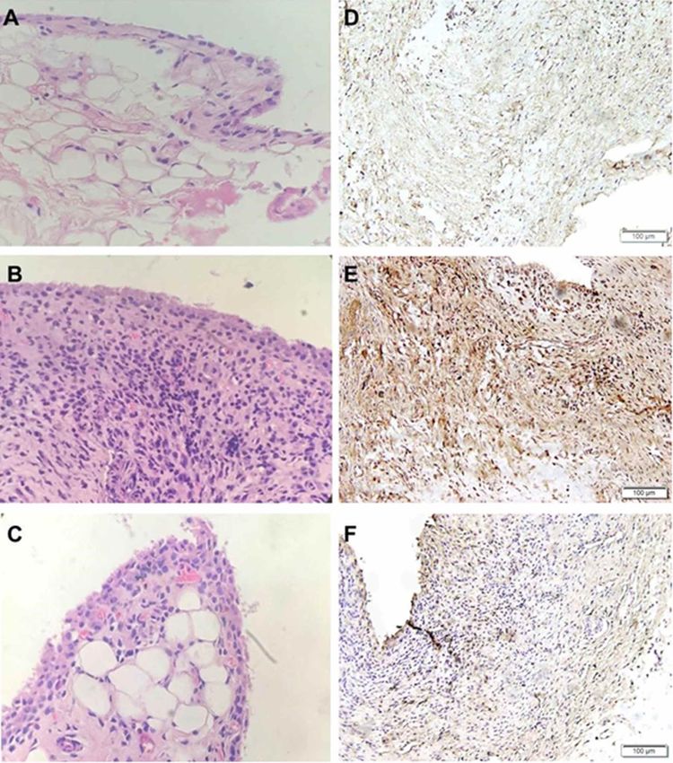
RESULTS demonstrated by severing synoviocyte hyperplasia and
infiltration of the inflammatory cells into the subsynovium
(Fig. 1B). TDZD-8 treatment (Fig. 1C) substantially
TDZD-8 partially inhibits COII-induced synovium preserved synovial tissue architecture. However, infiltrations
proliferation and tissue infiltration of inflammatory cells. of a few inflammatory cells were still be seen. Furthermore,
After 21 days on TDZD-8 following the completion of the immunohistochemical staining for CD45 in synovial tissue
immunization regimen, harvested synovial tissues were sections of the model group (COII) showed strong positive
stained with H&E and CD45 immunostaining and analysed CD45 stained cells (Fig. 1E) compared to negative to
by light microscopy. In comparison to unchanged tissue weakCD45 stained cells in the control group (Fig. 1D) that
architecture in the control groups (Fig. 1A), COII were considerably (pDAWOOD, A. F. TDZD-8 suppresses rheumatoid arthritis induced by collagen type ii in rats: Potential role of apoptosis and inflammation. Int. J. Morphol., 39(1):311-317, 2021.
TDZD-8 inhibits inflammation induced by COII increase in synovial TNF-α (Fig. 2A) and IL-6 (Fig. 2B) in
immunization. To investigate if the detected synovial tissue the model group (COII) compared with the control groups
damage caused by COII in our animal model of the disease (Control and TDZD-8) of rats. Treatment of COII rats with
was also linked with the induction of inflammatory TDZD-8 (COII+TDZD-8) for 21 days post-RA induction
biomarkers that are included in the RA pathogenesis significantly (pDAWOOD, A. F. TDZD-8 suppresses rheumatoid arthritis induced by collagen type ii in rats: Potential role of apoptosis and inflammation. Int. J. Morphol., 39(1):311-317, 2021.
To determine whether the treatment protocol by Correlation between rheumatoid factor and biomarkers
TDZD-8 used here can inhibit RF, which is a well-known of inflammation and apoptosis.The correlation between the
RA marker (Gavrila et al., 2016; Lu et al., 2018), we scoring of RF and biomarkers of inflammation and apoptosis
assessed, in the four groups of rats, the RF blood levels. As were evaluated to further promote TDZD-8 as a suitablec
shown in Figure 3B, immunization of rats with COII ompound in RA, and also to endorse the link between RF and
substantially (pDAWOOD, A. F. TDZD-8 suppresses rheumatoid arthritis induced by collagen type ii in rats: Potential role of apoptosis and inflammation. Int. J. Morphol., 39(1):311-317, 2021.
infiltration (CD45, Fig. 1E); (ii) inflammation, TNF-α (Fig. feración celular y la activación de la enzima glucógeno sintasa
2A) and IL-6 (Fig. 2B); and (iii) RF (Fig. 3B), which were quinasa-3b (GSK3β) Por lo tanto, probamos la hipótesis de que el
substantially treated by TDZD-8 (Figs. 1-4). Also, we inhibidor de GSK3β, TDZD-8, puede tratar el tejido sinovial hacia
demonstrate that COII inhibited the synovial protein el colágeno tipo II (COII) - AR mediada por inducción de apoptosis
y supresión de biomarcadores de inflamación. Se inmunizaron ra-
expression of the apoptotic biomarker, Bax, which was
tas Wistar con COII (el grupo modelo) durante 21 días. Se inyec-
significantly augmented by TDZD-8 to control levels (Fig. taron diariamente ratas emparejadas inmunizadas con TDZD-8 (1
3A). Therefore, ourdata are in agreement with our employed mg / kg; i.p) durante tres semanas adicionales (COII + TDZD-8).
hypothesis, in which TDZD-8 can substantially treat RA Después de 42 días de post-inmunización, se recolectó sangre y
induced by COII in rats. tejidos. Las imágenes de histología (H&E) e inmunohistoquímica
(CD45; antígeno común de leucocitos) mostraron que la AR indu-
The findings of this study confirm those previously cida por COII presentaba un daño profundo en el tejido sinovial e
published in literature and show that GSK3β inhibition infiltración de las células inflamatorias, las que mejoraron con
ameliorates several pathological manifestations connected TDZD-8. Además, la inmunización con COII provocó la induc-
ción de factor reumatoide (FR), factor de necrosis tumoral alfa
with COII-induced RA in human-derived cells and animal
(TNF-α), interleucina-6 (IL-6) e interleucina 1 beta (IL-1β) que
models (Pope; Cuzzocrea et al., 2006; Bao et al., 2007; Kwon fueron suprimidos por TDZD-8 de manera significativa (p < 0.05).
et al., 2014). For example, (i) levels of pro-inflammatory Considerando que TDZD-8 aumentó el biomarcador apoptótico,
mediators were reduced in fibroblast-like synoviocytes in la proteína X asociada a Bcl-2 (Bax), que fue mejorado (pDAWOOD, A. F. TDZD-8 suppresses rheumatoid arthritis induced by collagen type ii in rats: Potential role of apoptosis and inflammation. Int. J. Morphol., 39(1):311-317, 2021.
Gavrila˘, B. I.; Ciofu, C. & Stoica, V. Biomarkers in rheumatoid arthritis, Corresponding author:
what is new? J. Med. Life, 9(2):144-8, 2016. Amal F. Dawood
Guo, Q.; Wang, Y.; Xu, D.; Nossent, J.; Pavlos, N. J. & Xu, J. Rheumatoid Department of Basic Medical Sciences
arthritis: pathological mechanisms and modern pharmacologic
College of Medicine
therapies. Bone Res., 6:15, 2018.
Hu, X.; Paik, P. K.; Chen, J.; Yarilina, A.; Kockeritz, L.; Lu, T. T.; Woodgett,
Princess Nourah Bint AbdulrahmanUniversity
J. R. & Ivashkiv, L. B. IFN-gamma suppresses IL-10 production and Riyadh
synergizes with TLR2 by regulating GSK3 and CREB/AP-1 proteins. SAUDI ARABIA
Immunity, 24(5):563-74, 2006.
Kwon, Y. J.; Yoon, C. H.; Lee, S. W.; Park, Y. B.; Lee, S. K. & Park, M. C.
Inhibition of glycogen synthase kinase-3b suppresses inflammatory Email: amal_tawadros@yahoo.com
responses in rheumatoid arthritis fibroblast-like synoviocytes and
collagen-induced arthritis. Joint Bone Spine, 81(3):240-6, 2014.
Lu, T.; Zong, M.; Fan, S.; Lu, Y.; Yu, S. & Fan, L. Thioredoxin 1 is associated
with the proliferation and apoptosis of rheumatoid arthritis fibroblast-
Received: 17-08-2020
like synoviocytes. Clin. Rheumatol., 37(1):117-25, 2018. Accepted: 22-10-2020
Malemud, C. J. Intracellular signaling pathways in rheumatoid arthritis. J.
Clin. Cell. Immunol., 4:160, 2013.
Miao, C. G.; Yang, Y. Y.; He, X.; Li, X. F.; Huang, C.; Huang, Y.; Zhang,
L.; Lv, X. W.; Jin, Y. & Li, J. Wnt signaling pathway in rheumatoid
arthritis, with special emphasis on the different roles in synovial
inflammation and bone remodeling. Cell. Signal., 25(10):2069-78, 2013.
Myouzen, K.; Kochi, Y.; Okada, Y.; Terao, C.; Suzuki, A.; Ikari, K.; Tsunoda,
T.; Takahashi, A.; Kubo, M.; Taniguchi, A.; et al. Functional variants
in NFKBIE and RTKN2 involved in activation of the NF-kB pathway
are associated with rheumatoid arthritis in Japanese. PLoS Genet.,
8(9):e1002949, 2012.
Pope, R. M. Apoptosis as a therapeutic tool in rheumatoid arthritis. Nat.
Rev. Immunol., 2(7):527-35, 2002.
Scott, D. L.; Wolfe, F. & Huizinga, T. W. J. Rheumatoid arthritis. Lancet,
376(9746):1094-108, 2010.
Shi, M.; Cui, F.; Liu, A. J.; Li, J.; Ma, H. J.; Cheng, M.; Yang, J. & Zhang,
Y. Protection of chronic intermittent hypobaric hypoxia against
collagen-induced arthritis in rat through increasing apoptosis. Sheng
Li Xue Bao, 63(2):115-23, 2011.
Smolen, J. S.; Aletaha, D.; Barton, A.; Burmester, G. R.; Emery, P.; Firestein,
G. S.; Kavanaugh, A.; McInnes, I. B.; Solomon, D. H.; Strand, V.; et
al. Rheumatoid arthritis. Nat. Rev. Dis. Primers, 4:18001, 2018.
Steinbrecher, K. A.; Wilson 3rd, W.; Cogswell, P. C. & Baldwin, A. S.
Glycogen synthase kinase 3beta functions to specify gene-specific, NF-
kappaB-dependent transcription. Mol. Cell. Biol., 25(19):8444-55, 2005.
Yoshino, Y. & Ishioka, C. Inhibition of glycogen synthase kinase-3 beta
induces apoptosis and mitotic catastrophe by disrupting centrosome
regulation in cancer cells. Sci. Rep., 5:13249, 2015.
Zhou, H.; Liu, J.; Zeng, J.; Hu, B.; Fang, X. & Li, L. Inhibition of GSK-3b
alleviates collagen II-induced rheumatoid arthritis in rats. Med. Sci.
Monit., 22:1047-52, 2016.
317You can also read