The effects of melatonin on the healing of burn wounds in pinealectomized rats
←
→
Page content transcription
If your browser does not render page correctly, please read the page content below
EXPERIMENTAL STUDY

 The effects of melatonin on the healing of burn
 wounds in pinealectomized rats
       E. Çiğdem Karadağ Sarı, M.D.,1                   Nedim Savacı, M.D.2

 1
     Department of Plastic, Reconstructive and Aesthetic Surgery, Acıbadem University Faculty of Medicine, İstanbul-Turkey
 2
     Department of Plastic, Reconstructive and Aesthetic Surgery, Necmettin Erbakan University Meram Faculty of Medicine, Konya-Turkey

 ABSTRACT
 BACKGROUND: The present study aims to investigate the favorable effects of melatonin on burn wound healing in rats.
 METHODS: In this study, forty Wistar-albino-type male rats were divided into four groups. Group 1 was the control group, Group
 2 rats were treated using exogenous melatonin, Group 3 rats were pinealectomized, and Group 4 rats were pinealectomized then
 treated with exogenous melatonin. In all groups, a deep second-degree burn was created on the backs of the rats with a metal plate
 heated in boiling water. We monitored the progress of burn healing for seven days. At the end of them, we evaluated hydroxyproline
 levels, type III collagen, edema, inflammatory infiltration, congestion, vascular proliferation, fibrosis, the thickness of the zone of stasis
 and the epithelium to assess the progress of healing.
 RESULTS: The zone of stasis was less thick in Group 2 than the other groups (p=0.009). Type III collagen dyeing (p=0.031), fibrosis
 (p=0.011) and edema (p=0.031) were higher in Group 2 than the other groups. Congestion was higher in the control group than
 Group 4 (p=0.031). Other evaluated parameters showed no significant differences among the groups.
 CONCLUSION: In this study, it was noted that once total melatonin levels exceeded a certain threshold, a preventive effect was
 exerted on burn wound damage progression by reducing the zone of stasis. Melatonin may also prevent the development of hypertro-
 phic scarring. Melatonin may be a potential therapeutic option that can supplement traditional treatment in burn wounds; however,
 further studies with higher doses of exogenous melatonin administered over longer periods are needed to further evaluate the effects
 noted in this study.
 Keywords: Burn; endogenous melatonin; exogenous melatonin; pinealectomy; wound.

 INTRODUCTION                                                                       of progressive tissue injury in this zone is the systematic ac-
                                                                                    tivation of neutrophils due to the production of free oxygen
 In burn wound healing, the progression of the tissue inju-                         radicals[2] and studies report that antioxidants can prevent
 ry determines the mortality, morbidity and treatment out-                          this progressive effect.[3]
 comes. Jackson described burn wounds as having three dis-
 tinct zones based on the severity of destruction and blood                         Melatonin, an antioxidant hormone produced in the pineal
 flow alterations.[1] The zone of coagulation is the central zone                   gland, increases the activity of enzymes like glutathione perox-
 characterized by necrosis. The zone of coagulation is encir-                       idase, superoxide dismutase, and nitric oxide synthase. Mela-
 cled by the zone of stasis, which is indirectly affected by the                    tonin also directly affects free oxygen radicals[5] by scavenging
 trauma, and the zone of stasis is surrounded by a zone of                          free radicals more effectively than other antioxidants.[6]
 hyperemia characterized by increased blood flow. Salvaging
 the zone of stasis can prevent an increase in the depth and                        Given the role of free oxygen radicals in burn wound pro-
 the width of the burn injury area, thereby significantly reduce                    gression, exogenous melatonin may be an alternative to tra-
 the risks of mortality and morbidity.[2–4] The underlying cause                    ditional burn injury treatments. To our knowledge, there are

     Cite this article as: Karadağ Sarı EÇ, Savacı N. The effects of melatonin on the healing of burn wounds in pinealectomized rats. Ulus Travma Acil
     Cerrahi Derg 2021;27:395-401.
     Address for correspondence: E. Çiğdem Karadağ Sarı, M.D.
     Acıbadem Üniversitesi Tıp Fakültesi, Plastik, Rekonstrüktif ve Estetik Cerrahi Anabilim Dalı, İstanbul, Turkey
     Tel: +90 216 - 649 45 31 E-mail: doktorcigdem@yahoo.com
      Ulus Travma Acil Cerrahi Derg 2021;27(4):395-401 DOI: 10.14744/tjtes.2020.12247 Submitted: 23.08.2019 Accepted: 28.05.2020
      Copyright 2021 Turkish Association of Trauma and Emergency Surgery

 Ulus Travma Acil Cerrahi Derg, July 2021, Vol. 27, No. 4                                                                                                395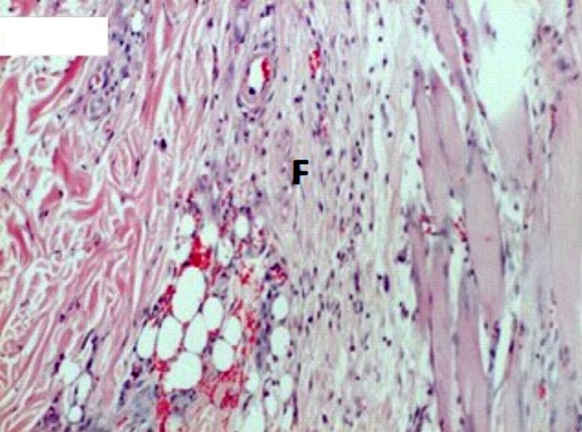
Karadağ Sarı et al. The effects of melatonin on the healing of burn wounds in pinealectomized rats
no published studies that examine the effects of endogenous backs of each rat for 15 seconds (without applying pressure)
melatonin and exogenous melatonin on burn healing. There- to produce deep second-degree burns. The reason for using
fore, in this study, we investigated the effects of the basal a circular metal plate was to assess the wound contraction
release of melatonin, i.e., endogenous melatonin levels, in our easily during healing. Burn wounds were left open for the
pinealectomy group, and whether explored exogenous mel- duration of this study, and no topical or other preparations
atonin has favorable effects on burn wound healing in the were applied.
groups administered exogenous melatonin.
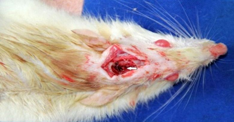
Pinealectomy
MATERIALS AND METHODS Pinealectomy was performed on rats in Groups 3 and 4 im-
mediately before burn-wound creation. Anesthetized rats
With the prediction of the difference in the medium effect were positioned on their abdomens and stabilized. Their
size accepted for animal experiments in groups as statisti- heads were cleaned gently with polyvinylpyrrolidone iodine,
cally significant, we determined our sample size as 24 rats and a 2-cm incision was made along the midline of the hairless
for 95% power at 0.05 alpha significance level. However, due scalp, right behind the eyes. We exposed the junction of the
to the increased mortality and morbidity risk of the pine- transverse and sagittal sutures and formed an 8-mm diameter
alectomy procedure, we increased the sample size to 40 an- circular bone flap using a reverse conic dental drill (Fig. 1a).
imals. Therefore, 40 Wistar-albino-type male rats with body We cut the dura bilaterally up to the sagittal sinus. The an-
weights ranging from 350 g to 500 g were used in this study. terior third of the sagittal sinus was doubly ligated with 8/0
polypropylene sutures (Fig. 1b). By folding the caudal portion
Five rats per cage were housed in a temperature-controlled of the ligated sagittal sinus posteriorly, the pineal gland was
room at 22°C ± 1°C and 50% ± 5% humidity on a 12-hour exposed beneath the junction of the transverse and sagittal
dark-light cycle with free access to water and food. Rats were sinuses (Fig. 1c). The gland was carefully removed, the bone
randomized into four groups (10 rats per group), and burn flap was replaced, and the skin was stitched closed. All ex-
wounds were created on the backs of all rats in all groups. cised pineal glands were confirmed by histopathological eval-
Group 1 was the control group that received no treatment. uation.
Group 2 rats received exogenous melatonin (ExM). Group
3 rats received pinealectomy (Px) and no treatment, and Melatonin Administration
Group 4 rats received Px+ExM. This study was approved Powdered melatonin (16.5 mg; Merck Schuchardt OHG, Ho-
by the local ethics committee of the university (project no: henbrunn, Germany) was dissolved in 0.5 cm3 100% ethanol;
2012/121518019). then diluted with 5 cm3 physiological saline to reduce the eth-
anol concentration to 10%. Melatonin was prepared freshly
Burn Wound Creation for each case in a dark environment and injected intraperito-
Before conducting this study, we reviewed the literature neally to the rats in Groups 2 and 4 at a dose of 10 mg/kg/day
and verified our chosen method with a pilot study in which between 16:00 and 17:00 hours for seven days.
we tested our method for burn wound creation on one
rat. This study was started only after we confirmed the Removal of Burn Wound Tissue
burn model and the degree of the burn using histopatho- On a postoperative day seven, burn wounds were excised
logical examination in this pilot application. In all rats, using full-thickness excision under general anesthesia to col-
burns were created under general anesthesia induced with lect the specimen. Each specimen was dissected into two
intraperitoneal ketamine hydrochloride (8 mg/100 g) and equal pieces: one piece was fixed in 10% buffered formalin
xylazine hydrochloride (1 mg/100 g). The right side of the for histopathological analysis, and the other was washed with
back skin was shaved in all rats. A circular metal plate with cold 0.9% NaCl and placed in Eppendorf tubes maintained at
a 1.5-cm-diameter was immersed in boiling water for five -80°C for biochemical analysis. All rats were sacrificed after
minutes measured at 100°C and then placed on the shaved this procedure.
(a) (b) (c)
Figure 1. (a) Circular craniotomy via reverse conic dental drill. (b) Ligation of the anterior third of superior sagittal sinus. (c) The pineal
gland made visible by reflecting the caudal portion of the ligated sagittal suture posteriorly (white arrow).
396 Ulus Travma Acil Cerrahi Derg, July 2021, Vol. 27, No. 4
Karadağ Sarı et al. The effects of melatonin on the healing of burn wounds in pinealectomized rats
Biochemical Analysis
Specimens were weighed and homogenized in 2N NaOH/g
wet tissue. Hydroxyproline levels of the specimens were
blind-measured according to the method described by Red-
dy and Enwermeka.[7] Results were recorded as µg/g wet
tissue.
Histopathological and Immunohistochemical
Analysis
Specimens were fixed in 10% buffered formalin and embed-
ded in paraffin media. Micron sections (5 µm) were depar-
affinized and processed to rehydration, followed by staining
with Hematoxylin-Eosin dye (Surgipath, 01562E, 01602E, Pe-
terborough, UK) for histopathological analysis.
Figure 2. Analysis of burn wound surface area using ImageJ soft-
ware.
For immunochemical staining, specimens were prepared using
the avidin-biotin peroxidase method (HRP, Thermo Scientific,
Evaluation UK). Collagen anti-type III rat monoclonal protein receptor
Photo Analysis antibody Thermo Scientific, Clone 1E7-D7 was raised for im-
Burn wounds were digitally photographed (Canon EOS60D munohistochemical analysis. Brown-colored staining of more
than 10% was considered positive. Sections stained with
SLR, 18 megapixels, Canon USA, Japan) on a daily basis, next
histopathological and immunohistochemical methods were
to a millimetric ruler in all groups under diethyl ether an-
examined using light microscopy (Olympus BX40 light micro-
esthesia. Surface areas of burn wounds were analyzed using
scope). Thicknesses of the zone of stasis and the epithelium
ImageJ software (NIH, Bethesda, Maryland, USA). We cali-
were measured from five different points using the Image An-
brated the wound area using the millimetric ruler, and the
alyzing System (BAB Bs 200proP; Fig. 3a, b). We determined
surface area was calculated in square millimeters (Fig. 2). The the average in microns.
burn wound margins for rats in each group were traced with
a fine-resolution computer mouse on day one and day seven, Vascular proliferation, edema, congestion, fibrosis and in-
and the burn wound surface area was calculated. Healing was flammatory infiltration were evaluated. Each parameter was
evaluated based on the progress observed in the burn wound scored as negative if none present, + (minimal) if present only
surface area between days one and seven. We also compared on one site, ++ (mild) if present on two sites and +++ (signif-
the mean progress of healing achieved during this period in icant) if present on three or more sites, to assess the extent
each group. Progress of healing was calculated based on the of modification.
difference between the day one and day seven measurements
of the burn wound area in each rat, and the mean healed The degree of immunohistochemical staining was semi-quan-
areas of the groups were compared. titatively evaluated. Type III collagen staining was scored neg-
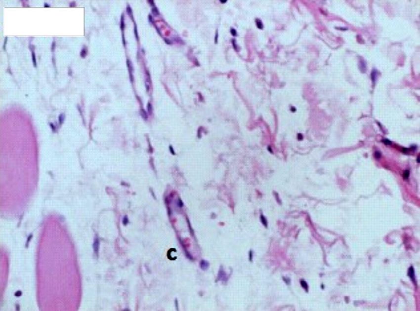
(a) (b)
Figure 3. (a) Measurement of the zone of stasis from five different points. (b) Measurement of epithelium thickness from five different
points.
Ulus Travma Acil Cerrahi Derg, July 2021, Vol. 27, No. 4 397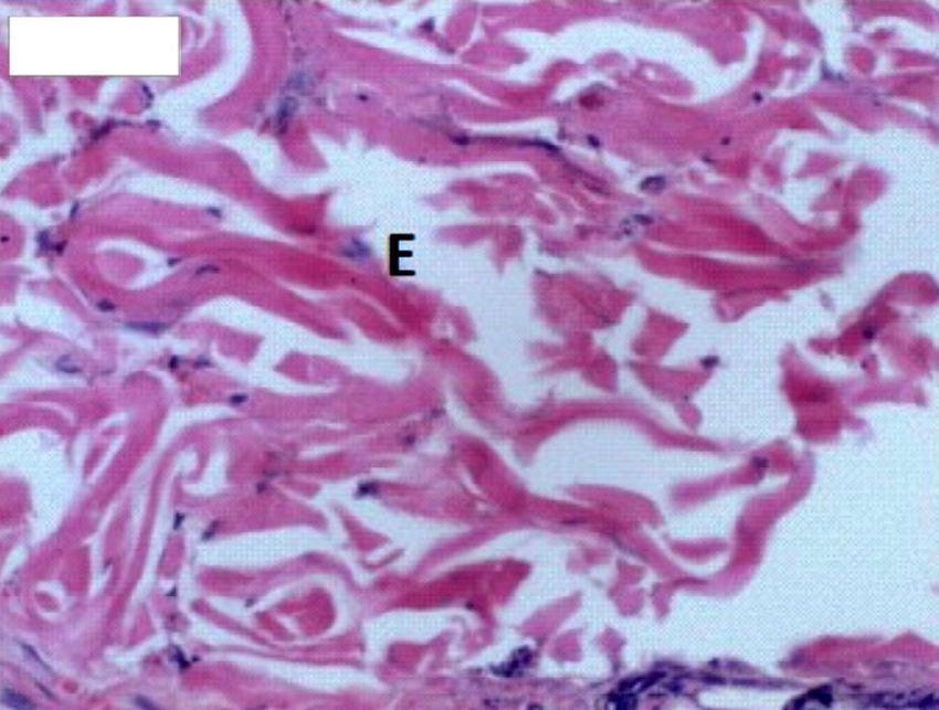
Karadağ Sarı et al. The effects of melatonin on the healing of burn wounds in pinealectomized rats

ative if 71%.
                                                                       Group                         n         Mean          Median (Min-Max)
Statistical Analysis
                                                                       Group 1 (Control)            10         2.00                2 (2–2)
Data were examined using the Shapiro-Wilk test. To investi-
gate the differences between the groups, the Kruskal-Wallis            Group 2 (ExM)                10         2.30                2 (2–3)
test was used for data outside of normal distribution; analysis        Group 3 (Px)                  8         2.00                2 (2–2)
of variance was used for data within the normal distribution.          Group 4 (ExM+Px)             10         2.00                2 (2–2)
In the nonparametric test, subgroup analysis was performed
                                                                       a
                                                                        Kruskal-Wallis Score = 8.880, p=0.031. Difference = Group 1 and Group 3 and
using Mann-Whitney U test and interpreted with Bonferroni
                                                                       Group 4 vs. Group 2. ExM: Exogenous melatonin; Px: Pinealectomy.
correction. P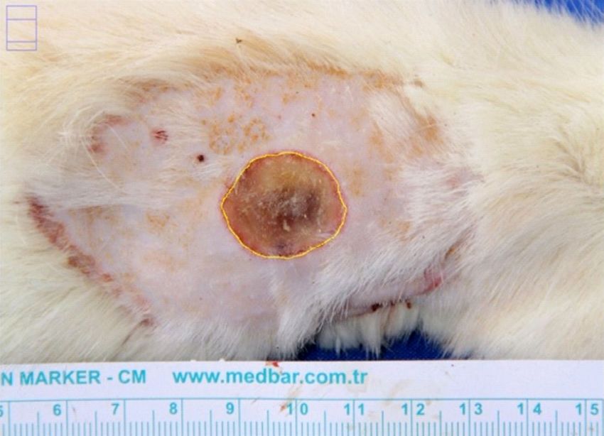
Karadağ Sarı et al. The effects of melatonin on the healing of burn wounds in pinealectomized rats
Table 2. Congestion scores (Kruskal-Wallis Test)a Table 4. ZS thickness (ANOVA)a
Group n Mean Median (Min-Max) Group n Mean Median (Min-Max)
Group 1 (Control) 10 2.70 3 (2–3) Group 1 (Control) 10 522.54 345.4–722.4
Group 2 (ExM) 10 2.40 2 (2–3) Group 2 (ExM) 10 521.30 340.8–903.4
Group 3 (Px) 8 2.38 2 (2–3) Group 3 (Px) 8 574.45 463.2–673.4
Group 4 (ExM+Px) 10 2.00 2 (1–3) Group 4 (ExM+Px) 10 700.12 493.0–960.4
a
Kruskal-Wallis Score = 8.180, p=0.042. Difference = Group 1 and Group 4. a
F=4.522, p=0.009, Difference = Group 1 and Group 2 vs. Group 4. ExM: Exog-
ExM: Exogenous melatonin; Px: Pinealectomy. enous melatonin; Px: Pinealectomy; ZS: Zone of stasis.
Table 3. Fibrosis scores (Kruskal-Wallis Test)a Table 5. Type III collagen staining score (Kruskal-Wallis Test)a
Group n Mean Median (Min-Max) Group n Mean Median (Min-Max)
Group 1 (Control) 10 2.20 2 (2–3) Group 1 (control) 10 1.20 1 (1–2)
Group 2 (ExM) 10 2.50 2.5 (2–3) Group 2 (ExM) 10 1.60 2 (1–2)
Group 3 (Px) 8 1.88 2 (1–2) Group 3 (Px) 8 1.00 1 (1–1)
Group 4 (ExM+Px) 10 1.90 2 (1–2) Group 4 (ExM+Px) 10 1.50 1.5 (1–2)
a
Kruskal-Wallis Score = 11.142, p=0.011. Difference = Group 2 vs. Group 3 and a
Kruskal-Wallis Score = 8.880, p=0.031, Difference = Group 2 vs. Group 3.
Group 4ExM: Exogenous melatonin; Px: Pinealectomy. ExM: Exogenous melatonin; Px: Pinealectomy.
4). However, there were no significant differences among the sion of damage in burn wounds by affecting the viability of the
groups concerning epithelium thickness. zone of stasis.[10]
The degree of type III collagen staining in the ExM group was In our literature review, we identified several experimen-
significantly higher than the Px group (p=0.031; Table 5). tal studies that report their investigations on antioxidants’
ability to prevent the progressive effects of free oxygen rad-
DISCUSSION icals in burn wounds. One study[11] reported that necrosis
Burn injuries are a major source of physical and physiologi- in the zone of stasis significantly reduced in groups treated
cal trauma worldwide. If second-degree deep burn wounds with N-acetylcysteine than controls. Zor et al.,[12] based on
are not treated promptly and appropriately, they may develop nuclear imaging and autoradiography, reported that glutathi-
into full-thickness burns due to progressive tissue damage, one helped salvage the zone of stasis. Singer et al.[13] found
leading to hypertrophic scars and contractures that require that curcumin reduced the progression of burn injury in a rat
surgical excision and graft repair.[3] Tissue damage in the zone comb burn model. Matsuda et al.[14] suggest that high-dose vi-
of stasis may be prevented with an appropriate treatment tamin C would reduce edema in burn wounds in the porcine
applied within two weeks from the initial injury.[8,9] Inflamma- burn model, and superoxide dismutase has a salvaging effect
tion, ischemia and free oxygen radicals increase the progres- on the zone of stasis.[15]
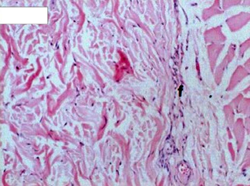
Group 2 Group 3
Figure 6. Fibrotic areas in Group 2 (F) are more significant (ExM) than Group 3 (f) (Px) (H&Ex20).
Ulus Travma Acil Cerrahi Derg, July 2021, Vol. 27, No. 4 399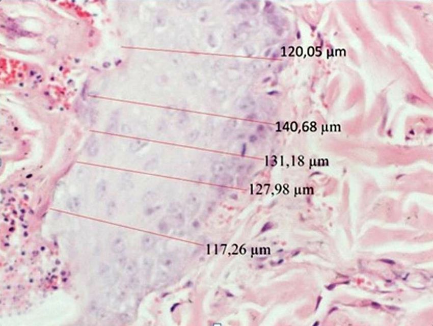
Karadağ Sarı et al. The effects of melatonin on the healing of burn wounds in pinealectomized rats

Melatonin’s free oxygen radical scavenging ability is more             thesis. Contraction of the wound surface area is a clinical in-
effective than other antioxidants.[6] There are few studies            dicator of healing. We found no significant differences among
that examine the effects of melatonin on burn healing. Most            the groups concerning the average healed areas after seven
melatonin studies have focused on non-burn wound healing.              days from the injury. We also noted similarities among the
Drobnik and Dabrowski[16] reported that pinealectomy en-               groups for epithelial thickness. Our study was designed to
hanced collagen deposition in the skin, and melatonin applica-         evaluate the early effects of melatonin on the healing of burn
tion reduced the pinealectomy-induced elevation of collagen            wounds in pinealectomized rats. A longer study is required to
levels. Bulbuller et al.,[17] on the other hand, indicated that        assess its macroscopic effects in the long-term.
exogenous melatonin reduced collagen synthesis and epitheli-
um proliferation and had negative effects on wound healing in          Additionally, because hydroxyproline levels of all collagens in
both normal and pinealectomized rats. Contrary to Bulbuller            the dermis did not show any significant differences among
et al.’s findings, we found the level of type III collagen staining    the groups, hydroxyproline was deemed to be less meaningful
to be significantly higher in the exogenous melatonin group            than the level of type III.
than the pinealectomy group. Our finding suggests that mel-
atonin, by enhancing the level of type III collagen in the burn        Our study had several limitations. The first limitation was
wound, can decrease the likelihood of a hypertrophic scar              the short duration of seven days. Longer studies are needed
and help burn wound healing since type III collagen deficien-          to assess the effects of melatonin on wound healing macro-
cy increases scar formation.[18] In our study, fibrosis levels in      scopically. While our study noted the early effects of mela-
the exogenous melatonin group were also significantly high-            tonin burn wound healing, this study design did not allow for
er than the pinealectomy groups—a finding that showed the              a longer-term evaluation of the effects of melatonin on burn
positive effect of melatonin on burn wound healing.                    wound healing.

Kayapınar et al.[10] assessed melatonin’s efficacy in saving the       While being a significant factor in the progression of burn
zone of stasis in rats. Kayapınar et al. divided their rats into       wound injury, total melatonin levels above a certain threshold
two groups, a control group and a melatonin-treated group.             will reduce the zone of stasis and exert a preventative effect
Their study did not, however, assess the effects of pinealec-          on burn wound damage progression. Additionally, melatonin
tomy on burn wound healing, nor did they analyze as many               may prevent the development of hypertrophic scarring by in-
parameters as our study (e.g., collagen levels, epithelial thick-      creasing fibrosis and type III collagen in burn wounds. The
ness, and hydroxyproline levels). They found most of the inter-        results of this study suggest that melatonin can be used as
spaced areas to be alive in the melatonin-treated group. We            an additional, supplemental treatment in burn wound heal-
found the thickness of the zone of stasis significantly less in        ing. Because our study aimed to evaluate the early effects
our ExM group (the group with the highest total melatonin lev-         of exogenous melatonin, further studies with higher doses
els). Given the correlation between the zone of stasis and the         administered over longer periods are warranted to assess
depth of the wound; hence, the progress of the burn injury, this       its macroscopic effects on burn wound healing. A study of
outcome proves that total melatonin levels have a profound             melatonin levels in serum or blood in each group would help
impact on reducing the progression of burn injuries. Further           determine an appropriate dosage of melatonin to affect burn
supporting this, the ExM+Px group rats that could not produce          wound healing. Despite promising experimental findings, fu-
endogenous melatonin presented with thicker zones of stasis            ture studies are needed to address three key areas: optimal
and were administered exogenous melatonin. These findings              dosages, administration timing and route of administration of
demonstrate that total melatonin production, once exceeding            exogenous melatonin for optimal outcomes in burn wound
a certain threshold, will exert a preventative effect on burn          healing.
wound damage progression by reducing the zone of stasis.
                                                                       Acknowledgements
Contrary to our findings, Kayapınar et al.[10] report higher
                                                                       I would like to thank Dr. Hatice Toy for the histopathological
edema levels in their controls than their melatonin-treat-
                                                                       and immunohistochemical analysis and Dr. Aysun Toker for
ed group. In our study, edema levels were higher in the
                                                                       the biochemical analysis.
ExM-treated group than controls, suggesting edema may be
a side effect of higher melatonin levels. Although we adminis-
                                                                       Ethics Committee Approval: Ethics Committee Approv-
tered exogenous melatonin in the ExM+Px group (Group 4),
                                                                       al: This study was approved by Necmettin Erbakan Universi-
we found no differences in edema scoring than the control
                                                                       ty Experimental Medicine Research and Application Center
group (Group 1) and the Px group (Group 3). This finding
                                                                       Animal Experiments Ethics Committee (Date: 30.07.2012,
may be due to the relatively low melatonin levels in these
                                                                       Decision No: 2012/121518019).
groups than the ExM group (Group 2).
                                                                       Peer-review: Internally peer-reviewed.
The main goal during the recovery period is to minimize the            Authorship Contributions: Concept: E.Ç.K.S., N.S.; De-
burn wound area by contraction and extracellular matrix syn-           sign: E.Ç.K.S.; Supervision: N.S.; Resource: E.Ç.K.S.; Materi-

400                                                                                 Ulus Travma Acil Cerrahi Derg, July 2021, Vol. 27, No. 4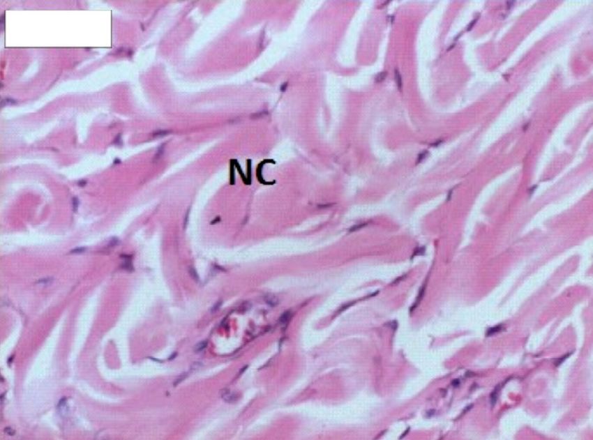
Karadağ Sarı et al. The effects of melatonin on the healing of burn wounds in pinealectomized rats

als: E.Ç.K.S.; Data: E.Ç.K.S.; Analysis: E.Ç.K.S., N.S.; Litera-                               following burn. Burns 2012;38:283–9. [CrossRef ]
ture search: E.Ç.K.S.; Writing: E.Ç.K.S.; Critical revision: N.S.                        9.    Nisanci M, Eski M, Sahin I, Ilgan S, Isik S. Saving the zone of stasis
                                                                                               in burns with activated protein C: an experimental study in rats. Burns
Conflict of Interest: None declared.
                                                                                               2010;36:397–402. [CrossRef ]
Financial Disclosure: This study was supported by Nec-                                   10.   Kayapınar M, Seyhan N, Avunduk MC, Savacı N. Saving the zone of sta-
mettin Erbakan University Scientific Research Projects Co-                                     sis in burns with melatonin: an experimental study in rats. Ulus Travma
ordination Unit.                                                                               Acil Cerrahi Derg 2015;21:419–24. [CrossRef ]
                                                                                         11.   Deniz M, Borman H, Seyhan T, Haberal M. An effective antioxidant
                                                                                               drug on prevention of the necrosis of zone of stasis: N-acetylcysteine.
                                                                                               Burns 2013;39:320–5. [CrossRef ]
REFERENCES
                                                                                         12.   Zor F, Ozturk S, Deveci M, Karacalioglu O, Sengezer M. Saving the zone
1.   Jackson DM. The diagnosis of the depth of burning. Br J Surg                              of stasis: is glutathione effective?. Burns 2005;31:972–6. [CrossRef ]
     1953;40:588–96. [CrossRef ]                                                         13.   Singer AJ, Taira BR, Lin F, Lim T, Anderson R, McClain SA, et al. Cur-
2.   Singh V, Devgan L, Bhat S, Milner SM. The pathogenesis of burn wound                      cumin reduces injury progression in a rat comb burn model. J Burn Care
     conversion. Ann Plast Surg 2007;59:109–15. [CrossRef ]                                    Res 2011;32:135–42. [CrossRef ]
3.   Shupp JW, Nasabzadeh TJ, Rosenthal DS, Jordan MH, Fidler P, Jeng JC.                14.   Matsuda T, Tanaka H, Shimazaki S, Matsuda H, Abcarian H, Reyes
     A review of the local pathophysiologic bases of burn wound progression.                   H, et al. High-dose vitamin C therapy for extensive deep dermal burns.
     J Burn Care Res 2010;31:849–73. [CrossRef ]                                               Burns 1992;18:127–31. [CrossRef ]
4.   Rizzo JA, Burgess P, Cartie RJ, Prasad BM. Moderate systemic hypother-              15.   Shalom A, Kramer E, Westreich M. Protective effect of human recombi-
     mia decreases burn depth progression. Burns 2013;39:436–44. [CrossRef ]                   nant copper-zinc superoxide dismutase on zone of stasis survival in burns
5.   Fujimoto T, Nakamura T, Ikeda T, Takagi K. Potent protective effects                      in rats. Ann Plast Surg 2011;66:607–9. [CrossRef ]
     of melatonin on experimental spinal cord injury. Spine (Phila Pa 1976)              16.   Drobnik J, Dabrowski R. Melatonin suppresses the pinealectomy-in-
     2000;25:769–75. [CrossRef ]                                                               duced elevation of collagen content in a wound. Cytobios 1996;85:51–8.
6.   Tan DX, Manchester LC, Esteban-Zubero E, Zhou Z, Reiter RJ. Mela-                   17.   Bulbuller N, Dogru O, Yekeler H, Cetinkaya Z, Ilhan N, Kirkil C. Ef-
     tonin as a Potent and Inducible Endogenous Antioxidant: Synthesis and                     fect of melatonin on wound healing in normal and pinealectomized rats. J
     Metabolism. Molecules 2015;20:18886–906. [CrossRef ]                                      Surg Res 2005;123:3–7. [CrossRef ]
7.   Reddy GK, Enwemeka CS. A simplified method for the analysis of hy-                  18.   Volk SW, Wang Y, Mauldin EA, Liechty KW, Adams SL. Diminished
     droxyproline in biological tissues. Clin Biochem 1996;29:225–9. [CrossRef ]               type III collagen promotes myofibroblast differentiation and increas-
8.   Eski M, Ozer F, Firat C, Alhan D, Arslan N, Senturk T, et al. Cerium                      es scar deposition in cutaneous wound healing. Cells Tissues Organs
     nitrate treatment prevents progressive tissue necrosis in the zone of stasis              2011;194:25–37. [CrossRef ]

 DENEYSEL ÇALIŞMA - ÖZ
   OLGU SUNUMU

      Pinealektomize sıçanlarda melatoninin yanık yara iyileşmesine etkisi
      Dr. E. Çiğdem Karadağ Sarı,1 Dr. Nedim Savacı2
      1
          Acıbadem Üniversitesi Tıp Fakültesi, Plastik, Rekonstrüktif ve Estetik Cerrahi Anabilim Dalı, İstanbul
      2
          Necmettin Erbakan Üniversitesi, Meram Tıp Fakültesi, Plastik, Rekonstrüktif ve Estetik Cerrahi Anabilim Dalı, Konya

      AMAÇ: Bu çalışmanın amacı, sıçanlarda melatoninin yanık yara iyileşmesine etkisini araştırmaktır.
      GEREÇ VE YÖNTEM: Bu çalışmada 40 adet Wistar-albino cinsi deneysel erkek sıçanlar dört gruba ayrıldı: Grup 1; kontrol grubu, Grup 2; eksojen
      melatonin verilen grup, Grup 3; pinealektomi uygulanan grup ve Grup 4; pinealektomi yapıldıktan sonra eksojen melatonin uygulanan grup idi. Sıçan-
      ların sırtlarına kaynar suda bekletilmiş metal plak ile ikinci derece derin yanık oluşturuldu. Yedi gün boyunca yanık alanlarının iyileşmesi takip edildi.
      Yedi gün sonunda eksize edilen yanık alan dokularında hidroksiprolin, Tip 3 kollajen, ödem, iltihabi infiltrasyon, konjesyon, vasküler proliferasyon,
      fibrozis düzeyleri, staz zonu ve epitel kalınlıkları değerlendirildi.
      BULGULAR: Staz zonu kalınlığı Grup 2’de diğer gruplara oranla daha azdı (p=0.009). Tip 3 kollajen boyanma (p=0.031), fibrozis (p=0.011) ve
      ödem (p=0.031) Grup 2’de diğer gruplara oranla daha fazlaydı. Konjesyon kontrol grubunda Grup 4’e göre daha fazlaydı (p=0.031). Değerlendirilen
      diğer parametrelerde, gruplar arasında anlamlı bir fark görülmedi.
      TARTIŞMA: Bu çalışmada, total melatonin düzeyinin belli eşik düzeyi geçmesi durumunda, staz zonunu azaltarak yanık yara hasarının ilerlemesin-
      de önleyici etkisi bulundu. Melatoninin, ayrıca hipertrofik skar gelişimini de önleyebildiği görüldü. Melatonin yanık yaralarında geleneksel tedaviyi
      destekleyebilecek potansiyel bir tedavi seçeneği olabilir. Bunun yanısıra, bu çalışmada belirtilen etkileri daha fazla değerlendirmek için daha uzun
      sürelerde ve daha yüksek dozlarda eksojen melatoninin uygulandığı çalışmalara ihtiyaç vardır.
      Anahtar sözcükler: Eksojen melatonin; endojen melatonin; pinealektomi; yanık; yara.

      Ulus Travma Acil Cerrahi Derg 2021;27(4):395-401           doi: 10.14744/tjtes.2020.12247

Ulus Travma Acil Cerrahi Derg, July 2021, Vol. 27, No. 4                                                                                                          401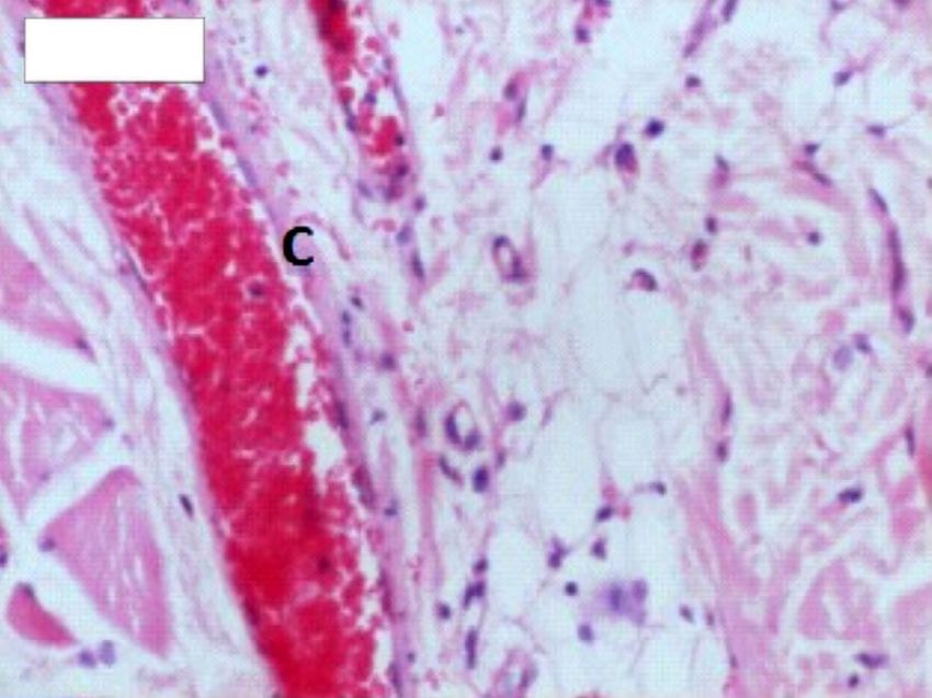
You can also read