International Journal of Veterinary Science
←
→
Page content transcription
If your browser does not render page correctly, please read the page content below
P-ISSN: 2304-3075; E-ISSN: 2305-4360

                       International Journal of Veterinary Science
                                              www.ijvets.com; editor@ijvets.com

Research Article
Pathological and Immunohistochemical Studies on the Ameliorating Effect of
Spirulina Platensis against Arsenic Induced Reproductive Toxicity in Female
Albino Rats
Reda MS Korany1*, Khaled S Ahmed2, Hanaa A El Halawany2 and Kawkab A Ahmed1
1
 Department of Pathology, Faculty of Veterinary Medicine, Cairo University, Giza, 12211, Egypt.
2
 Department of Pathology and Reproductive Diseases, Animal Reproduction Research Institute (A.R.R.I.), Giza
*Corresponding author: reda.pathology@yahoo.com

    Article History:     Received: March 07, 2019          Revised: March 12, 2019            Accepted: March 16, 2019

    AB S T RA C T
    The aim of this study was to determine the effect of sodium arsenate (Na 3AsO4) on the female reproductive organs,
    and to study the effect of Spirulina platensis (Sp) as an ameliorating agent in the arsenic-induced toxicity. 40 female
    Wistar albino rats were used. The rats were divided into four equal groups, 10 rats in each group; control group and
    three groups that received spirulina (Sp), sodium arsenate, and sodium arsenate plus spirulina respectively, for 2
    months. Results showed that the body weight in female rats was significantly reduced in arsenic-treated group
    compared to the control, while the co-treatment with Sp significantly restored the body weight. Arsenic significantly
    increased serum malondialdehyde (MDA) and significantly reduced serum glutathione (GSH) activities in comparison
    with the control group. Spirulina co-treatment significantly restored GSH levels and significantly reduced MDA in
    comparison to arsenic-treated group. Histopathologically, uterus and ovaries of arsenic-treated group showed different
    degenerative changes which improved with the Spirulina co-treatment. Immunohistochemistry determined the role of
    nuclear erythroid 2-related factor 2 (Nrf2) in the arsenic toxicity and Spirulina Platensis mechanism of protection in
    arsenic-induced toxicity. In conclusion, arsenic-induced toxicity in female rats could be ameliorated by Spirulina
    platensis co-administration through Nrf2 pathway.
    Key words: Sodium arsenate, Spirulina platensis, Uterus, Ovaries, Histopathology, Nrf2

                       INTRODUCTION                                 problems including developmental abnormalities,
                                                                    diabetes, hematological disorders, neurological and
     Arsenic is one of the most important worldwide                 reproductive problems and cancer (Rajesh et al, 2010).
environmental toxicants, which is found in air, water and           Arsenic may cause obvious damage in various organs,
soil. Arsenic is mainly distributed in the environment              including female reproductive system, as manifested by
through using pesticides and herbicide, burning coal and            disruption of the circulating levels of gonadotropins and
treated wood, mining wastes, smelting metal and glass.              estradiol, leading to degeneration of luminal epithelial,
The main exposure to arsenic is occupational and the most           stromal and myometrial cells of the uterus and abrogation
frequent reason of poisoning is the use of contaminated             of the estrogen-signaling pathway in female (Chatterjee
water (Goudarzia et al., 2018). Arsenic (AS) is widely              and Chatterji, 2010). Arsenic toxicity expedites generation
distributed in the environment due to its natural and               of reactive oxygen species (ROS). Mitochondrial
anthropogenic sources. High levels of inorganic arsenic             dysfunction contributes to enhanced intracellular reactive
are found in ground water in many regions of the world as           oxygen species (ROS) levels, which further elicit damage
a result of geochemical processes posing serious chronic            to the cells and mitochondria itself (Firdausa et al., 2018).
health risks to humans (Rajesh et al, 2010). Reports since          Arsenic exposure leads to the increased production of
early 19th century have confirmed a relationship between            reactive oxygen species (ROS) and other toxic
arsenic exposure and morbidities. Drinking water is the             intermediates via biotransformation of arsenic, which may
most common source of arsenic exposure (Firdausa et al.,            subsequently cause oxidative stress and alterations of
2018). Chronic arsenic exposure may affect a number of              cellular system. Glutathione peroxidase (GSH-Px) is
organs. Arsenic exposure has been associated with health            regarded as the first line to protect the membrane lipids

Cite This Article as: Korany RMS, Ahmed KS, El Halawany HA and Ahmed KA, 2019. Pathological and
immunohistochemical studies on the ameliorating effect of Spirulina platensis against arsenic induced reproductive
toxicity in female albino rats. Inter J Vet Sci, 8(2): 113-119. www.ijvets.com (©2019 IJVS. All rights reserved)

                                                              113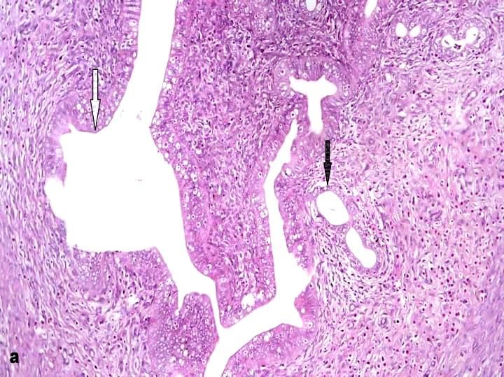
Inter J Vet Sci, 2019, 8(2): 113-119.

from oxidative damage. When this enzymatic scavenger                   approved by the institutional Animal care and Use
cannot counteract excessive ROS, lipid peroxidation will               Committee (CU-IACUC), Cairo university, Egypt
occur. One of the lipid peroxidation metabolites is                    (approval No. CU-II-F-37-18).
malondialdehyde (MDA), which arises primarily from
peroxidative cleavage of polyunsaturated fatty acids in                Experimental design
biological systems and is also used as an biomarker to                     Female rats were divided randomly into four equal
balance oxidative damage (Zhao et al., 2017). Chelation                groups. The first group served as a control group that
therapy using synthetic chelating agents is the only                   received standard laboratory chow and water ad libitum.
available therapeutic method for arsenicosis. However,                 The second group received Sp (300 mg/ kg bwt) dissolved
related adverse side-effects such as chelation of essential            in water by oral route daily. The third group daily
metals and arsenic redistribution in tissues mostly limited            received orally sodium arsenate (5 mg/kg bwt) dissolved
their clinical use. Also, dietary antioxidants are known for           in normal saline. Fourth group was given Sp (300 mg/kg
a long time for their effectiveness against oxidative stress-          body weight) and sodium arsenate (5 mg/kg body weight).
related complications. The correlation between arsenic                 The treatment was continued for two months. Rats were
toxicity and oxidative stress provides an indisputable                 euthanized by decapitation at the end of the experimental
platform for phytochemicals which may serve as a useful                period.
preventive/therapeutic approach as recently recommended
by World Health Organization (WHO) (Firdausa et al.,                   Body weights
2018). Microalgae have been known as food and animal                       The final body weight of rats was recorded by
feed; they grow in freshwater and marine (Vigani et al.,               weighing each rat in all groups for detection of body
2015). Spirulina is a photosynthetic cyanobacterium that               weight changes.
is used commercially as a dietary supplement and food
additive. The extensive production of Spirulina is due to              Lipid peroxidation and reduced glutathione
its     original     chemical      composition      [proteins,              After euthanizing the rats, blood samples were
polyunsaturated fatty acids, and vitamins]. Besides, it is a           collected from the retro-orbital venous plexus in a sterile
source of bioactive components like, phycocyanin, β-                   centrifuge tubes just before necropsy. Blood samples were
carotene, and allophycocyanin, which has anti-                         left to clot at room temperature and centrifuged at 3000
inflammatory and antioxidant properties (Wang et al.,                  rpm for 15 minutes and the sera were separated and stored
2007). The studies conducted in the past using Spirulina               at -20˚C as aliquots for further biochemical analysis. Lipid
platensis as a supplement have proved that it has an                   peroxidation was evaluated by measurement of serum
ability to counteract the toxicity caused by many                      malondialdehyde (MDA) (Ohkawa et al., 1979) based on
medications and chemicals (Banji et al., 2013).                        the formation of thiobarbituric acid reactive substances
                                                                       (TBARs) and expressed as the extent of malondialdehyde
Aim of work: This study was aimed to investigate the                   (MDA) production. The non-enzymatic antioxidant
effect of arsenic toxicity on the female reproductive                  biomarker, reduced glutathione (GSH) was assessed
system in albino rats and evaluating the ameliorative                  (Beutler et al., 1963).
potential of Spirulina platensis against the arsenic-
induced toxicity by its co-administration with arsenic.                Postmortem and histopathological examination
                                                                           After euthanizing, rats were subjected to careful
            MATERIALS AND METHODS                                      postmortem examination. Specimens from uterus and
                                                                       ovaries were collected and fixed in formal saline 10%
Chemicals                                                              then washed, dehydrated, cleared and embedded in
    Pure Spirulina Platensis powder was obtained from                  paraffin. The paraffin embedded blocks were sectioned at
Arab Academy for Science, Technology and Maritime                      4-5 micron thickness and stained with Hematoxylin and
Transportation, Alexandria, Egypt. Sodium arsenate                     Eosin (Bancroft et al., 2012).
(Na3AsO4) obtained from the representative of Fisons
Scientific Apparatus Ltd. UK in Egypt. All kits for                    Histomorphometric studies of uterine and ovarian
biochemical     analysis     were    purchased    from                 tissues
Biodiagnostics, Egypt. All other chemicals used in the                      The size of endometrial glands and thickness of
experiment were of analytical grade.                                   endometrium and myometrium were measured by light
                                                                       microscope (Olympus BX50, Japan). The number of
Animals                                                                different follicles from three sections of each ovary per /
     40 female Wistar albino rats weighing 125 ± 20 gm                 rat (n=10) were counted microscopically using TSView
were randomly divided into four equal groups: Control                  version 6.2.4.5 software.
and three treatment groups (As, Sp and As+Sp) (10 rats in
each group). Rats were obtained from Helwan Animal                     Immunohistochemistry
Colony belonging to VACSERA. The animals were                              Formalin-fixed, paraffin-embedded 4 µm sections
housed in standard cages, kept in a ventilated room under              were fixed into poly-L-lysine coated slides (Thermo
controlled laboratory conditions of normal light-dark cy-              Scientific, Karlsruhe, Germany). After deparaffinization
cle (12 hours light/dark) and temperature (25 ± 2˚C).                  and rehydration, the slides were immersed in buffer
Standard laboratory chow and water were provided ad                    Target Retrieval Solution, pH 9.0 (Dako, Glostrup,
libitum. Animals allowed to acclimatizing for two weeks                Denmark). Peroxidase Blocking Solution (Dako,
before starting the study. This experimental protocol was              Glostrup, Denmark) was used to block activity of

                                                                 114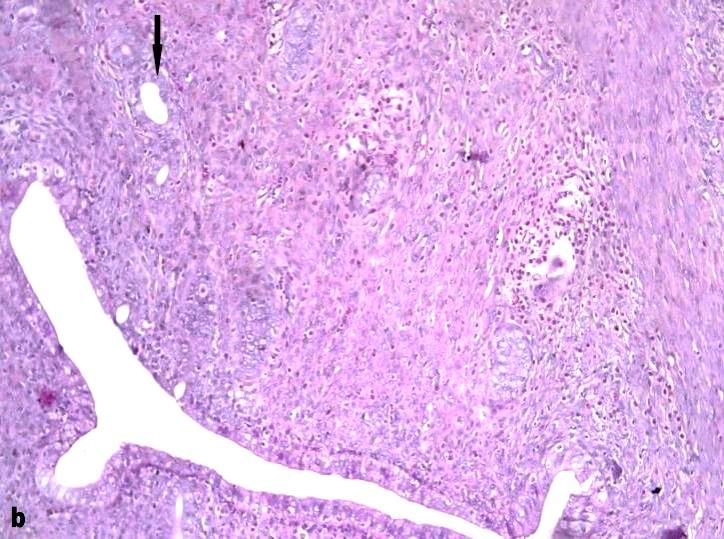
Inter J Vet Sci, 2019, 8(2): 113-119.

endogenous peroxidase. The slides were incubated with 1
mg/ml of the nuclear erythroid 2–related factor 2 (Nrf2)
Rabbit Anti- Human Polyclonal Antibody (LS-C118543 –
LSBio; LifeSpan Biosciences, Seattle, WA, USA) for 30
min at room temperature and immunostained with DAKO
RealTM EnvisionTM Detection System Peroxidase/DAB+,
HRP Rabbit/Mouse (Dako, Glostrup, Denmark).
Hematoxylin was used as a counterstain.

Statistical analysis
     All results were expressed as mean ± SD (Standard
Deviation). Statistical analyses were performed using the
SPSS version 24.0 statistical analysis package (SPSS,               Fig. 1: Female average body weight (g) among different
Inc., Chicago, IL, USA). The parametric test one-way                experimental groups. Sp: Spirulina; As: arsenic; As+Sp:
ANOVA was used for data analysis and comparison was                 Arsenic and Spirulina. The values are expressed as the means ±
done using Tukey post-hoc test. In all calculations, a              SD, where n=10. Superscript a refers to a significance from
difference at P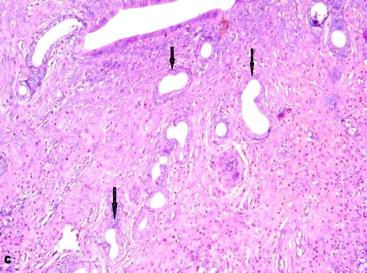
Inter J Vet Sci, 2019, 8(2): 113-119.
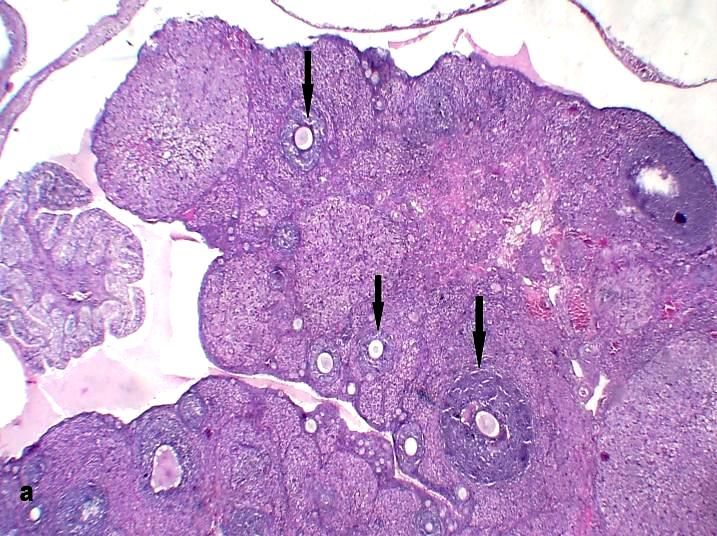
Fig. 3: Micrograph, uterus, female rat. Control untreated group
Fig. 4: Micrograph, ovary, female rat. Control untreated group
showing the normal histological structure of the uterus with
showing considerable number of ovarian healthy follicles
normal invagination of uterine lumen (white arrow) and normal
(arrows) (a). In arsenic treated group the ovary showing
uterine glands (black arrow) (a). In arsenic treated group the
dramatic decrease in the ovarian follicles (arrow) (b). In groups
uterus appeared with decreased size and invaginations of the
co-administered with spirulina, the ovary restored its function
uterine lumen with decreased endometrial glands number
(arrow) (b). Recovery was noted in sections of uterus co- with increased healthy ovarian follicles number (arrows) (c). (H
administered with spirulina as the number of uterine glands & E X40).
showed noticeable increase (arrows) (c). (H & E X100).
Histomorphometry of uterine and ovarian tissues
Concerning ovaries, examined sections from control Concerning uterus, the size of endometrial glands and
rats as well as spirulina treated rats revealed the normal thickness of endometrium and myometrium were
histological structure with normal number of healthy significantly reduced (P
Inter J Vet Sci, 2019, 8(2): 113-119.

Fig. 5: Immunostaining of Nrf2. Arsenic treated group showing strong positive cytoplasmic reaction in stromal cells of uterus (a) and
ovary (b). In spirulina co-treated group, the immunostaining reaction is weak in both uterus (c) and ovary (d). (H & E X400).

with spirulina significantly increased the number of ovarian               The present experiment was designed to assess the
follicles compared to arsenic treated group (Table 2).                deleterious effect of Sodium arsenate (Na3AsO4) on the
                                                                      female reproductive organs (uterus and ovaries) in female
Immunohistochemistry                                                  albino rats following oral exposure, and to evaluate the
Nrf2 Expression                                                       efficacy of Spirulina platensis on modulating the female
     Nrf2 expression in uterus and ovary revealed an                  reproductive and neurotoxic alterations induced by
intense reaction in stromal cells of the uterus and the               Sodium arsenate with determination of its pathways of
ovaries of As-treated group (Fig. 5a & b). This strong                protection.
reaction shown with As-treated rats is abated to less                      Effect of Sodium arsenate on animals’ body weight
intense reaction with the co-administration of Spirulina in           has been studied by many researchers (Elshawarby et al.
the As + Sp group revealing the tendency to return to the             2014; Khan et al., 2014 and Rodriguez et al., 2016). In
control expression of Nrf2 (Fig. 5c & d).                             this study, the adverse effect of As- induced toxicity on
                                                                      body weight was significantly (P< 0.01 and P< 0.001)
                       DISCUSSION                                     present. As-treated female rats showed a significant
                                                                      decrease in body weight and retardation of growth in
      Arsenic is a natural component of the environment               comparison with control group. Although (Souza et al.
and widely used in human activities. Thus, people are                 2016) reported that arsenic exposure did not affect the
inevitable to contact with pollutant of arsenic along with            total body weight in female rats in their study but this may
the increase of arsenic in the environment (Zhao et al.,              be due to the dose or duration of their studies or may be
2017).Sodium arsenate is the pentavalent form of                      the animal model used. Body weight of female As-treated
inorganic arsenic and is biotransformed quickly into                  rats was significantly recovered by spirulina co-
sodium arsenite, a trivalent form of arsenic. Numerous                administration. This improvement could be due to the
reports have substantially shown that arsenic exposure                unique nutrients that present in Sp, like B-complex
irrespective of the inorganic salt form leads to a                    vitamins, minerals, proteins, γ-linolenic acid and anti-
tremendous increase in the generation of free radicals                oxidants like β-carotene, vitamin E (Holman and Malau-
(Firdausa et al., 2018). The naturally occurring sources of           Aduli, 2012).
antioxidants have been used in various in vitro and in vivo                In this experiment, the serum antioxidant enzymes
studies and have demonstrated promising outcomes with                 GSH and MDA were measured and the results revealed
regard to their effects on metal-induced toxicity (Khalil et          significant reduction in the GSH in As-treated rats while
al., 2018).                                                           showed significant increase of MDA levels. The co-

                                                                117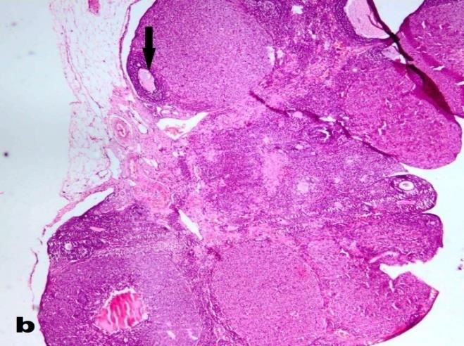
Inter J Vet Sci, 2019, 8(2): 113-119.

administration of Sp showed significant reverse of these            which are a critical source for the synthesis and release of
values towards the control levels. The changes in levels of         prostaglandin D2, reactive oxygen species (ROS),
various biochemical parameters in sodium arsenate-                  leukotrienes, and cytokines, such as TNF-α, which trigger
treated rats indicate various aspects of metabolism of              the release of interleukin (IL)–1β, IL–6, as well as block
animals. Sodium arsenate decreased the activities of                the phosphorylation of p38 mitogen-activated protein
antioxidant enzymes, thereby generating oxidative stress            kinases (MAPK), which in turn regulate the synthesis of
as evidenced by increased lipid peroxidation levels. The            cytokines. Furthermore, Spirulina diminishes nitrite
presence of antioxidant enzymes helps in combating free             generation, suppresses inducible nitric oxide synthase
radicals/oxygen-derived species generated during normal             expression, and lessens liver microsomal lipid
physiological process. Any alterations in the activities of         peroxidation (Khalil et al., 2018)
these enzymes change the redox status of the cells, thus                 In this study, immunohistochemistry was used to
altering the normal physiological processes. Arsenic                prove the role of oxidative stress in As- induced toxicity
exposure decreased the activities of antioxidant enzymes            and to assess the role of Sp in counteracting the arsenic
along with increase in lipid peroxidation. A continued              mode of action and ameliorating the As-mediated
oxidative stress as indicated by the increased MDA values           oxidative stress in As- induced toxicity. The results
usually causes inflammation, which in turn may lead to              revealed the overexpression of Nrf2 in the ovaries and
chronic diseases, including cancer (Mehta &Hundal,                  uterus of As-treated rats and weak expression of Nrf2 with
2016).                                                              Sp co- treatment. Numerous studies have shown that
     Histopathologically, female reproductive system                arsenic was an Nrf2inducer in several cell types. Exposure
showed also degenerative changes in both uterus and                 to arsenic or other exogenous stressors may activate the
ovaries in As-treated rats, and these findings collaborate          Nrf2 pathway to maintain cellular redox homeostasis and
with the previous findings of (Akram et al.,2010; Wares et          limit oxidative damage, Oxidative stressactivatesNrf2 by
al. 2013). Also,the size and number of endometrial glands           permitting its dissociation from Keap1and translocation
and thickness of endometrium and myometrium were                    into the nucleus where it binds to the antioxidant response
significantly reduced. Exposure to sodium arsenate                  element and leads to the expression of the target genes (Li
significantly decreased the number of ovarian follicles,            et al., 2015).
these changes could be attributed to that, arsenic is an                 In conclusion, sodium arsenate induced reproductive
endocrine disruptor influences sex hormones and induces             toxicity in female albino rats could be ameliorated by
inhibition of ovarian steroidogenesis and reproductive              Spirulina platensisco- treatment, with focusing on the role
disturbances (Sun et al. 2016). It is known that arsenic            of Nrf2 in arsenic-induced toxicity as Spirulina platensis
treatment is also associated with the hypertrophy of                counteract the arsenic-induced oxidative stress through
adrenal gland, and inhibition in gonadotropin secretion             activating the Nrf2 pathway.
(Chatterjee and Chatterji, 2011). The cellular degeneration
of the female sex organs in arsenic-exposed rats may have                                 REFERENCES
resulted from the low levels of plasma gonadotrophins
and estradiol as ovarian-cell proliferation and                     Akram Z, S Jalali, SA Shami, L Ahmad, S Batool and O
differentiation are controlled by gonadotrophins, while                 Kalsoom, 2010. Adverse effects of arsenic exposure
uterine weight and histoarchitecture are regulated by                   on uterine function and structure in female rat.
plasma estradiol level. Elevation in the number of atretic              Experim Toxicol Pathol, 62: 451-459.
follicles and diminution in the numbers of healthy                  Bancroft D, A Stevens and R Turner, 2012. Theory and
follicles may be due to low plasma levels of                            practice of histological technique, 4th edition, Churchill,
gonadotrophins and estradiol. In another way, these                     Livingstone, Edinburgh, London, Melbourne.
changes may be elucidated by arsenic-induced oxidative              Banji D, OJ Banji, NG Pratusha, AR Annamalai, 2013.
stress in ovary, which is supported by the diminution in                Investigation on the role of Spirulina platensis in
the activities of GSH along with overproduction of MDA                  ameliorating behavioral changes, thyroid dysfunction
levels. (Chattopadhyay and Ghosh, 2010).Uterine                         and oxidative stress in offspring of pregnant rats
endometrium degeneration is associated with the                         exposed to fluoride. Food Chem, 140: 321–331.
increased production of reactive oxygen species (ROS)               Beutler E, O Duron and BM Kelly, 1963. Improved
such as superoxide radicals, hydrogen peroxide, and                     method for the determination of blood glutathione. J
hydroxyl radicals. Endometrium may be a potential                       Lab Clin Med, 61: 882-890.
candidate site for superoxide anion generation in the               Chatterjee A and U Chatterji, 2010. Arsenic abrogates the
uterus because ROS are generated in the endometrium.                    estrogen-signaling pathway in the rat uterus. Reprod
Arsenic is known to produce oxidative stress by the                     Biol Endocrinol, 8:80.
promotion of ROS. Under normal conditions endometrial               Chatterjee A and U Chatterji, 2011. All-trans retinoic acid
stroma regulates the growth and function of endometrial                 protects against arsenic-induced uterine toxicity in
glands. Possibly disrupted endometrial stroma may affect                female Sprague-Dawley rats. Toxicol Appl
the growth and differentiation of endometrial glands                    Pharmacol, 257: 250-263.
(Akram et al., 2010). Co-administration of Sp revealed a            Chattopadhyay S and D Ghosh, 2010. Role of dietary
protection against these degenerative changes seen in the               GSH in the amelioration of sodium arsenite-induced
histopathology of As-treated rats and showed a normal                   ovarian and uterine disorders. Reprod Toxicol, 30:
uterine and ovarian structure.                                          481-488.
     Potency of spirulina may be attributed to c-                   Elshawarby AM, HA Saleh, AAM Attia and EA Negm,
phycocyanin, which may impact the function of mast cells                2014. Arsenic-induced toxicity in the endometrium of

                                                              118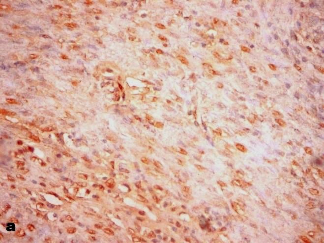
Inter J Vet Sci, 2019, 8(2): 113-119.

    adult albino rat and the possible role of human               Rajesh S, KS Rajendra, LS Madhu, KP Devendra, WA
    chorionic gonadotropin hormone: a histological                    Reyazet al., 2010. Neuroprotective effect of curcumin
    study. Egyptian J Histol, 37:327-338.                             in arsenic-induced neurotoxicity in rats. Neuro
Firdausa F, M Zafeer, M Waseem, R Ullah, M Ahmad, M                   Toxicol, 31: 533–539.
    Afzal, 2018. Thymoquinone alleviates arsenic                  Rodriguez KF, EK Ungewitter, Y Crespo-Mejias, C Liu,
    induced hippocampal toxicity and mitochondrial                    B Nicol, GE Kissling and HH Yao, 2016. Effects of
    dysfunction by modulating mPTP in Wistar rats.                    in utero exposure to arsenic during the second half of
    Biomed Pharmacother, 102: 1152–1160.                              gestation on reproductive end points and metabolic
Goudarzi M, S Amiric, A Nesarid, A Hosseinzadehe, E                   parameters in female CD-1 mice. Environm Health
    Mansourif, S Mehrzadig, 2018. The possible                        Perspectives, 124:336–343.
    neuroprotective effect of ellagic acid on sodium              Souza, AC, SC Marchesi, RP Ferraz, GD Lima, JA
    arsenateinduced neurotoxicity in rats. Life Sci, 198:             Oliveira and M Machado-Neves, 2016. Effects of
    38–45.                                                            sodium arsenate and arsenite on male reproductive
Holman, BW and AE Malau-Aduli, 20120.Spirulina as a                   functions in wistar rats. J Toxicol Environ Health,
    livestock supplement and animal feed. J Anim                      79:274-286.
    Physiol Anim Nutr, 97: 615-623.                               Sun HJ, P Xiang, J Luo, H Hong, H Lin, HB Li, LQ Ma,
Khalil SR, WM Elhady, YH Elewa, NE El-Hameed., SA                     2016. Mechanisms of arsenic disruption on gonadal,
    Ali, 2018. Possible role of Arthrospira platensis in              adrenal and thyroid endocrine systems in humans: A
    reversing oxidative stress-mediated liver damage in               review. Environ Int, 95:61–68.
    rats exposed to lead. Biomed. Pharmacother, 97:               Vigani M, C Parisi, E Rodríguez-Cerezo, MJ Barbosa, L
    1259-1268.                                                        Sijtsma, M Ploeg, C Enzing, 2015. Food and feed
Khan A, HI Hussain, A Sattar, MZ Khan and RZ Abbas,                   products from micro-algae: market opportunities and
    2014. Toxico-pathological aspects of arsenic in birds             challenges for the EU. Trends Food Sci Technol, 42:
    and mammals: A Review. Int J Agric Biol, 16: 1213‒                81–92.
    1224.                                                         Wang L, B Pan, J Sheng, J Xu, Q Hu, 2007. Antioxidant
Li S G, SZ Xu, Q Niu, YS Ding, LJ Pang, RL Ma, MX                     activity of Spirulina platensis extracts by supercritical
    Jing, K Wang, XM Ma, GL Feng, JM Liu, XF Zhang,                   carbon dioxide extraction. Food Chem., 105 (1): 36–
    HL Xiang and F Li, 2015. Lutein alleviates arsenic-               41.
    induced reproductive toxicity in male mice via Nrf2           Wares MA, MA Awal, SK Das and J Alam, 2013.
    signaling. Human Experim Toxicol, 35:491-500.                     Environmentally persistant toxicant arsenic affects
Mehta M and SS Hundal, 2016. effect of sodium arsenite                uterus grossly and histologically. Bangladesh Soc Vet
    on reproductive organs of female wistar rats. Arch                Med, 11:61-68.
    Environm Occupational Health, 71:16-25.                       Zhao P, Y Guo, W Zhang, H Chai, H Xing, M Xing,
Ohkawa H, N Ohishi and K Yagi, 1979. Assay tor lipid                  2017. Neurotoxicity induced by arsenic in Gallus
    peroxides in animal tissues by thiobarbituric acid                Gallus: Regulation of oxidative stress and heat shock
    reaction. Analytical Biochem, 95:351-358.                         protein response. Chemosphere, 166: 238e245.

                                                            119
You can also read