Morphometric Analysis of 3D Soft-Tissue for Sexual Dimorphism in Human Face - SciELO
←
→
Page content transcription
If your browser does not render page correctly, please read the page content below
Int. J. Morphol.,
38(2):367-373, 2020.

                        Morphometric Analysis of 3D Soft-Tissue
                        for Sexual Dimorphism in Human Face

         Análisis Morfométrico de Tejidos Blandos 3D de Dimorfismo Sexual en Rostro Humano

                Olalekan Agbolade1,2; Azree Nazri1,2; Razali Yaakob1; Abdul Azim Ghani3 & Yoke Kqueen Cheah4

AGBOLADE, O.; NAZRI, A.; YAAKOB, R.; GHANI, A. A. & CHEAH, Y. K. Morphometric analysis of 3D soft-tissue for sexual
dimorphism in human face. Int. J Morphol., 38(2):367-373, 2020.

         SUMMARY: Sexual dimorphism in Homo-sapiens is a phenomenon of a direct product of evolution by natural selection where
evolutionary forces acted separately on the sexes which brought about the differences in appearance between male and female such as in
shape and size. Advances in morphometrics have skyrocketed the rate of research on sex differences in human and other species.
However, the current challenges facing 3D in the acquisition of facial data such as lack of homology, insufficient landmarks to characterize
the facial shape and complex computational process for facial point digitization require further study in the domain of sex dimorphism.
This study investigates sexual dimorphism in the human face with the application of Automatic Homologous Multi-points Warping
(AHMW) for 3D facial landmark by building a template mesh as a reference object which is thereby applied to each of the target mesh
on Stirling/ESRC dataset containing 101 subjects (male = 47, female = 54). The semi-landmarks are subjected to sliding along tangents
to the curves and surfaces until the bending energy between a template and a target form is minimal. Principal Component Analysis
(PCA) is used for feature selection and the features are classified using Linear Discriminant Analysis (LDA) with an accuracy of 99.01
% which demonstrates that the method is robust.

        KEY WORDS: Sexual dimorphism; Facial landmark; 3D geometric morphometrics; Multi-point warping; LDA.

INTRODUCTION

       Identification of sexes plays a remarkable role when             Alvarez et al., 2013). Sliding semi-landmark was used in
it comes to social communication. This identification of                (Perez et al., 2006) to investigate craniofacial and dental
sexes by human beings is relatively easy and accurate.                  variation in human, by minimizing bending energy and
However, achieving the same result with classification                  Procrustes distance. Sliding semi-landmarks are used in
through machine remains a challenge in computer vision.                 the study of surfaces and curves on meshes. To circumvent
As the face is the part that hosts the most crucial sensory             the problem of asymmetry caused by manual semi-
organs and acts as the central interface for appearance,                landmarks, sliding semi-landmarks that are relaxed against
communication, expression, and identification (Peng et al.,             a symmetrized mean using bending energy minimization
2013).                                                                  was proposed in (Schlager & Rüdell, 2015), in the
                                                                        investigation of nasal soft tissue reconstruction. To evaluate
       Morphometric examines shape variation, group                     difference software packages for semi-landmark, Botton-
differences in shape, central tendency of shape, and                    Divet et al. (2015) used sliding semi-landmark to analyze
associations of shape with extrinsic factors (Slice, 2007).             the workflow complexity and time consumption to com-
In morphometry, sliding semi-landmarks have been used                   plete the sliding task. However, analyzing facial variation
in the study of bone surface such as articular and the                  in soft-tissue for sexual dimorphism in human from sliding
diaphysis (Fabre et al., 2014) and curves, providing                    semi-landmark is not prevalent in the three dimensional
descriptors of outlines and crests (De Groote et al., 2010;             model.

1
  Department of Computer Science, Faculty of Computer Science & IT, Universiti Putra Malaysia, Selangor, Malaysia.
2
  Institute of Bioscience, Universiti Putra Malaysia, Selangor, Malaysia.
3
  Department of Software Engineering, Faculty of Computer Science & IT, Universiti Putra Malaysia, Selangor, Malaysia.
4
  Department of Biomedical Science, Faculty of Medicine and Health Sciences, Universiti Putra Malaysia, Selangor, Malaysia.
  FUNDING: This work was supported by Fundamental Research Grant Scheme, Ministry of Higher Education, Malaysia (MOHE)–FRGS Code: 5524959.
And Putra German UPM-Code:9538100.

                                                                                                                                      367
AGBOLADE, O.; NAZRI, A.; YAAKOB, R.; GHANI, A. A. & CHEAH, Y. K. Morphometric analysis of 3D soft-tissue for sexual dimorphism in human face. Int. J Morphol., 38(2):367-373, 2020.
This work aims to investigate whether geometric Multi-Point Warping Approach. The template mesh is
morphometric analyses of soft-tissue landmarks using multi- created by manually locating sixteen anatomical points on the
point warping is reliable to assess sex differences in the 3D face (Fig. 1) called anchor points according to 3D facial
human face. This is done by projecting the surface semi- landmark standard in (Caple & Stephan, 2016) with little
landmarks from the template object to the target objects and modification (details in Table I).
iteratively slides the semi-landmarks to a point relaxed. Here
we used six iterations to ensure convergence and optimum The anchor landmarks are not subjected to sliding, but
smoothness. This method is not new, in analyzing shape are used for establishing the warping fields that will be used
variation in geometry morphometric, but its application to for minimizing the bending energy. Due to the easy detection,
the analysis of shape variation for soft-tissue three-dimen- pose correction and invariance to facial expression of nose
sional sexual dimorphism in human face is novel and the tip, the nose tip (pronasale) was selected as the most robust
simplicity of the workflow requires to performing the semi- and prominent landmark point. Since the nose tip area can be
landmark sliding task in Viewbox 4.0. The results are further approximated as a semi-sphere of the human face. This is
used to investigate size and shape variation in Stirling dataset where the sliding points begin to spread across the facial
to identify the features that are most dimorphic in male and surface. Using this anchor point (pronasale), 484 semi-
female faces; as the features responsible for dimorphism in landmarks were automatically generated overlapping on each
humans are still under study (Samal et al., 2007). other at the pronasale region showing in blue color. These are
uniformly and randomly distributed on the selected facial
surface with 1.5 mm radius to accommodate all 500 points
MATERIAL AND METHOD using method in (Zelditch et al., 2012). And the landmarks
sliding and acquisition is implemented in Viewbox 4.0 soft-
ware (Halazonetis, 2018).
Dataset and description. The dataset is acquired from
Stirling/ESRC 3D Face Database captured by a Di3D camera The semi-landmarks are allowed to slide on the curve
system (Stirling-ESRC, 2018). The image format is in and surface mesh of each target using TPS warping of the
wavefront obj file containing 101 subjects (male = 47, female template. This positions the reference points on the target fa-
= 54) of 3D facial scans in neutral expression were randomly cial mesh by minimizing the bending energy. Because warping
selected which are intended to facilitate research in sexual may result in points that do not lie directly on the facial surface
dimorphism, face recognition, expression recognition, and on the target mesh (Figs. 2A,B), the transferred points are
perception. The dataset is being used as a test set for a projected on the closest point on the mesh surface using ICP
competition on 3D face reconstruction from 2D images, with method (Creusot et al., 2010) which aims to iteratively
the 3D scans acting as 'ground truth' in IEEE conference. minimize the mean square error between two point sets. If the
distance between the two points is within an acceptable
threshold, then the closest point is determined as the
corresponding point. During the relaxation of the spline, the
semi-landmarks slide along the surface and the curve tangent
structures and not on the surfaces or the curves which reduces
the computational effort, as the minimization problem became
linear. This is because the sliding along the tangents lets the
semi landmarks slip off the data and the target surface mesh is
then considered homologous (Figs. 2C, D).
In assessing error, six subjects (three males and three
females) from the sample are randomly selected; each one
belonging to a different individual, distinct from the template
subject. Each was digitized twice following the same method
to account for digitization error. The results are analyzed using
Procrustes ANOVA. This is done by the minimization of the
squared sum of the distance of all objects and the consensus
configuration (Fruciano, 2016).
Fig. 1 A three-dimensional mesh template showing 16 PCA and LDA. Due to a large number of facial
fixed anatomical landmarks. landmarks, landmark coordinates were decomposed into
368AGBOLADE, O.; NAZRI, A.; YAAKOB, R.; GHANI, A. A. & CHEAH, Y. K. Morphometric analysis of 3D soft-tissue for sexual dimorphism in human face. Int. J Morphol., 38(2):367-373, 2020.
Table I. Anchor anatomical points and descriptions.
No Anchor Landmarks 3D Notation Description
Eyes Region
Left most medial point of the palpebral fissure, a t the inner commissure
1 Endocanthion left Enl
of the eye
Left most lateral point of the palpebral fissure, at the outer commissure
2 Exocanthion left Exl
of the eye
Right most lateral point of the palpebral fissure, a t the outer
3 Exocanthion right Exr
commissure of the eye
Right most medial point of the palpebral fissure, at the inner
4 Endocanthion right enr
commissure of the eye
5 Sellion se Deepest midline point of the nasofrontal angle
Nasal Region
6 Pronasale pr The most anteriorly protruded point of the apex nasi
Median point at the junction between the lower margin of the nasal
7 subnasale su
septum and the philtrum area
8 Alare left all Left most lateral point on the nasal ala
9 Alare right alr Right most lateral point on the nasal ala
Mouth Region
Left outer corners of the mouth where the outer edges of the upper and
10 Cheilion lef t chl
lower vermilions meet
Right outer corners of the mouth where the outer edges of the upper
11 Cheilion right chr
and lower vermilions meet
12 Labiale superius ls Midpoint of the vermilion margin of the upper lip
13 Labiale inferius li Midpoint of the vermilion margin of the lower lip
Chin-Cheek Region
14 Gnathion gn Median point halfway between pogonion and menton
Left median point where the sagittal suture intersec ts with a transverse
15 Obelion left obl
line connecting parietal foramina
Right median point where the sagittal suture intersec ts with a
16 Obelion right obr
transverse line connecting parietal foramina
their principal components and computed to account for
the proportion of variation and its significance. The first
seven principal components accounting for over 80 %
of the variation and the PCAs are used to characterize
the features of shape variation. To compare sizes, CS
(Centroid Size) between sexes, values were log-
transformed and Mann-Whitney U test was used for
significant differences in log CS medians between sexes
and the Kolmogorov-Smirnov D tests for overall equal
distribution of both sexes as CS does not assume a nor-
mal distribution. To correct the effect of size on the fa-
cial shape (Klingenberg & McIntyre, 1998), allometry
effect is explored to compute multivariate regression of
shape on log centroid size in MorphoJ. Subsequently,
differences in effects and size are examined by
computing non-parametric analysis of variance
Fig. 2 Sliding point warped on target facial surface. (A) Male partial
sliding on target mesh. (B) Female partial sliding on target mesh.
(C) Male complete and homologous warping on target mesh. (D)
Female complete and homologous warping on target mesh.
369AGBOLADE, O.; NAZRI, A.; YAAKOB, R.; GHANI, A. A. & CHEAH, Y. K. Morphometric analysis of 3D soft-tissue for sexual dimorphism in human face. Int. J Morphol., 38(2):367-373, 2020.
(MANOVA) in terms of Wilks’ lambda in PAST software. calculating the amount of displacement from the average
Using sex as a group and size as the covariate, the sex by position calculated from all digitization and the variation
size interaction term is performed. The MANOVA is accounts for the smallest portion of the total variation using
recomputed after removing the interaction term (sex by size) Procrustes ANOVA. The digitization error accounts for only
and the sex effect tests difference in regression intercept. 0.025 from the total variation (Table II).
The significant level for all tests is accepted at p < 0.05. To
predict the classification accuracy of the sexes, we apply To visualize the shape changes with landmark
LDA in PAST software. displacement after Procrustes superimposition, a lollipop
graph is plotted where the landmark positions of the starting
shape are denoted by dots (candy) and the shifts of the
RESULTS landmark to the target shape are denoted as lines (stick)
(Klingenberg, 2013). This visualization is sometimes
cumbersome in a three-dimensional context which has been
Significance of Landmark. The overall landmarks are tested applied in many morphometrics studies such as in (Rohlf,
using ANOVA to see the significance of the variation on the 1993). Because lollipop graph provides little information
overall landmarks in each sex group. Male: F=3045, df = underlying anatomical structure, more sophisticated relative
1391, p-value = 0.00001; Female: F = 3638, df = 1391, p- warped graphs are plotted after Procrustes fit to provide more
value = 0.00001. Furthermore, we conduct PERMANOVA information of the position of shape changes using PCs as a
(Non-Parametric MANOVA) which is a non-parametric test 3D vector from the mean configuration (Dryden, 2014).
of the significant difference between the sex groups based
on the distance measured (Anderson, 2001) with F = 17.33, The first 7 PCs of the PCA explain more than 80 %
p = 0.00001 and r = 0.98. The large positive of F value of the variance; PC1 explains 37.25 of the variance followed
indicates that there is a significant difference between the by PC2 with 11.91 % variance. The log centroid size for
sex groups. multivariate regression is only slightly different between the
sexes (U =1134, p = 0.25) (Fig. 3A), therefore not significant,
Error Assessment, Size and Shape Variation. For the though the test for equal distribution is significant (D = 2.08,
assessment of digitization errors of the overall landmarks; p = 0.024). Inter-individual allometry explains only 2.65 %
the deviations of each landmark is obtained by simply of shape differences according to size (Fig. 3B) and therefore
is not significant (p = 0.11). The regression results
Table II. Procrustes ANOVAs for facial shape indicate that there is some weak evidence against the
Effect SS MS DF F P null hypothesis of independence. Given the sample size
Sex 0.011884 7.96E-06 1493 3.22AGBOLADE, O.; NAZRI, A.; YAAKOB, R.; GHANI, A. A. & CHEAH, Y. K. Morphometric analysis of 3D soft-tissue for sexual dimorphism in human face. Int. J Morphol., 38(2):367-373, 2020.
The pattern of major facial shape variation occurs in
nasal region (alare), mouth region (labiale superius, labiale
interius and chelion) and chin-cheek region (gnathion and
obelion) and the sticks tell us which way things change along
the principal components (Fig. 4). In the nasal region, the
direction of the stick is longer in the female group than that of
male, but the nasal width in male is wider than that of the
female. In chin-cheek region, the female group has a more
curved gnathion to obelion but shorter than that of the male
group. In the mouth region, the stick is longer from labiale
superius to labiale interius in female group and chelion is wider
in the male group; which indicates that female group has ticker
lips than that of the male group, although the male group has
wider lips. There is no noticeable shape difference in the eyes
region. These results demonstrate that although male facial
tissues are generally larger than that of female, the difference
is not generally isometric and the relative warp of the princi-
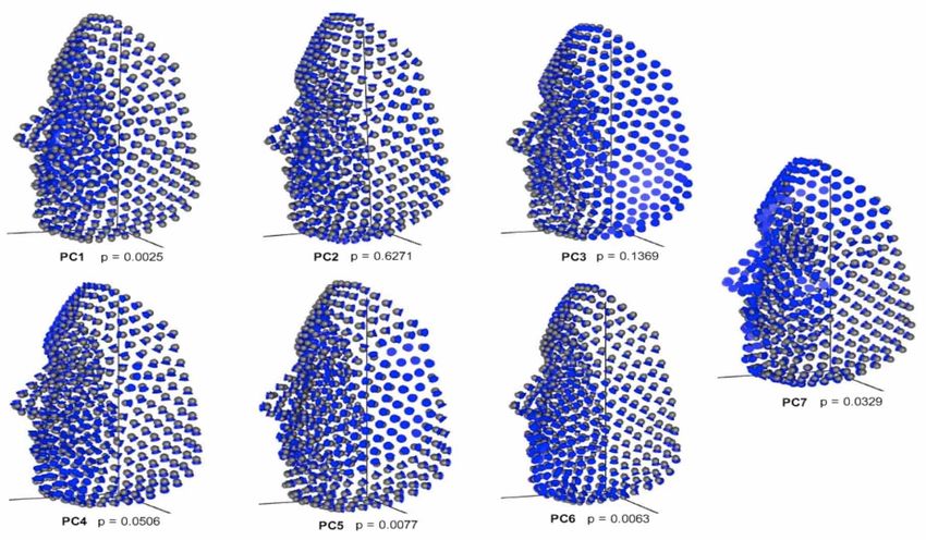
Fig. 4. Lollipop Graphs visualizing face shape changes. A (male), pal components (PC1-PC7) (Fig. 5) gives us more information
B (female). and helps us to identify those regions where the facial shapes
are more dimorphic between the sexes. The images are
The interaction term (test for slopes) is statistically normally plotted horizontally but we flipped vertically to have
significant (Table III). When the size effect is removed and a better profile view for proper presentation.
the MANOVA is repeated, the result is still statistically
significant. This suggests that the effect of size on shape for The relative warp scores are subjected to LDA, testing
both slope and intercept is strong and not similar in the sex for differences in face shape between sexes in PAST software,
group. which are treated as independent variables. And a multivariate
function was defined such that males and females were
maximally discriminated (Nacua et al., 2010). The confusion
Table III. MANOVA results in terms of Wilks’ Lambda. matrix in Table IV shows the actual and predicted values of
Effect Wilk df1 df2 f P-value the sex groups. No performance report is given as LDA does
Sex x CS 0.301 8 91 26.42AGBOLADE, O.; NAZRI, A.; YAAKOB, R.; GHANI, A. A. & CHEAH, Y. K. Morphometric analysis of 3D soft-tissue for sexual dimorphism in human face. Int. J Morphol., 38(2):367-373, 2020.
DISCUSSION The female generally has elongated chin height (li-
gn) than the male group, though male chin height is more
curved than that of the female. The nasal width (all-alr) is
The approach in this algorithm uses sliding semi- wider in the female than that of male, though mere looking
landmark, starting at the pronasale with respect to the arbitrary at one face may confuse the viewer, but the average data
template model where the bending energy between all subjects gathered justifies the reports. Cheek length (gn-obr) in male
is minimized by six cycles iterative sliding. This is important is longer and more curved than that of the female, though
because manual semi-landmarks are not appropriate for the that of female steep downward than that of the male. The
comparison of forms and shapes when the curves and surfaces nasal bridge length (se-pr) in male is a little longer than that
are not homologous among the targets. Because the point of the female. The female nasal tip (pr-su) is a little bit more
homology across specimens experienced by morphometrics protuberant than that of the male but wider in male group.
measurement of semi-landmarks on curves and surfaces Biocular width (enl-enr) is wider in male than that of the
manually is no longer guaranteed due to the biological female and upper lip height (su-ls) is longer in female.
meaninglessness and un-interpretable of sample averages and Morphological characteristics of male and female faces vary
variances (Mitteroecker et al., 2013). in different races or datasets, therefore presenting a consensus
may not be scientifically acceptable.
The Procrustes ANOVA suggests a modest but
appreciable variation in facial shape. Shape differences are Generally, the male face shape is bigger than the
statistically significant even after averaging faces within sex. female face shape. We further predict the sexes by employing
Small measurement error shows that the landmarks can be one of the supervised learning techniques, LDA; and the
annotated with precision using the proposed method. Though, classifier classified the sex groups with an accuracy of 99.01
many approaches are available in addressing measurement %. Though, one male was misclassified as female by the
error. Discussing such at length is beyond the scope of this classifier. To the best of our knowledge, there is currently
study, more and extended details can be found in Fruciano. no facial landmark annotation analysis or sexual dimorphism
performed using Stirling/ESRC dataset.
Allometry in shape is tested by examining the
statistical correlation between size and shape. This
characterizes the expected shape changes per (centroid size) CONCLUSIONS
unit increase size. The statistical significance of the
association between shape and size is tested statistically
based on Goodall (1991) F statistic. When the factors other This method combines pragmatic solutions to con-
than size, have effects on shape variation such as sexual figure an optimized pipeline for high-throughput
dimorphism, the plots are not optimal. To avoid this, a homologous multi-points facial signature in three dimen-
computation on regression score is performed by projecting sional to the application of sexual dimorphism. The
data points in shape space unto the axis in the direction of landmarks accuracy is measured using deviation from the
regression vector (Drake & Klingenberg, 2008). The tests reference surface tothe target surface with Procrustes
for intercept and slope using MANOVA are both statistically distance after superimposition and the error rate through
significant. This suggests that the effect of size on shape is Procrustes ANOVA is minimal. The dimorphic regions are
strong and not similar in the sex group. As it is expected identified and visualized using PCA. Though regression
since sex has a large phenotypic variation, the allometric results indicate weak evidence of allometry yet the tests for
trajectories are largely aligned with the vector of mean shape slope and intercept for the effect of size on shape are
differences. significant. Such a high-throughput phenotypic facial data
with good classification accuracy like this is not only
Morphological differences associated with the prin- valuable for sexual dimorphism but also in forensic studies
cipal components are linked to sexual dimorphism to explain of human facial morphology, anthropology, disease diag-
more anatomical details. Only PC1 (p = 0.0025), PC5 (p = nosis and prediction, statistical shape or image analysis, face
0.0077), PC6 (p = 0.0063) and PC7 (p = 0.0329) are recognition, age estimation, facial expression recognition,
significant; whereas PC2 (p = 0.6271), PC3 (p = 0.1369) etc. This study is based on Stirling/ERSC dataset which is
and PC4 (p = 0.0506) are not significant. The PCs also reveal the European population, therefore the methods and results
sexual dimorphic regions: PC1 and PC2 (mouth region), PC3 presented here should be tested in other populations. Finally,
and PC5 (cheek region), PC4 and PC6 (chin region) and implementation with deep learning may yield better perfor-
PC7 (nasal region and upper-head region), though upper- mance and robust result in the feature with respect to
head region is not considered in this study. ethnicity and moderate changes in facial features.
372AGBOLADE, O.; NAZRI, A.; YAAKOB, R.; GHANI, A. A. & CHEAH, Y. K. Morphometric analysis of 3D soft-tissue for sexual dimorphism in human face. Int. J Morphol., 38(2):367-373, 2020.
ACKNOWLEDGMENTS Creusot, C.; Pears, N. & Austin, J. 3D Face Landmark Labelling. Proceedings of
the ACM Workshop on 3D Object Retrieval, 2010.
De Groote, I.; Lockwood, C. A. & Aiello, L. C. Technical note: A new method for
measuring long bone curvature using 3D landmarks and semi-landmarks.
We acknowledge Stirling/ESRC (University of Am. J. Phys. Anthropol., 141(4):658-64, 2010.
Drake, A. G. & Klingenberg, C. P. The pace of morphological change: historical
Stirling) for prompt agreement to use their datasets. transformation of skull shape in St Bernard dogs. Proc. Biol. Sci.,
Furthermore, credit goes to Computer Laboratory of the 275(1630):71-6, 2008.
Faculty of Computer Science & Information Technology, Dryden, I. L. Shape Analysis. Wiley StatsRef: Statistics Reference Online, 2014.
doi: 10.1002/9781118445112.stat05087
Universiti Putra Malaysia.
Fabre, A. C.; Goswami, A.; Peigné, S. & Cornette, R. Morphological integration
in the forelimb of musteloid carnivorans. J. Anat., 225(1):19-30, 2014.
Fruciano, C. Measurement error in geometric morphometrics. Dev. Genes Evol.,
AGBOLADE, O.; NAZRI, A.; YAAKOB, R.; GHANI, A. A. & 226(3):139-58, 2016.
Goodall, C. Procrustes methods in the statistical analysis of shape. J. R. Stat. Soc.
CHEAH, Y. K. Análisis morfométrico de tejidos blandos 3D de
Series B Methodol., 53(2):285-321, 1991.
dimorfismo sexual en rostro humano. Int. J. Morphol., 38(2):367- Halazonetis, D. Viewbox 4 - Cephalometric Software. 2018. Available from: http:/
373, 2020. /dhal.com/viewboxindex.htm
Klingenberg, C. P. & McIntyre, G. S. Geometric morphometrics of developmental
RESUMEN: El dimorfismo sexual en el Homo-sapiens instability: Analyzing patterns of fluctuating asymmetry with procrustes
methods. Evolution, 52(5):1363-75, 1998.
es un fenómeno directo de la evolución por selección natural, don-
Klingenberg, C. P. Visualizations in geometric morphometrics: how to read and
de las fuerzas evolutivas actuaron por separado en los sexos, lo how to make graphs showing shape changes. Hystrix Ital. J. Mammal.,
que provocó las diferencias en la apariencia entre hombres y mu- 24(1):15-24, 2013.
jeres, tal como la forma y tamaño. Los avances en el área de la Klingenberg, C. P.; Barluenga, M. & Meyer, A. Shape analysis of symmetric
morfometría, han generado un aumento significativo de las inves- structures: Quantifying variation among individuals and asymmetry.
tigaciones en las diferencias de sexo en humanos y otras especies. Evolution, 56(10):1909-20, 2002.
Mitteroecker, P.; Gunz, P.; Windhager, S. & Schaefer, K. A brief review of shape,
Sin embargo, los desafíos actuales que enfrenta el 3D en el análisis form, and allometry in geometric morphometrics, with applications to human
de datos faciales, como la falta de homología, puntos de referencia facial morphology. Hystrix Ital. J. Mammal., 24(1):59-66, 2013.
insuficientes para caracterizar la forma facial y la complejidad del Nacua, S. S.; Torres, M. A. J. & Demayo, C. G. Landmark-based geometric
proceso computacional para la digitalización de puntos faciales, morphometrics in visualizing body shape dimorphism in the endemic cyprinid,
requiere un estudio adicional en el área del dimorfismo sexual. Puntius tumba (Herre, 1924), from Lake Lanao, Philippines. 2010
International Conference on Environmental Engineering and Applications
Este estudio investiga el dimorfismo sexual en el rostro humano
(ICEEA), 2010.
con la aplicación de la deformación automática de múltiples pun- Peng, S.; Tan, J.; Hu, S.; Zhou, H.; Guo, J.; Jin, L. & Tang, K. Detecting genetic
tos homólogos para el hito facial 3D, mediante la elaboración de association of common human facial morphological variation using high
una malla de plantilla como objeto de referencia, y se aplica en density 3D image registration. PLoS Comput. Biol., 9(12):e1003375, 2013.
cada una de las mallas objetivas en el conjunto de datos Stirling / Perez, S. I.; Bernal, V. & Gonzalez, P. N. Differences between sliding semi-
landmark methods in geometric morphometrics, with an application to human
ESRC que contiene 101 sujetos (hombre = 47, mujer = 54). Los
craniofacial and dental variation. J. Anat., 208(6):769-84, 2006.
semi-puntos de referencia se deslizan a lo largo de las tangentes a Rohlf, F. J. Relative Warp Analysis and an Example of its Application to Mosqui-
las curvas y superficies hasta que la energía de flexión entre una to Wings. New York, State University of New York, 1993.
plantilla y una forma objetivo es mínima. El análisis de compo- Samal, A.; Subramani, V. & Marx, D. Analysis of sexual dimorphism in human
nentes principales (PCA) se utiliza para la selección de caracterís- face. JoJ. Vis. Commun. Image Represent., 18(6):453-63, 2007.
ticas y las características se clasifican mediante el análisis discri- Schlager, S. & Rüdell, A. Analysis of the human osseous nasal shape--population
differences and sexual dimorphism. Am. J. Phys. Anthropol., 157(4):571-81,
minante lineal (ADL) con una precisión del 99,01 %, lo que de- 2015.
muestra la validez del método. Slice, D. E. Geometric morphometrics. Ann. Rev. Anthropol., 36(1):261-81, 2007.
Stirling-ESRC. Stirling-ESRC 3D Face Database, 2018. Available from: http://
PALABRAS CLAVE: Dimorfismo sexual; Punto de re- pics.stir.ac.uk/ESRC/3d_images.htm
ferencia facial; Morfometría geométrica 3D; Deformación Zelditch, M. L.; Swiderski, D. L. & Sheets, H. D. Geometric Morphometrics for
Biologists: A Primer. Amsterdam, Elsevier Academic Press, 2012.
multipunto; LDA.
Corresponding author:
Dr. Azree Nazri
REFERENCES Department of Computer Science
Faculty of Computer Science & IT
Universiti Putra Malaysia
Alvarez, A.; Ercoli, M. D. & Prevosti, F. J. locomotion in some small to medium-
sized mammals: a geometric morphometric analysis of the penultimate lum-
Selangor - MALAYSIA
bar vertebra, pelvis and hindlimbs. Zoology (Jena), 116(6):356-71, 2013.
Anderson, M. J. A new method for non-parametric multivariate analysis of
variance. Austral Ecol., 26(1):32-46, 2001. Email: azree@upm.edu.my
Botton-Divet, L.; Houssaye, A.; Herrel, A.; Fabre, A. C. & Cornette, R. Tools for
quantitative form description; an evaluation of different software packages
for semi-landmark analysis. PeerJ, 3:e1417, 2015.
Caple, J. & Stephan, C. N. A standardized nomenclature for craniofacial and Received: 29-08-2019
facial anthropometry. Int. J. Legal Med., 130(3):863-79, 2016. Accepted: 14-10-2019
373You can also read