The Experimental Infections of the Human Isolate of Strongyloides Stercoralis in a Rodent Model (The Mongolian Gerbil, Meriones Unguiculatus) - MDPI
←
→
Page content transcription
If your browser does not render page correctly, please read the page content below
Article
The Experimental Infections of the Human Isolate of
Strongyloides Stercoralis in a Rodent Model (The
Mongolian Gerbil, Meriones Unguiculatus)
Sarit Charuchaibovorn 1, Vivornpun Sanprasert 2,3 and Surang Nuchprayoon 2,3,*
 1 Medical Sciences Program, Faculty of Medicine, Chulalongkorn University, Bangkok 10330, Thailand;
   sarit.src@gmail.com
 2 Lymphatic Filariasis and Tropical Medicine Research Unit, Chulalongkorn Medical Research Center,

   Faculty of Medicine, Chulalongkorn University, Bangkok 10330, Thailand; vivornpun@chula.md
 3 Department of Parasitology, Faculty of Medicine, Chulalongkorn University, Bangkok 10330, Thailand

 * Correspondence: fmedstt@gmail.com; Tel.: +66-2-256-4387

 Received: 9 January 2019; Accepted: 31 January 2019; Published: 5 February 2019

 Abstract: Strongyloidiasis is life-threatening disease which is mainly caused by Strongyloides
 stercoralis infection. Autoinfection of the parasite results in long-lasting infection and fatal
 conditions, hyperinfection and dissemination (primarily in immunosuppressed hosts). However,
 mechanisms of autoinfection and biology remain largely unknown. Rodent models including mice
 and rats are not susceptible to the human isolate of S. stercoralis. Variations in susceptibility of the
 human isolate of S. stercoralis are found in dogs. S. ratti and S. venezuelensis infections in rats are an
 alternative model without the ability to cause autoinfection. The absence of appropriate model for
 the human isolate of strongyloidiasis hampers a better understanding of human strongyloidiasis.
 We demonstrated the maintenance of the human isolate of the S. stercoralis life cycle in the
 Mongolian gerbil (Meriones unguiculatus). The human isolate of S. stercoralis caused a patent
 infection in immunosuppressed gerbils, more than 18 months. The mean number of recovery adult
 parasitic worms were 120 ± 23 (1.2% of the initial dose) and L1s were 12,500 ± 7,500 after day 28
 post-inoculation (p.i.). The prepatent period was 9–14 days. Mild diarrhoea was found in gerbils
 carrying a high number of adult parasitic worms. Our findings provided a promising model for
 studying biology and searching new alternative drugs against the parasites. Further studies about
 the hyperinfection and dissemination would be performed.

 Keywords: Strongyloidiasis; Strongyloides stercoralis; Mongolian gerbil (Meriones unguiculatus),
 Hyperinfection; Dissemination

1. Introduction
      Strongyloidiasis is a parasitic disease, caused by Strongyloides spp. infections. Strongyloides
stercoralis is a major pathogenic species which is capable of infecting humans, dogs and non-human
primates. S. stercoralis infection causes significant health problems throughout the world, particularly
in Sub-Saharan Africa, South-America and Southeast Asia [1,2]. Approximately, 370 million people
are infected with the parasite [3–5]. The most unique characteristic of the parasite is the ability to
multiply and complete their life cycle within hosts as a result of so-called “autoinfection.” In
immunocompetent hosts, S. stercoralis can cause a long-lasting infection, up to 75 years [6,7]. In
immunocompromised hosts, the cycle of autoinfection can dramatically increase resulting in
potentially life-threatening conditions, including hyperinfection and dissemination. The
autoinfective larvae (L3a) can penetrate through the intestinal wall into other organs and cause death
due to organ failure and sepsis. Mortality rates of the hyperinfection and dissemination syndrome

Pathogens 2019, 8, 21; doi:10.3390/pathogens8010021                          www.mdpi.com/journal/pathogens
Pathogens 2019, 8, 21 2 of 11
are 34.6–87% [8–11]. Moreover, fatal hyperinfection cases are also found, even in immunocompetent
hosts [12,13]. The mortality rate of hyperinfection in immunocompetent hosts is 22% [13]. The
mechanisms of autoinfection and biology of S. stercoralis are largely unknown. To obtain a better
understanding of S. stercoralis infection in a human, an appropriate animal model is required.
In the 1980s, dogs and Patas monkeys were used as a model for strongyloidiasis studies [14–18].
However, they are unsatisfactory to be a laboratory model, due to a few disadvantages, such as
difficulty of handling and high maintenance costs. Moreover, there are variabilities in susceptibility
of dogs to the human isolate of S. stercoralis [14,19–21]. Cats and ferrets are also demonstrated for S.
stercoralis infection, however, they are not able to develop a chronic infection as found in humans.
Most rodent models, including mice, rats, guinea pigs are not susceptible to S. stercoralis [22].
Although SCID mice is susceptible to S. stercoralis, only 0.02–0.3% of infective larvae can develop into
adult parasitic worms, even in a high initial dose of infective larvae [23]. Although S. ratti and S.
venezuelensis infections in rats are used as an alternative model for strongyloidiasis study, these
parasites lack the ability to cause the autoinfection which is the unique feature of S. stercoralis
infection in humans [24–26]. The Mongolian gerbil (Meriones unguiculatus) is an important model for
strongyloidiasis study. Gerbils are able to develop a chronic infection, up to 131 days. Moreover,
complicated conditions (hyperinfection and dissemination) are developed in immunosuppressed
animals [27]. However, most of the recent studies of the gerbil model used the dog isolate S. stercoralis
[28–30], while human isolate S. stercoralis is less developed in gerbils. Although marmoset is able to
develop uncomplicated and complicated strongyloidiasis, using human isolate S. stercoralis, they are
less than ideal laboratory host [31].
In this study, we developed the gerbil model for the human isolate of S. stercoralis. The human
isolate of S. stercoralis developed a chronic infection, more than 18 months, in immunosuppressed
gerbils. The findings of this study provide a useful model for the study of biology and novel drug
research of strongyloidiasis.
2. Results
2.1. Immunosuppressed gerbils were susceptible to the human isolate of S. stercoralis.
Thirteen Mongolian gerbils were immunosuppressed with 2 mg methylprednisolone (MPA) and
inoculated by subcutaneous injection with 1,000 (group 1, N = 7) or 10,000 (group 2, N = 6) of S.
stercoralis infective stage larvae (iL3s) that were obtained from human faecal cultures (Table 1). The
susceptibility of infections was observed daily by faecal direct smear and agar plate after day 7 post-
inoculation (p.i.). In group 1, two of five male gerbils were positive for S. stercoralis infection by agar
plates after day 9 p.i, while two female gerbils died unexpectedly on day 3 p.i. (Table 1). The direct
smear was negative in all gerbils (Table 1). After day 28 p.i., a few adult parasitic worms (2–4 worms)
were recovered from intestines of the two male gerbils (Table 1 and Figure 1A). No L1s and L3a were
found from the infected animals. With higher inoculation dose (group 2), all 4 male gerbils were
positive for S. stercoralis on direct smear and agar plates after day 9 p.i., while the 2 female gerbils
were negative. After day 28 p.i., three infected gerbils were euthanized, while the remaining gerbil
(no. 11) was used as the source of iL3s for the later experiment (group 3–8) and still alive at 18 months.
Adult parasitic worms were found in small and large intestines. The number of recovered adult
parasitic worms from infected male gerbils varies from 22 to 188 worms and L1s were between 3,500
and 22,000 worms (Table 1 and Figure 1). A few L1s were found in the lungs of one gerbil (gerbil no.
8). No L3a were recovered from infected animals. Mild diarrhoea was noted in infected gerbils
carrying a high number of adult parasitic worms (gerbil no. 8).Pathogens 2019, 8, 21 3 of 11
Table 1. Recovery of S. stercoralis adult parasitic worms and L1s 28 days after initial infection of gerbils
with S. stercoralis iL3s.
Recovered
Dose
Source Dose of Direct Agar adult
Group No. Gender of Recovered L1s
of iL3s iL3s smear plate parasitic
MPA
worms
1 − + 4 NF
2 − + 2 NF
3 Male 1,000 2mg − − NF NF
1 4 − − NF NF
5 − − NF NF
6 Death
Female 1,000 2mg
7 Death
Human
8 + +++ 188 22,000
9 + ++ 22 4,800
Male 10,000 2mg
10 + ++ 55 3,500
2 11 + +++ ND * ND *
12 − − NF NF
Female 10,000 2mg
13 − − NF NF
14 − + 7 66
3 Male 4,000 1mg
15 − + 2 500
16 − + 20 110
4 Male 5,000 1mg
17 − + 2 140
18 + ++ 1 ND **
5 Male 10,000 1mg
19 + ++ 55 ND **
20 + ++ 20 110
6 Male 3,000 2mg
Gerbil 21 − − NF NF
22 Death
7 Male 5,000 2mg
23 Death
24 + +++ 143 20,000
25 Male 10,000 2mg + +++ 97 5,000
8 26 + +++ ND * ND *
27 − ++ 2 NF
Female 10,000 2mg
28 − ++ 44 2,000
Note: −: No tracks of migration and/or larvae were found; +: Only tracks of migration were found; ++:
Free-living adults were first seen on day 4-5; +++: Numerous free-living adults were first seen on day
3; *: Animals still alive; **: The number of L1s could not be counted and estimated, due to a presence
of numerous of stool faecal debris in intestines; NF: not found; ND: no data.Pathogens 2019, 8, 21 4 of 11
(A) (B)
Figure 1. Recovery of S. stercoralis from gerbils infected with the human isolate of S. stercoralis
(obtained from human faecal cultures). The number of adult parasitic worms (A) and L1s (B) were
represented.
2.2. The human isolate of S. stercoralis life cycle could be maintained by gerbil-to-gerbil transfer.
To maintain S. stercoralis life cycle by gerbil-to-gerbil transfer, we suspected that gerbils were
more susceptible to human isolate S. stercoralis which were obtained from infected gerbils obtained
from strongyloidiasis patient. We decreased doses of MPA and varied initial doses of iL3s. Six gerbils
were immunosuppressed with 1 mg MPA and two each inoculated with 4,000 (group 3), 5,000 (group
4) or 10,000 (group 5) S. stercoralis iL3s that were obtained from gerbil No.11 faecal cultures. All gerbils
faeces were positive for S. stercoralis by agar plates, while group 3 and 4 were negative by direct smear
after day 9 p.i. (Table 1). The number of recovered adult parasitic worms varies from 1 to 55 without
relationship with inoculation dosage (Table 1 and Figure 2A). Similarly, the number of recovered L1s
varies from 66 to 500 without relationship with inoculation dosage (Table 1 and Figure 2B).
Unfortunately, the number of L1s in gerbil group 5 could not be counted or estimated, due to the
presence of numerous of the stool faecal debris in intestines. No L3a were found from infected
animals.
The experiment was repeated with higher degree of immunosuppression. Nine gerbils were
immunosuppressed with 2 mg MPA and two each were inoculated with 3,000 (group 6), 5,000 (group
7) and 5 gerbils with 10,000 (group 8) S. stercoralis iL3s that were obtained from gerbil No.11 faecal
cultures. One gerbil in group 6 and three gerbils in group 8 were positive for S. stercoralis by direct
smear and agar plates after day 9 p.i., while rest was negative (Table 1). Two female gerbils in group
8 were also positive by agar plates. Two gerbils in group 7 died unexpectedly on day 3 and 7 p.i. One
gerbil from the lowest inoculation (group 6) did not get infected. After day 28 p.i., the infected gerbil
from group 6, 2 male and 2 female gerbils from group 8 were euthanized. The remaining male gerbil
in group 8 still alive for 8 months. The number of recovered adult parasitic worms varies from 2 to
143 without relationship with inoculation dosage (Table 1 and Figure 2A). The number of recovered
L1s varies from 110 to 20,000 without relationship with inoculation dosage (Table 1 and Figure 2B).
A few L1s were found in the lungs of a gerbil in group 8 (gerbil no. 26). A few L3a were recovered in
the lungs (gerbil no. 26), representing an autoinfection.(A) (B)
Figure 2. Recovery of S. stercoralis from gerbils infected with the human isolate of S. stercoralis
(obtained from gerbil faecal cultures) (1 mg of MPA versus 2 mg of MPA). The number of adult
parasitic worms (A) and L1s (B) were represented.
3. Discussion
The absence of appropriate laboratory model for the human isolate of strongyloidiasis hampers
a better understanding of S. stercoralis infection in humans. The previous studies showed that the
Mongolian gerbil is susceptible to the dog isolate of S. stercoralis [32–34]. However, the dog isolate of
S. stercoralis might be different from the human isolate of S. stercoralis. Here, we demonstrate that the
immunosuppressed gerbils is a suitable laboratory animal to maintain the life cycle of the human
isolate of S. stercoralis.
Although linear relationship between the inoculation dosage and adult worm recovery from
gerbil is not consistent, higher inoculation dosage of iL3s and MPA result in a high yield of worms
and larvae from gerbils (Figure 2 and Figure 3). Unfortunately, the differences were not statistically
significant. It might be due to small size of animals per group. We found that male gerbils were more
susceptible to S. stercoralis infection more than female gerbils, (Table 1). This observation is consistent
with the results using the dog isolate of S. stercoralis. [27] Moreover, the source of iL3s (obtained from
strongyloidiasis patients and infected gerbils) did not influence the number of recovered adult
parasitic worms in gerbils.
Our percentage of recovery adult parasitic worms (1.2%) were slightly higher than recovery
worms in immunocompetent gerbils (0.29%) [34]. The percentage of recovered adult parasitic worms
of the human isolate of S. stercoralis were lower than the using the dog isolate of S. stercoralis
(7%).[27,34] The recovered L1s in this study was higher than the number of recovery worms in
immunocompetent gerbils up to ~ 20 folds [34]. The fecundity (L1s/adult ratio) was ~ 104.1, while the
fecundity of the dog isolate of S. stercoralis was 4.0–21.7 [27]. The result of coproparasitological
examination by agar plates showed that the L1s, excreted in faeces, were active and numerous,
consistent with the number of larvae recovered from the gut.
The human isolate of S. stercoralis can cause a persistent infection in MPA-treated gerbils as
found in humans (S. stercoralis, obtained from strongyloidiasis patients: for at least 18 months and S.
stercoralis, obtained from infected gerbils: for at least 8 months). The prepatent period was 9–14 days.
Mild diarrhoea (common clinical sign of human strongyloidiasis) was found in infected gerbils
carrying a high number of adult parasitic worms (> 100 worms/gerbil). Focal lesions were also
presented at the larval or MPA injection sites of a high number of adult parasitic worms, as previously
found in a previous study [32]. Other clinical symptoms were not found.
The most important characteristic of human strongyloidiasis is that the disease can be fatal in
immunocompromised hosts, due to hyperinfection and dissemination. The two fatal conditions arose
from increasing of autoinfection rates in which the L1s in the host’s intestines become the L3a and
Pathogens 2019, 8, 21; doi:10.3390/pathogens8010021 www.mdpi.com/journal/pathogensPathogens 2019, 8, 21 2 of 11
complete their parasitic life cycle in the host, especially in immunocompromised hosts. However, the
small number of L3a were found in 2 mg MPA-treated gerbils, even L1s were numerous in the
intestines. The results were not consistent with the results from using dog isolate of S. stercoralis
[27,34]. Either the characteristics of the parasites or the animal hosts affected the susceptibility. We
suspected that the discrepancies might be due to several factors. The autoinfection of dog isolate of
S. stercoralis in gerbils was induced by two factors, including treatment with MPA and injection with
a high dose of iL3s [27,34].
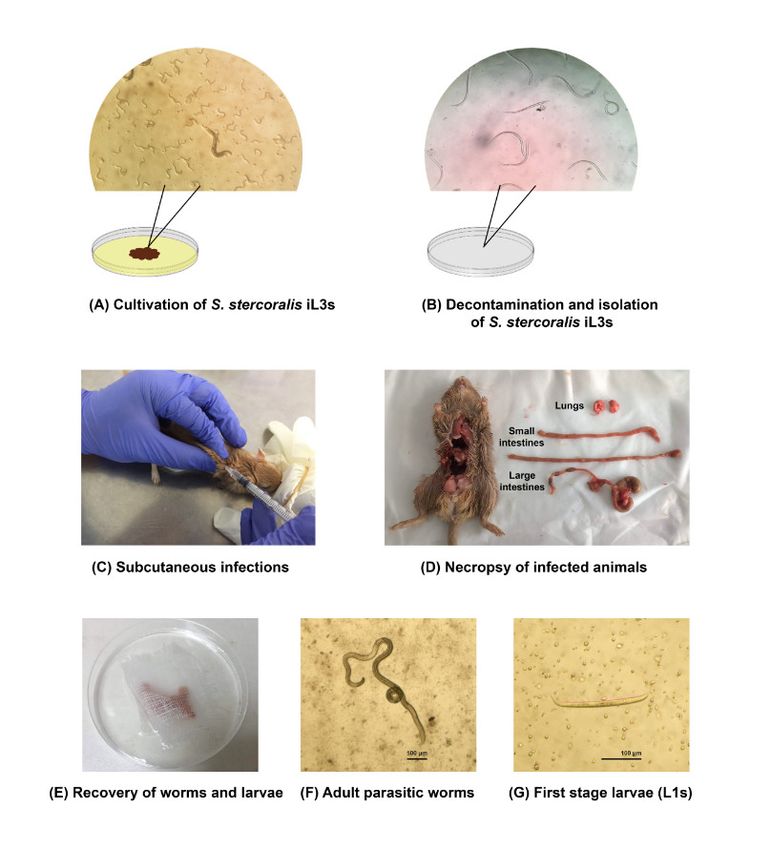
Figure 3. The process of experimental infections of human isolate of S. stercoralis in gerbils.
The corticosteroid drug is a major trigger for the development of autoinfection [8,9,35]. The
minimum dose of MPA to induce the autoinfection of the dog isolate of S. stercoralis in gerbils was 1
mg (Few L3a were found.). However, the number of L3a dramatically increased 400 fold when
increased to a dose of 2 mg MPA [27]. It is probably that using the higher dose of MPA (more than 2
mg) may induce autoinfection of human isolate of S. stercoralis in gerbils.
The genetic factors might influence the susceptibility of gerbils to the human isolate of S.
stercoralis. There were variabilities in the susceptibility of the human isolate of S. stercoralis in dogsPathogens 2019, 8, 21 3 of 11
[14,19–21,36]. The human isolate of S. stercoralis is usually incapable to develop a patent infection in
dogs. The genetic analysis between humans and dog isolate of S. stercoralis showed the variations in
Cox1, 18S rDNA and 28S rDNA genes [37–42]. Further studies about the genetics of humans and dog
isolate of S. stercoralis could be performed to get a better understanding about the susceptibility to
gerbils. Moreover, a different isolate of S. stercoralis might also influence the efficiency of infection.
There was a variability of S. ratti infection in rats. The usual dose to initiate an infection in the rat is
500 iL3s, however, there was a severe case of using 500 iL3s of S. ratti wild isolate in the rat [26].
In summary, we demonstrated the use of immunosuppressed gerbils as a laboratory model to
maintain the human isolate of S. stercoralis life cycle. In this study, we showed that the human isolate
of S. stercoralis is able to develop a patent infection in MPA-treated gerbils, more than 18 months,
mimicking the chronicity of human strongyloidiasis. Interestingly, a number of recovered L1s from
this study were comparable to recovered L1s from gerbils infected with the dog isolate S. stercoralis
when the autoinfection occurred [27,34], even though the number of recovered adult parasitic worms
from this study were lower than using the dog isolate of S. stercoralis [27,34]. Further studies aim to
induce the autoinfection in gerbils infected with the human isolate of S. stercoralis by increasing doses
of MPA and/or initial doses of iL3s. Moreover, the histopathology of the human isolate of S. stercoralis
would be demonstrated. Our findings provided a promising model for studying biology and
searching new alternative drugs against the parasites. Using gerbils has advantages in ease of
handling and breeding and cost.
4. Materials and Methods
4.1. Animals
The Mongolian gerbils (Meriones unguiculatus) were purchased from the Zooya farm (Bangkok,
Thailand) at about 4 weeks of age. The animals were screened negative for bacterial-, viral and
parasitic infections prior to entering into the facility. They were kept 2–4 animals/cage under
controlled air and temperature at 25 ± 2°C. Gerbils were bred together in captivity for two
generations. The F2 generation was used in experimental infections. The animals were provided with
food and water ad libitum. All animal experimental protocols were carried out under the approval by
the Chulalongkorn University Animal Care and Use Committee, Faculty of Medicine, Chulalongkorn
University, Bangkok, Thailand (CU-ACUC approval no: 022/2560).
4.2. Immunosuppression of animals
To induce the autoinfection in gerbils, 6–12 weeks old gerbils were subcutaneously injected with
200 µl of 10–20 mg/ml MPA (total 1–2 mg, depending on the experiments) at day −3, 0, 7, 14 and 21
p.i.
4.3. The human isolate of S. stercoralis infective stage (iL3s)
The human isolate of S. stercoralis was harvested from faecal specimens of strongyloidiasis
patients from the King Chulalongkorn Memorial Hospital, Bangkok, Thailand. Agar plate cultures
were set up to obtain iL3s for the experimental infections [43]. Approximately, 1–3 grams of stool
specimens were placed on the centre of agar plates and incubated at 25ºC for 5 days (Figure 3A). The
iL3s were collected from the surface of positive agar plates by washing with the buffer saline solution
(BU saline) (22 mM KH2PO4, 50 mM Na2HPO4, 70 mM NaCl). The iL3s were decontaminated by using
low melting point agarose and then axenized in BU saline with 100 U/ml penicillin, 10
µg/ml streptomycin and 12.5 µg/ml tetracycline for 3 hours (Figure 3B).
4.4. Experimental infections
Immunosuppressed gerbils (6–12 weeks old) were subcutaneously injected with a suspension of
1,000–10,000 iL3s (depending on the experiment) in 200 µl of inoculation volume on day 0 p.i. (Figure
3C). Each gerbil was separated for 1 animal/cage under controlled air and temperature 25°C ± 2. TherePathogens 2019, 8, 21 4 of 11
were 8 groups of experimental infections (Table 1). Group 1 and 2 aimed to test whether
immunosuppressed gerbils were susceptible to the human isolate of S. stercoralis, gerbils in group 1
and 2 were immunosuppressed with 2 mg MPA and injected with 1,000 and 10,000 of S. stercoralis
iL3s (obtained from human faecal cultures), respectively. Group 3–8 aimed to test whether S.
stercoralis could be maintained by gerbil-to-gerbil transfer, gerbil no. 11 from group 2 were used as a
source of iL3s for experimental infections in group 3–8. Gerbils in group 3–5 were
immunosuppressed with 1 mg MPA and injected with 4,000, 5,000 and 10,000 of S. stercoralis iL3s
(obtained from gerbil faecal cultures), respectively. Gerbils in group 6-8 were immunosuppressed
with 2 mg MPA and injected with 3,000, 5,000 and 10,000 of S. stercoralis iL3s (obtained from gerbil
faecal cultures), respectively. S. stercoralis infection in gerbils was confirmed by the presence of larvae
in stool and parasitic adult worms in the intestines.
4.5. Coproparasitological examination
To determine the susceptibility of infections and observe the prepatent period of the infections,
a semi-quantitative estimation of the number of L1s from infected animals were performed.
Approximately, 2 grams of faecal specimens were collected and cultured daily by using agar plates,
after day 7 p.i. The agar plates were observed daily for 5 days under a stereo microscope. The faecal
culture results were categorized by convention as: – (No tracks of migration and/or larvae were
found.); + (Only tracks of migration were found.); ++ (Free-living adults were first seen on day 4–5.);
+++ (Numerous free-living adults were first seen on day 3.).
4.6. Necropsy and recovery of S. stercoralis
On day 28 p.i., gerbils were anesthetized and then euthanized by cervical dislocation. The
intestines and lungs of infected animals were slit longitudinally (Figure 3D) and individually
wrapped with gauze (Figure 3E). The organs were soaked in BU saline and incubated at 37ºC for 3
hours. After incubation, the contents of the intestines and lungs were examined for the presence of
adult worms and larvae under a stereo microscope. The morphology of adult parasitic worms and
L1s were represented in Figure 3F and Figure 3G, respectively.
4.7. Statistics analysis
All data were represented in the text and figures as a mean ± standard error. Averages between
groups were compared and tested for statistical significance by using the unpaired t-test. A P value
of < 0.05 was considered statistically significant.
Author Contributions: Conceptualization, methodology, investigation, writing - original draft, S.C.;
conceptualization, methodology, resources, writing—review and editing, V.S.; conceptualization, project
administration, supervision, resources, writing—review and editing, funding acquisition, S.N.; all authors read
and agreed with the final version of this the paper.
Funding: This research was funded by the 100th Anniversary Chulalongkorn University Fund for Doctoral
Scholarship.
Acknowledgments: We appreciated the staff members of the Lymphatic Filariasis and Tropical Medicine
Research Unit, Department of Parasitology, Faculty of Medicine, Chulalongkorn University for their technical
assistance in the laboratory work. We would like to thank staff members of laboratory animal centre, Faculty of
Medicine, Chulalongkorn University for their assistance in animal care. We would like to thank Prof. Issarang
Nuchprayoon for English language editing.
Conflicts of Interest: The authors declare no conflict of interest.
References
1. Krolewiecki, A.J.; Lammie, P.; Jacobson, J.; Gabrielli, A.F.; Levecke, B.; Socias, E.; Arias, L.M.; Sosa, N.;
Abraham, D.; Cimino, R.; et al. A public health response against Strongyloides stercoralis: Time to look at
soil-transmitted helminthiasis in full. PLoS Negl. Trop. Dis. 2013, 7, e2165, doi:10.1371/journal.pntd.0002165.Pathogens 2019, 8, 21 5 of 11
2. Schar, F.; Trostdorf, U.; Giardina, F.; Khieu, V.; Muth, S.; Marti, H.; Vounatsou, P.; Odermatt, P.
Strongyloides stercoralis: Global Distribution and Risk Factors. PLoS Negl. Trop. Dis. 2013, 7, e2288,
doi:10.1371/journal.pntd.0002288.
3. Bisoffi, Z.; Buonfrate, D.; Montresor, A.; Requena-Mendez, A.; Munoz, J.; Krolewiecki, A.J.; Gotuzzo, E.;
Mena, M.A.; Chiodini, P.L.; Anselmi, M.; et al. Strongyloides stercoralis: A plea for action. PLoS Negl. Trop.
Dis. 2013, 7, e2214, doi:10.1371/journal.pntd.0002214.
4. Farthin, M; Albonico, M.; Bisoffi, Z.; Buonfrate, D.; Katelaris, P.; Kelly, P.; Savioli, L.; Le Mair, A.
Management of Strongyloidiasis. Available online: http://www.webcitation.org/75si8tJzH. (accessed on 2
Febuary 2019).
5. Beknazarova, M.; Whiley, H.; Judd, J.A.; Shield, J.; Page, W.; Miller, A.; Whittaker, M.; Ross, K. Argument
for Inclusion of Strongyloidiasis in the Australian National Notifiable Disease List. Trop. Med. Infect. Dis.
2018, 3, doi:10.3390/tropicalmed3020061.
6. Gill, G.V.; Beeching, N.J.; Khoo, S.; Bailey, J.W.; Partridge, S.; Blundell, J.W.; Luksza, A.R. A British Second
World War veteran with disseminated strongyloidiasis. Trans. R. Soc. Trop. Med. Hyg. 2004, 98, 382–386,
doi:10.1016/j.trstmh.2003.11.002.
7. Prendki, V.; Fenaux, P.; Durand, R.; Thellier, M.; Bouchaud, O. Strongyloidiasis in man 75 years after initial
exposure. Emerg. Infect. Dis. 2011, 17, 931–932, doi:10.3201/eid1705.100490.
8. Marcos, L.A.; Terashima, A.; Canales, M.; Gotuzzo, E. Update on strongyloidiasis in the
immunocompromised host. Curr. Infect. Dis. Rep. 2011, 13, 35–46, doi:10.1007/s11908-010-0150-z.
9. Buonfrate, D.; Requena-Mendez, A.; Angheben, A.; Munoz, J.; Gobbi, F.; Van Den Ende, J.; Bisoffi, Z. Severe
strongyloidiasis: A systematic review of case reports. BMC Infect. Dis. 2013, 13, 78, doi:10.1186/1471-2334-
13-78.
10. Kim, J.H.; Kim, D.S.; Yoon, Y.K.; Sohn, J.W.; Kim, M.J. Donor-Derived Strongyloidiasis Infection in Solid
Organ Transplant Recipients: A Review and Pooled Analysis. Transplant. Proc. 2016, 48, 2442–2449,
doi:10.1016/j.transproceed.2015.11.045.
11. Vazquez Guillamet, L.J.; Saul, Z.; Miljkovich, G.; Vilchez, G.A.; Mendonca, N.; Gourineni, V.; Lillo, N.;
Pinto, M.; Baig, A.; Gangcuangco, L.M. Strongyloides Stercoralis Infection Among Human
Immunodeficiency Virus (HIV)-Infected Patients in the United States of America: A Case Report and
Review of Literature. Am. J. Case Rep. 2017, 18, 339–346.
12. Myint, A.; Chapman, C.; Almira-Suarez, I.; Mehta, N. Strongyloides hyperinfection syndrome in an
immunocompetent host resulting in bandemia and death. BMJ Case Rep. 2017, 2017, doi:10.1136/bcr-2016-
217911.
13. Chan, F.L.Y.; Kennedy, B.; Nelson, R. Fatal Strongyloides hyperinfection syndrome in an
immunocompetent adult with review of the literature. Intern. Med. J. 2018, 48, 872–875,
doi:10.1111/imj.13940.
14. Grove, D.I.; Northern, C. Infection and immunity in dogs infected with a human strain of Strongyloides
stercoralis. Trans. R. Soc. Trop. Med. Hyg. 1982, 76, 833–838.
15. Genta, R.M.; Harper, J.S., 3rd; Gam, A.A.; London, W.I.; Neva, F.A. Experimental disseminated
strongyloidiasis in Erythrocebus patas. II. Immunology. Am. J. Trop. Med. Hyg. 1984, 33, 444–450.
16. Harper, J.S.; Genta, R.M.; Gam, A.; London, W.T.; Neva, F.A. Experimental disseminated strongyloidiasis
in Erythrocebus patas. I. Pathology. Am. J. Trop. Med. Hyg. 1984, 33, 431–443.
17. Schad, G.A.; Hellman, M.E.; Muncey, D.W. Strongyloides stercoralis: Hyperinfection in immunosuppressed
dogs. Exp. Parasitol. 1984, 57, 287–296.
18. Barrett, K.E.; Neva, F.A.; Gam, A.A.; Cicmanec, J.; London, W.T.; Phillips, J.M.; Metcalfe, D.D. The immune
response to nematode parasites: Modulation of mast cell numbers and function during Strongyloides
stercoralis infections in nonhuman primates. Am. J. Trop. Med. Hyg. 1988, 38, 574–581.
19. Sandground, J.H. Some studies on susceptibility, resistance, and acquired immunity to infection with
Strongyloides stercoralis (Nematoda) in dogs and cats. Am. J. Epidemiol. 1928, 8, 507–538.
20. Augustine, D.L.D., D.G. Observations on a natural infection with Strongyloides in the dog. J. Parasitol. 1939,
25, 117–119.
21. Galliard, H. Pathogenesis of Strongyloides. Helminthol. Abstr. 1967, 36, 247–260.
22. Dawkins, H.J.; Grove, D.I. Attempts to establish infections with Strongyloides stercoralis in mice and other
laboratory animals. J. Helminthol. 1982, 56, 23–26.Pathogens 2019, 8, 21 6 of 11
23. Rotman, H.L.; Yutanawiboonchai, W.; Brigandi, R.A.; Leon, O.; Nolan, T.J.; Schad, G.A.; Abraham, D.
Strongyloides stercoralis: Complete life cycle in SCID mice. Exp. Parasitol. 1995, 81, 136–139,
doi:10.1006/expr.1995.1101.
24. Lok, J.B.; Shao, H.; Massey, H.C.; Li, X. Transgenesis in Strongyloides and related parasitic nematodes:
Historical perspectives, current functional genomic applications and progress towards gene disruption and
editing. Parasitology 2017, 144, 327–342, doi:10.1017/S0031182016000391.
25. Breloer, M.; Abraham, D. Strongyloides infection in rodents: Immune response and immune regulation.
Parasitology 2017, 144, 295–315, doi:10.1017/S0031182016000111.
26. Viney, M.; Kikuchi, T. Strongyloides ratti and S. venezuelensis—Rodent models of Strongyloides infection.
Parasitology 2017, 144, 285–294, doi:10.1017/S0031182016000020.
27. Nolan, T.J.; Megyeri, Z.; Bhopale, V.M.; Schad, G.A. Strongyloides stercoralis: The first rodent model for
uncomplicated and hyperinfective strongyloidiasis, the Mongolian gerbil (Meriones unguiculatus). J. Infect.
Dis. 1993, 168, 1479–1484.
28. Li, X.; Massey, H.C., Jr.; Nolan, T.J.; Schad, G.A.; Kraus, K.; Sundaram, M.; Lok, J.B. Successful transgenesis
of the parasitic nematode Strongyloides stercoralis requires endogenous non-coding control elements. Int.
J. Parasitol. 2006, 36, 671–679, doi:10.1016/j.ijpara.2005.12.007.
29. Junio, A.B.; Li, X.; Massey, H.C., Jr.; Nolan, T.J.; Todd Lamitina, S.; Sundaram, M.V.; Lok, J.B. Strongyloides
stercoralis: Cell- and tissue-specific transgene expression and co-transformation with vector constructs
incorporating a common multifunctional 3′ UTR. Exp. Parasitol. 2008, 118, 253–265,
doi:10.1016/j.exppara.2007.08.018.
30. Albarqi, M.M.; Stoltzfus, J.D.; Pilgrim, A.A.; Nolan, T.J.; Wang, Z.; Kliewer, S.A.; Mangelsdorf, D.J.; Lok,
J.B. Regulation of Life Cycle Checkpoints and Developmental Activation of Infective Larvae in
Strongyloides stercoralis by Dafachronic Acid. PLoS Pathog. 2016, 12, e1005358,
doi:10.1371/journal.ppat.1005358.
31. Mati, V.L.; Raso, P.; de Melo, A.L. Strongyloides stercoralis infection in marmosets: Replication of
complicated and uncomplicated human disease and parasite biology. Parasit Vectors 2014, 7, 579,
doi:10.1186/s13071-014-0579-2.
32. Kerlin, R.L.; Nolan, T.J.; Schad, G.A. Strongyloides stercoralis: Histopathology of uncomplicated and
hyperinfective strongyloidiasis in the Mongolian gerbil, a rodent model for human strongyloidiasis
[corrected]. Int. J. Parasitol. 1995, 25, 411–420.
33. Sithithaworn, P.; Fujimaki, Y.; Mitsui, Y.; Prasanthong, R.; Yutanawiboonchai, W.; Aoki, Y. Efficacy of
ivermectin against Strongyloides stercoralis infection in jirds (Meriones unguiculatus). Exp. Parasitol. 1998,
89, 205–212, doi:10.1006/expr.1998.4278.
34. Nolan, T.J.; Bhopale, V.M.; Rotman, H.L.; Abraham, D.; Schad, G.A. Strongyloides stercoralis: High worm
population density leads to autoinfection in the jird (Meriones unguiculatus). Exp. Parasitol. 2002, 100, 173–
178.
35. Croker, C.; Reporter, R.; Redelings, M.; Mascola, L. Strongyloidiasis-related deaths in the United States,
1991–2006. Am. J. Trop. Med. Hyg. 2010, 83, 422–426, doi:10.4269/ajtmh.2010.09-0750.
36. Genta, R.M. Strongyloides stercoralis: Loss of ability to disseminate after repeated passage in laboratory
beagles. Trans. R. Soc. Trop. Med. Hyg. 1989, 83, 539–541.
37. Neefs, J.M.; Van de Peer, Y.; De Rijk, P.; Chapelle, S.; De Wachter, R. Compilation of small ribosomal
subunit RNA structures. Nucleic Acids Res. 1993, 21, 3025–3049.
38. Ramachandran, S.; Gam, A.A.; Neva, F.A. Molecular differences between several species of Strongyloides
and comparison of selected isolates of S. stercoralis using a polymerase chain reaction-linked restriction
fragment length polymorphism approach. Am. J. Trop. Med. Hyg. 1997, 56, 61–65.
39. Hasegawa, H.; Hayashida, S.; Ikeda, Y.; Sato, H. Hyper-variable regions in 18S rDNA of Strongyloides spp.
as markers for species-specific diagnosis. Parasitol. Res. 2009, 104, 869–874, doi:10.1007/s00436-008-1269-9.
40. Hasegawa, H.; Sato, H.; Fujita, S.; Nguema, P.P.; Nobusue, K.; Miyagi, K.; Kooriyama, T.; Takenoshita, Y.;
Noda, S.; Sato, A.; et al. Molecular identification of the causative agent of human strongyloidiasis acquired
in Tanzania: Dispersal and diversity of Strongyloides spp. and their hosts. Parasitol. Int. 2010, 59, 407–413,
doi:10.1016/j.parint.2010.05.007.
41. Schar, F.; Guo, L.; Streit, A.; Khieu, V.; Muth, S.; Marti, H.; Odermatt, P. Strongyloides stercoralis genotypes
in humans in Cambodia. Parasitol. Int. 2014, 63, 533–536, doi:10.1016/j.parint.2014.01.010.Pathogens 2019, 8, 21 7 of 11
42. Nagayasu, E.; Aung, M.; Hortiwakul, T.; Hino, A.; Tanaka, T.; Higashiarakawa, M.; Olia, A.; Taniguchi, T.;
Win, S.M.T.; Ohashi, I.; et al. A possible origin population of pathogenic intestinal nematodes,
Strongyloides stercoralis, unveiled by molecular phylogeny. Sci. Rep. 2017, 7, 4844, doi:10.1038/s41598-017-
05049-x.
43. Koga, K.; Kasuya, S.; Khamboonruang, C.; Sukhavat, K.; Ieda, M.; Takatsuka, N.; Kita, K.; Ohtomo, H. A
modified agar plate method for detection of Strongyloides stercoralis. Am. J. Trop. Med. Hyg. 1991, 45, 518–
521.
© 2019 by the authors. Licensee MDPI, Basel, Switzerland. This article is an open access
article distributed under the terms and conditions of the Creative Commons Attribution
(CC BY) license (http://creativecommons.org/licenses/by/4.0/).You can also read