The first study on the effect of crocodile oil from Crocodylus siamensis on hepatic mitochondrial function for energy homeostasis in rats
←
→
Page content transcription
If your browser does not render page correctly, please read the page content below
Veterinary World, EISSN: 2231-0916                                                                        RESEARCH ARTICLE
Available at www.veterinaryworld.org/Vol.15/April-2022/24.pdf                                                   Open Access

  The first study on the effect of crocodile oil from Crocodylus siamensis
    on hepatic mitochondrial function for energy homeostasis in rats
Kongphop Parunyakul1       , Krittika Srisuksai1 , Pitchaya Santativongchai2 , Urai Pongchairerk3      , Sumate Ampawong4       ,
                                         Phitsanu Tulayakul5 and Wirasak Fungfuang1

    1. Department of Zoology, Faculty of Science, Kasetsart University, Bangkok, Thailand; 2. Bio-Veterinary Sciences
   (International Program), Faculty of Veterinary Medicine, Kasetsart University, Bangkok, Thailand; 3. Department of
 Anatomy, Faculty of Veterinary Medicine, Kasetsart University, Bangkok, Thailand; 4. Department of Tropical Pathology,
Faculty of Tropical Medicine, Mahidol University, Bangkok, Thailand; 5. Department of Veterinary Public Health, Faculty of
                           Veterinary Medicine, Kasetsart University, Nakhon Pathom, Thailand.
                          Corresponding author: Wirasak Fungfuang, e-mail: fsciwsf@ku.ac.th
  Co-authors: KP: kongphop.pa@ku.th, KS: krit_ti_ka@hotmail.com, PS: pitchaya.sant@ku.th, UP: fveturp@ku.ac.th,
                                   SA: am_sumate@hotmail.com, PT: fvetpnt@ku.ac.th
                     Received: 14-01-2022, Accepted: 11-03-2022, Published online: 19-04-2022

doi: www.doi.org/10.14202/vetworld.2022.986-997 How to cite this article: Parunyakul K, Srisuksai K,
Santativongchai P, Pongchairerk U, Ampawong S, Tulayakul P, Fungfuang W (2022) The first study on the effect of
crocodile oil from Crocodylus siamensis on hepatic mitochondrial function for energy homeostasis in rats, Veterinary
World, 15(4): 986-997.

                                                            Abstract
Background and Aim: Consumption of fatty acids (FA) can alter hepatic energy metabolism and mitochondrial function
in the liver. Crocodile oil (CO) is rich in mono-and polyunsaturated FAs, which have natural anti-inflammatory and healing
properties. In rat livers, we investigated the effect of CO on mitochondrial function for energy homeostasis.
Materials and Methods: Twenty-one male Sprague-Dawley rats were divided into three groups at random. Group 1 rats were
given sterile water (RO), Group 2 rats were given CO (3% v/w), and Group 3 rats were given palm oil (PO) (3% v/w). For
7 weeks, rats were given sterile water, CO, and PO orally. The researchers looked at body weight, food intake, liver weight,
energy intake, blood lipid profiles, and mitochondria-targeted metabolites in the liver. The liver’s histopathology, mitochondrial
architecture, and hydrolase domain containing 3 (HDHD3) protein expression in liver mitochondria were studied.
Results: Body weight, liver weight, liver index, dietary energy intake, and serum lipid profiles were all unaffected by
CO treatment. The CO group consumed significantly less food than the RO group. The CO group also had significantly
higher levels of oxaloacetate and malate than the PO group. CO treatment significantly ameliorated hepatic steatosis, as
evidenced by a greater decrease in the total surface area of lipid particles than PO treatment. CO administration preserved
mitochondrial morphology in the liver by upregulating the energetic maintenance protein HDHD3. Furthermore, chemical-
protein interactions revealed that HDHD3 was linked to the energy homeostatic pathway.
Conclusion: CO may benefit liver function by preserving hepatic mitochondrial architecture and increasing energy
metabolic activity.
Keywords: crocodile oil, energy metabolism, liver, mitochondria, rat.
Introduction                                                       by an increase in adipose lipolysis, which increases
      The liver is a metabolic biotransformation hub               hepatic de novo lipogenesis, impairs the synthesis
and plays an important role in homeostasis. Among                  and/or secretion of very-low-density cholesterol, and
its many functions, the liver is in charge of physio-              causes triacylglycerol esterification dysfunction, or
logical processes such as bile production, energy gen-             impaired mitochondrial β‐oxidation [3]. Mitochondria
eration, as well as carbohydrate, proteins, and lipids             are unique organelles that metabolize nutrients and
metabolism [1]. Mitochondrial dysfunction is a broad               are responsible for energy metabolism, free radicals
term that encompasses changes in various metabolic                 generation, calcium homeostasis, cell survival, and
pathways and mitochondrial component damage.                       cell death [4]. Metabolic activities are supported by
Furthermore, mitochondrial dysfunction can lead to                 hepatic mitochondria, which also contribute to the
various negative outcomes, including oxidative stress,             pathophysiology of insulin resistance and diabe-
energy deficiency, accumulation of triglycerides (ste-             tes [5,6]. The mitochondria’s primary function is to
atosis), and cell death [2]. Liver steatosis is caused             synthesize adenosine triphosphate (ATP) through oxi-
                                                                   dative phosphorylation in accordance with metabolite
Copyright: Parunyakul, et al. Open Access. This article is         oxidation through the tricarboxylic acid (TCA) cycle
distributed under the terms of the Creative Commons Attribution
4.0 International License (http://creativecommons.org/licenses/    and β-oxidation of fatty acids (FA). A previous study
by/4.0/), which permits unrestricted use, distribution, and        found that a high-fat diet alters rat hepatocyte energy
reproduction in any medium, provided you give appropriate credit
to the original author(s) and the source, provide a link to the
                                                                   metabolism by inhibiting mitochondrial oxidative
Creative Commons license, and indicate if changes were made.       phosphorylation [7].
The Creative Commons Public Domain Dedication waiver (http://
creativecommons.org/publicdomain/zero/1.0/) applies to the data
                                                                         Over the last few decades, public concerns about
made available in this article, unless otherwise stated.           the interaction of health and nutrition have grown
Veterinary World, EISSN: 2231-0916                                                                                          986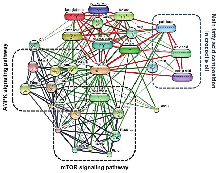
Available at www.veterinaryworld.org/Vol.15/April-2022/24.pdf

rapidly. Fat is a necessary macronutrient in the human     ethics committee of Kasetsart University Research and
diet, and vegetable oils are now the most widely con-      Development Institute, Kasetsart University, Thailand,
sumed fat in the world. Recently, attention has been       approved this study (Approval No. ACKU61-VET-088).
given to the association between the fatty liver con-      Study period and location
dition and the type of dietary fat consumed; the pre-           This study was conducted from October 2019 to
vious research has linked high-saturated FA (SFA)          May 2020 at the Department of Zoology, Faculty of
intake to the development of hepatic steatosis [8-10],     Science, Kasetsart University, Bangkok, Thailand.
a loss of whole-body fat oxidation [11-13], and bal-
                                                           Animal care, diets, and experimental design
looned cristae, as well as condensed matrix structures
of hepatic mitochondria [14]. However, certain edi-              In brief, Nomura Siam International Co. Ltd.,
ble cooking oils, such as olive oil, soybean oil, and      Samutprakan province, Thailand, provided 21
palm oil (PO), are consumed in greater quantities          Sprague-Dawley male rats (age: 7 weeks). This
than other higher-quality oils. PO has higher ratios       study used only male rats to prevent the effect of the
of SFA to mono-unsaturated FA (MUFA) and poly-             estrous cycle on consumption behavior. The animals
unsaturated FA (PUFA) than other cooking oils [15].        were individually housed in controlled environments
Previously, Li et al. [16] investigated the effects of     (25±2°C on a 12-h light/12-h dark cycle). Throughout
a PO diet versus a low-fat diet on the expression of       the study, rats had ad libitum access to food and water
lipid breakdown-related genes and discovered that          and were randomly assigned to one of three groups
PO significantly reduced hepatic peroxisome prolif-        (n=7/group). Rats in Group 1 were given sterile water
erator-activated receptor alpha (PPAR-α) expression        (RO), those in Group 2 were given CO (3% v/w), and
levels. When FAs inhibit PPARα, FA oxidation (FAO)         those in Group 3 were given commercial PO (3% v/w)
and the resulting ATP production are reduced. Many         (PO). For 7 weeks, the animals were given sterile
previous studies have also linked SFA consumption to       water, CO, and PO orally.
secondary diseases such as glucose intolerance, insulin    CO preparation
resistance, dyslipidemia, cardiovascular disease, and            When the meat was trimmed and prepared, CO
hepatic steatosis [17-19]. Consumption of SFA-rich         was extracted using the wet cold-pressed oil method
oils, such as PO, has been linked to the development       described by Santativongchai et al. [25]. Abdominal fat
of liver dyslipidemia, leading to mitochondrial dys-       samples were obtained as a waste product from slaugh-
function progression. Compared to other animal oils,       tered C. siamensis (age: 3-5 years) collected from a
crocodile oil (CO), extracted from the fatty tissues       crocodile farm in Nakhon Pathom Province, Thailand.
of Crocodylus siamensis, has a high concentration of             The samples were pressed through two layers
MUFA and PUFA [20]. CO has been used for centuries         of filter cloth with distilled water in a 1:1 (w/v) pro-
by traditional practitioners and has been very effective   portion. Following that, the solution was left undis-
in treating various ailments ranging from skin condi-      turbed until the mixture separated. The upper clear oil
tions to cancer [21]. CO has been used to treat skin       fraction was then collected, evaporated, and stored at
rashes and promote wound healing in many previous          room temperature in a sealed container.
studies [21,22]. Furthermore, a hamsters study found       Measurement of body weight, food intake, and
that a low MUFA and low PUFA to SFA ratio induced          energy intake of animals
weight gain and body fat accumulation. In contrast,              Each rat’s food intake was measured by weighing
a high MUFA and high PUFA/SFA ratio prevented              the remaining chow daily between 11:00 and 11:30 h,
white adipose tissue accumulation [23]. It has been        and this was used to calculate energy intake. The daily
demonstrated that PUFA increases the expression of         energy intake per rat (kcal/day) was calculated as (food
proteins involved in FAO while decreasing the expres-      intake per rat*ME from standard chow) + ME from
sion of proteins involved in lipid synthesis [24].         the treatment (ME is the total energy of the rat diets,
      However, the mechanism by which CO regulates         which is 3.040 kcal/g in standard chow, 12 kcal in CO
hepatic mitochondrial-related energy metabolism in a       per day, and 13 kcal in PO per day). Throughout the
rat model is largely unknown. We hypothesized that         experiment, body weight was measured every week.
CO could be linked to changes in mitochondrial func-
                                                           Sample collection
tion in energy metabolic pathways in rat livers. As per
our knowledge, this is the first report of the effect of        After the experiment, all animals were sacrificed
CO on energy homeostasis via a hepatic mitochon-           with a lethal dose of pentobarbital sodium. Blood sam-
drial function. The present study aimed to look into       ples were drawn through cardiac puncture and centri-
the effect of CO on hepatic energy metabolism and          fuged at 2200 g for 15 min at 4°C. The serum was kept
mitochondrial function in rats.                            at −20°C until further testing. The serum lipid profile
                                                           was determined using a Hitachi 7080 analyzer (Hitachi,
Materials and Methods                                      Tokyo, Japan) and included triglycerides, cholesterol,
Ethical approval                                           high-density lipoprotein, and low-density lipoprotein.
     The research conducted adhered to the Guidelines           While performing a standard protocol for
for the Care and Use of Laboratory Animals. The            mitochondrial extraction, liver specimens were
Veterinary World, EISSN: 2231-0916                                                                            987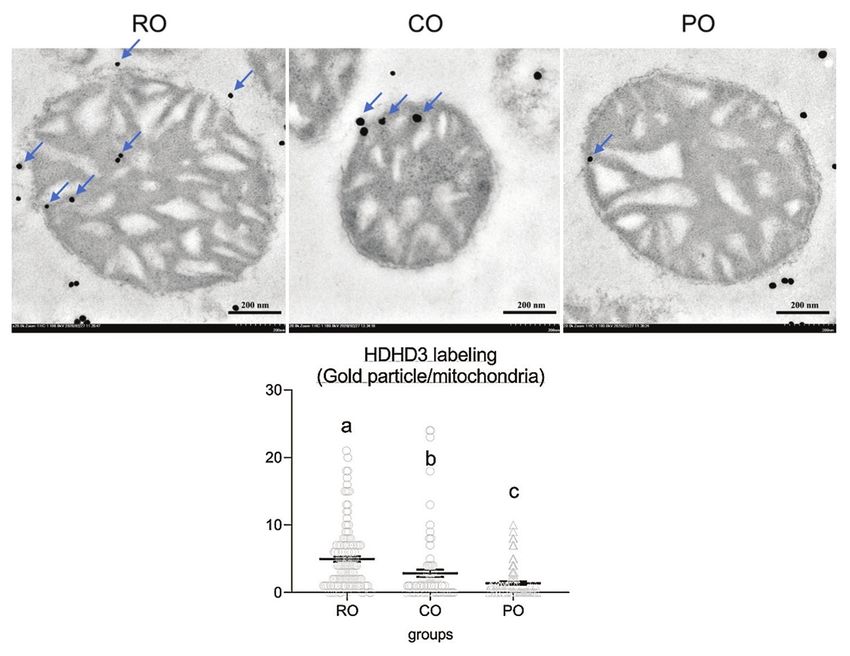
Available at www.veterinaryworld.org/Vol.15/April-2022/24.pdf
immediately collected, weighed to determine their in homogenate buffer several times. The pellets were
index (organ weight/body weight), and stored in ice- resuspended in an ice-cold final equilibrated buffer
cold homogenate buffer (0.32 M sucrose, 1 mM eth- (250 mM sucrose, 5 mM KH2PO4, 10 mM Tris-HCl,
ylenediaminetetraacetic acid, and 10 mM Tris-HCL; and 2 mg/mL bovine serum albumin [BSA]; pH 7.2).
pH 7.4). Pieces of liver tissue were also separated Subsequently, 200 μL of the resuspended pellet was
for histopathology analysis and stored in a 10% neu- fixed in 2.5% glutaraldehyde in 0.1 M sucrose phos-
tral buffer formalin solution. Further liver samples phate buffer for electron microscopy analysis. The
were collected and homogenized in ice-cold phos- protein content of mitochondria was determined
phate-buffered saline (PBS) (20% w/v) before centrif- using a spectrophotometer (NanoDrop-1000, Thermo
ugation at 2000 × g for 20 min at 4°C; the supernatants Scientific, Wilmington, DE, USA) and a protein assay
were stored at −80°C until further analysis. (Bio-Rad®, Hercules, CA, USA).
Mitochondrial energy-related intermediates analysis Conventional electron microscopy
The frozen supernatants were mixed with The ultrastructure of liver mitochondria was
methanol in a ratio of 2:8 (v/v). After centrifuga- studied using electron microscopy. Each group’s
tion (20,000 × g for 20 min at 4°C), the superna- liver specimens were fixed with 1% osmium tetrox-
tants were evaporated in a −80°C freeze dryer. After ide, dehydrated in graded ethanol, infiltrated in a
that, the metabolites were redissolved in 500 µL of series of LR white resin (EMS®, USA), embedded
high-performance liquid chromatography (HPLC) in pure LR white (EMS®), polymerized at 60°C for
buffer. HPLC analysis was performed on each 5 µL 48 h, cut into 100-nm-thick sections, and stained
sample. Chromatography was carried out in the fol- with lead citrate and uranyl acetate. A transmis-
lowing manner: The injection volume was set to 5 µL sion electron microscope (model HT7700, Hitachi)
and the column was kept at 40°C. An InertSustain was used to examine the ultrastructure of the liver.
C18 (150×4.6 mm) measuring 5 µm was used to sepa- Hepatocytes with unimpaired or intact mitochondria
rate the mobile phase, which contained 8% 1 N sulfu- were counted (50 cells per group) and compared with
ric acids. Gradient elution at a flow rate of 1 mL/min other treatment groups.
was used. Metabolite-protein and protein-protein interaction
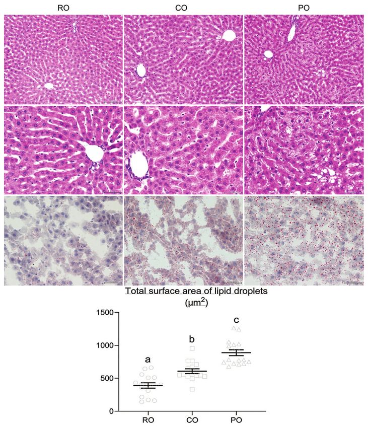
Hepatic histopathology and fat accumulation analysis We used STITCH v. 5.0 (European Molecular
Hepatocellular steatosis (macrovesicular and Biology Laboratory, Heidelberg, Germany) (http://
microvesicular) is a type of hepatotoxicity character- stitch.embl.de/) to predict interactions between
ized by an abnormal accumulation of fat droplets in expected metabolites and proteins that interact with
hepatocytes. Fixed livers were kept at room tempera- the targeted mitochondrial energy-maintenance pro-
ture (24-25°C) for 24 h in 10% formalin. To determine teins (haloacid dehalogenase-like hydrolase domain
hepatocellular steatosis, the samples were embedded containing 3: HDHD3), such as the energy-related
in paraffin, section, ed at 5-μm thickness, and stained proteins (Prkaa1, Prkaa2, Prkab1, Prkab2, Prkag1,
with hematoxylin and eosin (H&E). Oil Red O (ORO) Prkag2, Akt1, Mtor, and Raptor) (Table-1) and mito-
staining was used to determine the size and position of chondrial energy-related metabolites (lactate, pyru-
the fat droplets. Liver cryosections (5 μm thick) were vate, alpha-ketoglutarate, oxaloacetate, citrate, and
fixed in PBS with 10% neutral-buffered formalin for malate). STITCH also provides a confidence score
20 min, and then incubated in freshly prepared ORO for each reported interaction, with values ranging
solution for 10 min before being counterstained with from 0.40-0.70, 0.70-0.90, and 0.90-1.00, indicating
hematoxylin for 20 s. A light microscope with a mag- medium, high, and highest confidence levels, respec-
nification of 200× was used to examine the sections. tively. Our study predicted the interaction with the
The number and total surface area of lipid particles required confidence thresholds (scores) of 0.40 or
were measured. higher.
Liver mitochondrial extraction Immunogold labeling technique
To avoid cellular damage, liver mitochondrial The immunogold labeling technique was used
extraction was carried out within 1-2 h. Each group’s to compare HDHD3 (mitochondrial energy marker)
liver samples were pooled, weighed, homogenized, expression and localization of mitochondria between
and washed in homogenate buffer. In a glass Potter- groups. The primary antibody marker (MyBioSource,
Elvehjem tissue grinder, liver specimens were homog- USA) was rabbit polyclonal anti-HDHD3.
enized with an appropriate volume of the homogenate The mitochondrial pellet from the pooled liver
buffer (4 mL homogenate buffer/1 g of the liver spec- extract in each group was secondary fixed, and tissue
imen). Several up and down strokes were performed processing was carried out as previously described.
with a motor-driven Teflon pestle at 600 rpm during For 30 min, the tissue sections were blocked with
this setup. The homogenates were then centrifuged at 50 mM glycine in PBS, followed by 5% BSA (EMS®)
1000 × g for 5 min at 4°C. The supernatants were col- in PBS. They were then incubated for 1 h with 1:50
lected and centrifuged at 15,000 × g for 2 min at 4°C. diluted primary antibodies before being treated with
The mitochondrial pellets were collected and washed goat anti-rabbit immunoglobulin G conjugated with
Veterinary World, EISSN: 2231-0916 988Available at www.veterinaryworld.org/Vol.15/April-2022/24.pdf
10-nm gold particles (EMS®). Sections were washed Results
several times with 0.1% BSA in PBS between steps. Effect of dietary CO on final body weight, body weight
After rigorously washing the tissue sections with dis- gain, liver index, food intake, and energy intake
tilled water, a silver enhancement kit (Aurion R-Gent As shown in Table-2, CO- and PO-treated rats
SE-EM kit; EMS®) was used to improve the contrast consumed significantly less food (15.84±0.28 and
of the gold particle labeling. Finally, the sections were 14.98±0.31 g/day, respectively) than the RO group
stained with lead citrate and uranyl acetate before (18.82±0.25 g/day). However, there were no signif-
transmission electron microscopy. The number of icant differences between groups in terms of final
labeled gold particles was counted to determine the body weight, body weight gain, liver index, or energy
intact stage of liver mitochondria (50 mitochondria/ intake.
group was evaluated). Effect of dietary CO on blood lipid profiles and liver
Statistical analysis mitochondrial energy-related metabolites
To better understand the biological processes, Figure-1 depicts serum lipid profiles. The
the targeted energy-related proteins (Prkaa1, Prkaa2, results revealed no statistically significant differences
Prkab1, Prkab2, Prkag1, Prkag2, Akt1, Mtor, and between the three groups. Interestingly, when com-
Raptor) were identified using the NCBI and UniProt pared to the RO group (138.25±30.14 mg/dL), both the
databases. CO (90.40±15.14 mg/dL) and PO (83.68±6.44 mg/dL)
The data are presented as mean±standard error of groups showed a decreasing trend in triglyceride
the mean. Statistical analysis was performed by one- levels.
way analysis of variance followed by Tukey’s post hoc Figure-2 shows that the CO-treated group
test in the R project statistical computing package (R had significantly higher hepatic oxaloacetate lev-
core team, 2019). pAvailable at www.veterinaryworld.org/Vol.15/April-2022/24.pdf
(Figure-2d). Meanwhile, there was no difference histologic examination revealed microvesicular depo-
in hepatic malate levels between the CO and RO sition (numerous small vesicles of fat in hepatocytes)
groups (246.92±6.02 and 258.03±6.72 mg/L, respec- in CO- and PO-treated rats. The size of fat droplets in
tively), but the CO group had a significantly higher CO, on the other hand, was clearly smaller than in PO.
malate level than the PO group (204.41±6.61 mg/L) Furthermore, macrovesicular steatosis (a single large
(Figure-2f). On the other hand, CO administration did vacuole of fat filling up the hepatocyte) was found
not affect lactate, pyruvate, citrate, or alpha-ketogluta- in the PO group, whereas the CO group showed had
rate levels in the liver (Figure-2a-c, and e). microvesicular steatosis (Figure-3a-f). ORO staining
Effect of dietary CO on hepatic lipid accumulation was used to examine the effect of CO supplementation
H&E staining was used to visualize hepatic ste- on intracellular lipid levels in hepatocytes (Figure-
atosis in liver sections at 200× and 400× magnifica- 3g-i). The number and size of hepatic fat droplets were
tions. Compared to the livers of RO-treated rats, liver lower in the RO group than in the CO and PO groups.
Meanwhile, as shown in Figure-3j, the CO group had
a significantly lower total surface area of lipid drop-
lets in the liver than the PO group (608.40±35.67 and
889.23±44.77 µm2, respectively). The total lipid par-
ticle area seen in CO-treated rats, on the other hand,
was significantly greater than that seen in RO-treated
rats (391±40.67 µm2).
Effect of CO on mitochondrial architecture and
expression of HDHD3 in liver mitochondria
After 7 weeks of CO administration, electron
a b microscopy analysis was performed. The CO-treated
rats had a similar percentage of intact mitochondria
(65.71%) as the control RO group (64.45%), but sig-
nificantly higher than the PO-treated rats (43.96%)
(Figure-4). The expression of HDHD3, an ener-
gy-maintenance protein, was chosen to test their
activity in the intact stage of mitochondria. The CO
group had significantly higher levels of mitochondrial
HDHD3 expression (2.86±0.52 gold particle/mito-
c d chondria) than the PO group (1.39±0.23 gold particle/
Figure-1: Effect of dietary crocodile oil on serum mitochondria). Meanwhile, when compared with the
(a) cholesterol, (b) triglyceride, (c) low-density cholesterol,
and (d) high-density cholesterol levels of rats for 7 weeks.
CO and PO groups, the expression of HDHD3 protein
Data are expressed as the mean±standard error of the was significantly higher in the RO group (4.98±0.40
mean. gold particle/mitochondria) (Figure-5).
a b c
d e f
Figure-2: Effect of dietary crocodile oil on liver mitochondrial energy related-metabolites (a) lactate, (b) pyruvate,
(c) citrate, (d) oxaloacetate, (e) alpha-ketoglutarate, and (f) malate levels. Data are expressed as the mean±standard
error of the mean. Different letters indicate statistically significant differences between groups (pAvailable at www.veterinaryworld.org/Vol.15/April-2022/24.pdf
a b c
d e f
g h i
j
Figure-3: Histological analysis of liver lipid accumulation after 7 weeks of crocodile oil administration. (a-c) Hematoxylin
and eosin (H&E) staining (200×), and (d-f) H&E staining (400×) with morphological forms of hepatic steatosis showing the
hepatic steatosis in rat liver section; microvesicular steatosis was visible in crocodile oil (CO) group, whereas macrovesicular
steatosis was visible in palm oil (PO) group. (g-i) Oil Red O staining showing the adipose deposition in rat liver section
(400×); both oil-treated groups caused liver lipid deposits. PO showed more severe fat accumulation in the liver than CO
group, and CO treatment exhibited more fat deposits than RO group. (j) The bar graph indicates the total surface area of
the lipid droplets; the total surface area of fat droplets was significantly higher in the PO group than in the CO group. Data
are represented by mean±standard error of the mean. Different letters indicate statistically significant differences between
groups (pAvailable at www.veterinaryworld.org/Vol.15/April-2022/24.pdf
a b c
d
Figure-4: Comparison of electron micrographs of hepatic mitochondria conformation among the RO (a), crocodile oil
(b), and palm oil (c) groups. The bar graph indicates the percentage of intact mitochondria (d), data are represented by
mean±standard error of the mean. Different letters indicate statistically significant differences between groups (pAvailable at www.veterinaryworld.org/Vol.15/April-2022/24.pdf
Figure-6: The chemical-protein and protein-protein interaction network of HDHD3 and the three main fatty acids of
crocodile oil on the energy metabolic pathway in rat livers, as analyzed by STITCH v. 5.0.
other mammals is PUFA. According to a previous study, However, we propose no difference in food ingestion
PUFAs play an important role in the composition of all responses between types of dietary fat; this finding
cell membranes, where they maintain homeostasis for necessitates additional research into the mechanism by
proper membrane protein function and influence mem- which saturation level can regulate food consumption
brane fluidity, thereby regulating cell signaling pro- and body weight management. On the other hand, the
cesses, cellular functions, and gene expression [26]. In results showed that both types of dietary fat increased
this regard, we agreed that CO consumption could have hepatic lipid accumulation compared to the RO group.
a protective effect on energy mitochondrial function. It is widely accepted that dietary FA composition is an
First, our findings showed that an intake of dietary important determinant of hepatic lipid metabolism. In
oil (CO and PO) was associated with lower food intake this context, a previous study found that increasing FAs
and higher fat accumulation in hepatocytes when in the diet, particularly SFA, played a significant role
compared to the RO group. A previous study looked in the development of hepatic fat accumulation [31].
at dietary FAs (SFA, MUFA, or PUFA) and found PO, which is rich in SFA like palmitic acid,
that they had a physiological effect on energy expen- increased fat accumulation in hepatocytes and
diture, energy intake, and body weight control [27]. induced mitochondrial dysmorphology in this study.
The previous research on the effect of fat saturation The previous studies have linked abnormalities in
on satiety and food intake reports that unsaturated mitochondrial morphology and function to fatty liver
FA-containing fats reduce food intake more than stea- accumulation [32,33]. This point could contribute to
ric acid-containing fats [28]. Furthermore, the current the decrease in energy metabolism and mitochondrial
study found that PO, which is rich in SFA (palmitic function. PO is currently the most widely used edi-
acid), reduced food intake compared to the RO group. ble oil in the food industry, although its relationship
Many previous studies agree that SFA sources con- with liver energy metabolism remains unknown. Our
taining palmitic acid (coconut oil and palm kernel oil) current findings of H&E and ORO staining of lipid
provide greater satiety than other fat sources [29,30]. droplets in hepatocytes of rats fed PO revealed large
Veterinary World, EISSN: 2231-0916 993Available at www.veterinaryworld.org/Vol.15/April-2022/24.pdf
lipid droplet size as macrovesicular steatosis and sig- TCA cycle in obesity; they discovered that PUFA may
nificantly increased surface area of intrahepatic tri- alleviate obesity by affecting mitochondrial function
glycerides compared with the RO and CO groups, and restoring TCA cycle homeostasis, specifically the
with CO exhibiting higher levels of liver content than transcription and translation of TCA cycle enzymes
the RO group. We propose that while the CO diet such as citrate synthase, succinate dehydrogenase
influences fat accumulation more than the normal subunits A (SDHA), fumarate hydratase, and malate
rat diet, it may also prevent the development of lipid dehydrogenase 2 (MDH2) in HepG2 cells. Meanwhile,
accumulation in the liver compared with the PO diet. a recent study on the effect of SFA supplementation
The previous research has suggested that SFA may on the architecture and protein expression patterns of
play a role in lipid metabolic toxicity, such as oxida- the murine heart discovered that protein expression
tive stress, and mitochondrial dysfunction [34]. PO patterns in animals fed a high-SFA diet show impair-
altered hepatic metabolism and caused lipid accumu- ment of the TCA cycle and ATP synthesis [43]. Thus,
lation via disturbed hepatocyte transcription, accord- PUFA-enriched diets could stimulate energy metabolic
ing to the previous study [35]. A subsequent study activity in rats by activating genes involved in the pro-
found that SFA sources containing palmitic acid can duction of energy mitochondrial-related intermediates.
activate NOD-like receptor family pyrin containing We observed the liver mitochondrial morphol-
3 (NLRP3) inflammasomes, which are linked to the ogy and expression levels of proteins related to energy
development of liver steatosis [36]. Meanwhile, Li homeostasis to investigate the possible mechanisms of
et al. [16] investigated the effects of PO and a low- CO for the improvement of liver mitochondrial func-
fat diet on the expression of lipid breakdown-related tion. The current study’s findings revealed that PO had
genes. The PO-treated group had significantly lower fewer healthy mitochondria than the CO and RO treat-
levels of expression of the PPAR-α (the gene involved ments. According to a previous study, a high-SFA diet
in FAO), FA transport proteins, and their derivatives caused hepatic fat accumulation which was consistent
entering into the β-oxidation pathway to control with impaired mitochondrial function and a dysreg-
energy homeostasis [37]. However, previous research ulated expression profile of mitochondrial dynamic
has shown that olive oil, which is rich in MUFA, proteins [44]. Furthermore, a high-SFA diet high in
reduces liver fat accumulation by improving insulin lard was linked to lower energy expenditure and the
resistance and increasing triglyceride release from expression of genes controlling lipid metabolism and
the liver [38]. A subsequent study found that PUFA mitochondrial function [45]. Furthermore, the previous
can reduce intracellular triglyceride deposition in the study in skeletal muscle cells has found that palmitic
liver [39], and act as a potent inhibitor of hepatic lipo- acid, the main SFA in PO, has a proclivity to induce
genesis [40]. As previously stated, a dietary CO, rich mitochondrial DNA damage [46], as well as changes
in MUFA (OA) and PUFA (LA), or PO, rich in SFA in mitochondrial morphology and function [47]. As a
(palmitic acid) has a distinct effect on liver metabo- result, PO may contribute to mitochondrial form and
lism and hepatic fat accumulation because PO causes a function changes, leading to the development and pro-
significant increase in liver fat when compared to CO. gression of energy metabolic dysfunction in the liver. In
PO, we believe, triggers energy metabolic deficiency contrast, according to our findings, CO-treated rats had
by downregulating the transcription of energy homeo- a significantly higher percentage of intact mitochondria
static genes and causes liver damage by impairing than the PO-treated group. Although there was little
mitochondrial function. It should come as no surprise evidence of the effect of CO on mitochondrial function,
that mitochondrial dysfunction plays a significant role studies on the effect of MUFA and PUFA on mitochon-
in the development of excessive hepatic fat accumula- drial function were moderately published. According to
tion following PO administration. a recent study on the effect of OA (the main MUFA of
Mitochondrial energy-related-metabolite levels CO) on mitochondrial homeostasis at a cellular level,
reflect liver cell energy homeostasis. Our findings MUFA modulates mitochondrial function and lipid
show that PO administration causes a decrease in some metabolism [48,49], resulting in increased mitochon-
key energy-related metabolite levels. In contrast, CO drial ATP production. Furthermore, a previous study
administration causes a significant increase in oxalo- discovered that MUFA and PUFA inhibit the pro-in-
acetate and malate levels in the TCA cycle compared flammatory action of excess palmitic acid and coun-
to PO administration. The TCA cycle is the cell’s cen- teract the morphological and functional changes in
tral metabolic hub and an important source of precur- mitochondria [50]. Importantly, animals studies and a
sors for energy supply. However, the effects of CO on steatotic hepatocyte model support our hypothesis that
energy metabolism are not completely understood. PUFA improves mitochondrial morphology and has a
Interestingly, according to a previous study, MUFA beneficial effect on mitochondrial function recovery by
(OA) and PUFA (LA) are broken down in mitochon- regulating the mammalian target of the rapamycin com-
dria to produce acetyl-CoA through FAO, a metab- plex 1 (mTORC1) pathway [51,52]. Previous research
olite product that enters the TCA cycle to maintain indicates that mTOR primarily acts through mTORC1,
ATP [41]. Furthermore, Liu et al. [42] investigated the reducing oxidative capacity and altering mitochondrial
mechanism by which omega-3 PUFA influences the morphology [53]. Many studies indicate that MUFA
Veterinary World, EISSN: 2231-0916 994Available at www.veterinaryworld.org/Vol.15/April-2022/24.pdf
and PUFA intake have beneficial effects on mitochon- mitochondrial function. Our findings add to our
drial dysmorphic features; these findings support our understanding of the long-term effect of CO consump-
findings that CO restores liver energy metabolism by tion on hepatic energy metabolism and mitochon-
maintaining mitochondria ultrastructure and function. drial function. Compared to the normal range, CO
Our current findings also confirmed CO’s protec- treatment reduced food intake and some key hepatic
tive effect on energy homeostasis; we discovered that energy metabolite levels. It also slowed the progres-
CO not only maintains mitochondrial morphology but sion of hepatic steatosis, improved mitochondrial
also increases energy production in the liver by upreg- morphology, and played an important role in energy
ulating mitochondrial HDHD3 protein. HDHD3 has metabolism maintenance by upregulating HDHD3
been identified as a mitochondrial protein involved in expression. Through the expression of protein-asso-
cell energy maintenance [54]. The protein HDHD3 was ciated metabolic homeostasis, our findings may have
found to be downregulated in PO-treated rats, whereas clinical and therapeutic implications. CO has the
it was found to be upregulated in the CO-treated potential to be a viable dietary fat substitute as well
group. The interaction network between the HDHD3 as a good choice for an economical therapeutic agent
and other predictable proteins involved in energy for treating metabolic energy disorders in the future.
metabolism (Akt1, Prkaa1, Prkaa2, Prkab1, Prkab2,
Authors’ Contributions
Prkag1, Prkag2, Mtor, and Raptor) were examined
using the STITCH database (Figure-6). HDHD3 pro- KP: Performed the study, analyzed the data,
teins were discovered to be associated with the protein and wrote the manuscript. KS and PS: Performed the
kinases AMP-activated non-catalytic subunit gamma experiments. UP: Performed histological analysis.
1 (Prkag1), protein kinases AMP-activated non-cata- SA: Provided and analyzed mitochondrial function,
lytic subunit gamma 2 (Prkag2), and ATP production. and edited the manuscript. PT: Resource, conceptu-
Prkag1 and Prkag2 encode two isoforms of the AMPK alization and revised the manuscript. WF: Conceived,
γ chain. AMPK is a key energy-sensing enzyme that designed, supervised the study and edited the man-
monitors cellular energy status and has dual effects on uscript. All authors read and approved the final
mitochondrial function and structure [55]. Consuming manuscript.
PUFA-rich oil may increase hepatic AMPK activity
Acknowledgments
and influence the regulation of hepatic lipid metabo-
lism and gene expression [56]. This study was funded by the Department of
Similarly, a previous study on the effects of krill Zoology, Faculty of Science and partially supported
oil (enriched with PUFA) on liver function found by the Faculty of Veterinary Medicine, Kasetsart
that the oil regulates genes and pathways involved University, Thailand. The Science Achievement
in hepatic energy metabolism [57]. AMPK activa- Scholarship of Thailand supported the scholarship
tion also causes mitochondrial biogenesis [58-60]. and fund for laboratory equipment without the grant
Mitochondrial biogenesis occurs in response to mito- number. A preprint has been published on the research
chondrial dysfunction in the liver to maintain cellu- square [65].
lar homeostasis against oxidative stress and injury Competing Interests
by forming new mitochondria [61]. Surprisingly, our
protein network result revealed a strong relationship The authors declare that they have no competing
between HDHD3, rapamycin, and an intermediate of interests.
mitochondrial energy metabolism. Rapamycin is an Publisher’s Note
effective inhibitor of the mTOR protein kinase, which
functions as a key integrator of nutrient signaling path- Veterinary World remains neutral with regard to
ways [62]. mTOR promotes anabolic processes such as jurisdictional claims in published institutional affiliation.
protein synthesis and is a key regulator of energy pro- References
duction in mitochondria [63], particularly TCA cycle 1. Vargas-Mendoza, N., Madrigal-Santillán, E., Morales-
intermediate production of mitochondrial metabolism. González, A., Esquivel-Soto, J., Esquivel-Chirino, C.,
Furthermore, previous research found that inhibiting García-Luna Y González-Rubio, M., Gayosso-de-Lucio, J.A.
mTOR decreased levels of five key TCA cycle interme- and Morales-González, J.A. (2014) Hepatoprotective effect
diates [63,64]. We believe that consuming PUFA-rich of silymarin. World J. Hepatol., 6(3): 144-149.
2. Begriche, K., Massart, J., Robin, M.A., Borgne-Sanchez, A.
oil has a one-of-a-kind ability to improve energy met- and Fromenty, B. (2011) Drug-induced toxicity on mitochon-
abolic associated mitochondrial structure and function. dria and lipid metabolism: Mechanistic diversity and delete-
Finally, this research revealed that CO could improve rious consequences for the liver. J. Hepatol., 54(4): 773-794.
liver energy homeostasis and mitochondrial contents 3. Kawano, Y. and Cohen, D.E. (2013) Mechanisms of hepatic
by activating the mitochondrial HDHD3 protein. triglyceride accumulation in non‐alcoholic fatty liver dis-
ease. J. Gastroenterol., 48(4): 434-441.
Conclusion 4. Kim, J.A., Wei, Y. and Sowers, J.R. (2008) Role of mito-
chondrial dysfunction in insulin resistance. Circ. Res.,
As per our knowledge, this is the first report of 102(4): 401-414.
the effect of CO on energy homeostasis via a hepatic 5. Jelenik, T. and Roden, M. (2013) Mitochondrial plasticity
Veterinary World, EISSN: 2231-0916 995Available at www.veterinaryworld.org/Vol.15/April-2022/24.pdf
in obesity and diabetes mellitus. Antioxid. Redox Signal., 23. Liao, F.H., Liou, T.H., Shieh, M.J. and Chien, Y.W. (2010)
19(3): 258-268. Effects of different ratios of monounsaturated and polyun-
6. Hesselink, M.K., Schrauwen-Hinderling, V. and Schrauwen, P. saturated fatty acids to saturated fatty acids on regulating
(2016) Skeletal muscle mitochondria as a target to prevent or body fat deposition in hamsters. Nutrition, 26(7-8): 811-817.
treat Type 2 diabetes mellitus. Nat. Rev. Endocrinol., 12(11): 24. Clarke, S.D. (2001) Non-alcoholic steatosis and steato-
633-645. hepatitis. I. Molecular mechanism for polyunsaturated
7. Vial, G., Dubouchaud, H., Couturier, K., fatty acid regulation of gene transcription. Am. J. Physiol.
Cottet-Rousselle, C., Taleux, N., Athias, A., Galinier, A., Gastrointest. Liver. Physiol., 281(4): G865-G869.
Casteilla, L. and Leverve, X.M. (2011) Effects of a high-fat 25. Santativongchai, P., Fungfuang, W., Boonyawiwat, V.,
diet on energy metabolism and ROS production in rat liver. Pongchairerk, U. and Tulayakul, P. (2020) Comparison of
J. Hepatol., 54(2): 348-356. physicochemical properties and fatty acid composition of croc-
8. Sobrecases, H., Lê, K.A., Bortolotti, M., Schneiter, P., odile oil (Crocodylus siamensis) extracted by using various
Ith, M., Kreis, R., Boesch, C. and Tappy, L. (2010) Effects of extraction methods. Int. J. Food Properties, 23(1): 1465-1474.
short-term overfeeding with fructose, fat and fructose plus 26. Das, U.N. (2006) Essential fatty acids: Biochemistry, phys-
fat on plasma and hepatic lipids in healthy men. Diabetes iology and pathology. Biotechnol. J., 1(4): 420-439.
Metab., 36(3): 244-246. 27. Wang, D.D. and Hu, F.B. (2017) Dietary fat and risk of
9. Rosqvist, F., Iggman, D., Kullberg, J., Cedernaes, J., cardiovascular disease: Recent controversies and advances.
Johansson, H.E., Larsson, A., Johansson, L., Ahlström, H., Annu. Rev. Nutr., 37: 423-446.
Arner, P., Dahlman, I. and Risérus, U. (2014) Overfeeding 28. Maljaars, J., Romeyn, E.A., Haddeman, E., Peters, H.P.
polyunsaturated and saturated fat causes distinct effects on and Masclee, A.A. (2009) Effect of fat saturation on sati-
liver and visceral fat accumulation in humans. Diabetes, ety, hormone release, and food intake. Am. J. Clin. Nutr.,
63(7): 2356-2368. 89(4): 1019-1024.
10. Rosqvist, F., Kullberg, J., Ståhlman, M., Cedernaes, J., 29. Alfenas, R.C. and Mattes, R.D. (2003) Effect of fat sources
Heurling, K., Johansson, H.E., Iggman, D., Wilking, H., on satiety. Obes. Res., 11(2): 183-187.
Larsson, A., Eriksson, O., Johansson, L., Straniero, S., 30. Kozimor, A., Chang, H. and Cooper, J.A. (2013) Effects of
Rudling, M., Antoni, G., Lubberink, M., Orho-Melander, M., dietary fatty acid composition from a high fat meal on sati-
Borén, J., Ahlström, H. and Risérus, U. (2019) Overeating ety. Appetite., 69: 39-45.
saturated fat promotes fatty liver and ceramides compared 31. Leamy, A.K., Egnatchik, R.A. and Young, J.D. (2013)
with polyunsaturated fat: A randomized trial. J. Clin. Molecular mechanisms and the role of saturated fatty acids
Endocrinol. Metab., 104(12): 62076219. in the progression of non-alcoholic fatty liver disease. Prog.
11. Jones, P.J., Pencharz, P.B. and Clandinin, M.T. (1985) Lipid. Res., 52(1): 165-174.
Whole body oxidation of dietary fatty acids: Implications 32. Rovés, P.M. and Fernandez-Checa, J.C. (2013) Mitochondrial
for energy utilization. Am. J. Clin. Nutr., 42(5): 769-77. dysfunction in non-alcoholic fatty liver disease and insulin
12. Jones, P.J. and Schoeller, D.A. (1988) Polyunsaturated: resistance: Cause or consequence? Free Radic. Res., 47(11):
Saturated ratio of diet fat influences energy substrate utili- 854-868.
zation in the human. Metabolism, 37(2): 145-151. 33. Begriche, K., Massart, J., Robin, M.A., Bonnet, F. and
13. DeLany, J.P., Windhauser, M.M., Champagne, C.M. and Fromenty, B. (2013) Mitochondrial adaptations and dys-
Bray, G.A. (2000) Differential oxidation of individual dietary functions in non-alcoholic fatty liver disease. Hepatology,
fatty acids in humans. Am. J. Clin. Nutr., 72(4): 905-911. 58(4): 1497-1507.
14. Simões, I.C.M., Fontes, A., Pinton, P., Zischka, H. and 34. Tariq, Z., Green, C.J. and Hodson, L. (2014) Are oxidative
Wieckowski, M.R. (2018) Mitochondria in non-alcoholic stress mechanisms the common denominator in the pro-
fatty liver disease. Int. J. Biochem. Cell. Biol., 95: 93-99. gression from hepatic steatosis towards non-alcoholic ste-
15. Dubois, V., Breton, S., Linder, M., Fanni, J. and atohepatitis (NASH)? Liver. Int., 34(7): e180-e190.
Parmentier, M. (2007) Fatty acid profiles of 80 vegetable 35. Sales, R.C., Medeiros, P.C., Spreafico, F., de Velasco, P.C.,
oils with regard to their nutritional potential. Eur. J. Lipid Gonçalves, F., Martín-Hernández, R., Mantilla-
Sci. Technol., 109(7): 710-732. Escalante, D.C., Gil-Zamorano, J., Peres, W., de Souza, S.,
16. Li, Y., Yu, X., Xu, Y.J., Li, J., Du, L., Su, Q., Cao, P. and Liu, Y. Dávalos, A. and Tavares do Carmo, M.G. (2018) Olive oil,
(2020) Effects of polar compounds in fried palm oil on liver palm oil, and hybrid palm oil distinctly modulate liver tran-
lipid metabolism in C57 mice. J. Food Sci., 85(6): 915-1923. scriptome and induce NAFLD in mice fed a high-fat diet.
17. Fabbrini, E., Sullivan, S. and Klein, S. (2010) Obesity and Int. J. Mol. Sci., 20(1): 8.
non-alcoholic fatty liver disease: Biochemical, metabolic, 36. Zhu, W., Feng, P.P., He, K., Li, S.W. and Gong, J.P. (2018)
and clinical implications. Hepatology, 51(2): 679-689. Liraglutide protects non-alcoholic fatty liver disease via
18. Heinrichsen, E.T., Zhang, H., Robinson, J.E., Ngo, J., inhibiting NLRP3 inflammasome activation in a mouse
Diop, S., Bodmer, R., Joiner, W.J., Metallo, C.M. and model induced by high-fat diet. Biochem. Biophys. Res.
Haddad, G.G. (2013) Metabolic and transcriptional response Commun., 505(2): 523-529.
to a high-fat diet in Drosophila melanogaster. Mol. Metab., 37. Feingold, K.R., Wang, Y., Moser, A., Shigenaga, J.K. and
3(1): 42-54. Grunfeld, C. (2008) LPS decreases fatty acid oxidation and
19. McArdle, M.A., Finucane, O.M., Connaughton, R.M., nuclear hormone receptors in the kidney. J. Lipid. Res.,
McMorrow, A.M. and Roche, H.M. (2013) Mechanisms 49(10): 2179-2187.
of obesity-induced inflammation and insulin resistance: 38. Hussein, O., Grosovski, M., Lasri, E., Svalb, S., Ravid, U.
Insights into the emerging role of nutritional strategies. and Assy, N. (2007) Monounsaturated fat decreases hepatic
Front. Endocrinol. (Lausanne), 4: 52. lipid content in non-alcoholic fatty liver disease in rats.
20. Gunstone, F.D. and Russell, W.C. (1954) Animal fats. The World. J. Gastroenterol., 13(3): 361-368.
component acids of crocodile fat. Biochem. J., 57(3): 462-465. 39. Di Minno, M.N., Russolillo, A., Lupoli, R., Ambrosino, P.,
21. Buthelezi, S., Southway, C., Govinden, U., Bodenstein, J. Di Minno, A. and Tarantino, G. (2012) Omega-3 fatty acids
and du Toit, K. (2012) An investigation of the antimicro- for the treatment of non-alcoholic fatty liver disease. World.
bial and anti-inflammatory activities of crocodile oil. J. J. Gastroenterol., 18(41): 5839-5847.
Ethnopharmacol., 143(1): 325-330. 40. Jump, D.B. (2011) Fatty acid regulation of hepatic lipid metab-
22. Li, H.L., Chen, L.P., Hu, Y.H., Qin, Y., Liang, G., olism. Curr. Opin. Clin. Nutr. Metab. Care, 14(2): 115-120.
Xiong, Y.X. and Chen, Q.X. (2012) Crocodile oil enhances 41. Han van der Kolk, J.H., Gross, J.J., Gerber, V. and
cutaneous burn wound healing and reduces scar formation Bruckmaier, R.M. (2017) Disturbed bovine mitochondrial
in rats. Acad. Emerg. Med., 19(3): 265-273. lipid metabolism: A review. Vet. Q., 37(1): 262-273.
Veterinary World, EISSN: 2231-0916 996Available at www.veterinaryworld.org/Vol.15/April-2022/24.pdf
42. Liu, R., Chen, L., Wang, Z., Zheng, X., Hou, Z., Zhao, D., 53. Risson, V., Mazelin, L., Roceri, M., Sanchez, H.,
Long, J. and Liu, J. (2021) Omega-3 polyunsaturated fatty Moncollin, V., Corneloup, C., Richard-Bulteau, H.,
acids prevent obesity by improving tricarboxylic acid cycle Vignaud, A., Baas, D., Defour, A., Freyssenet, D., Tanti, J.F.,
homeostasis. J. Nutr. Biochem., 88:108503. DOI.10.1016/j. Le-Marchand-Brustel, Y., Ferrier, B., Conjard-Duplany, A.,
jnutbio.2020.108503 Romanino, K., Bauché, S., Hantaï, D., Mueller, M.,
43. Lepczyński, A., Ożgo, M., Michałek, K., Dratwa-Chałupnik, A., Kozma, S.C., Thomas, G., Rüegg, M.A., Ferry, A.,
Grabowska, M., Herosimczyk, A., Liput, K.P., Poławska, E., Pende, M., Bigard, X., Koulmann, N., Schaeffer, L. and
Kram, A. and Pierzchała, M. (2021) Effects of three-month Gangloff, Y.G. (2009) Muscle inactivation of mTOR causes
feeding high fat diets with different fatty acid composition on metabolic and dystrophin defects leading to severe myopa-
myocardial proteome in mice. Nutrients, 13(2): 330. thy. J. Cell Biol., 187(6): 859-874.
44. Lionetti, L., Mollica, M.P., Donizzetti, I., Gifuni, G., 54. Giguere, V. (2008) Transcriptional control of energy
Sica, R., Pignalosa, A., Cavaliere, G., Gaita, M., De homeostasis by the estrogen-related receptors. Endocr. Rev.,
Filippo, C., Zorzano, A. and Putti, R. (2014) High-lard and 29(6): 677-696.
high-fish-oil diets differ in their effects on function and 55. Zhang, J., Wang, Y., Liu, X., Dagda, R.K. and Zhang, Y.
dynamic behaviour of rat hepatic mitochondria. PLoS One, (2017) How AMPK and PKA interplay to regulate mito-
9(3): e92753. chondrial function and survival in models of ischemia and
45. Choi, M.S., Kim, Y.J., Kwon, E.Y., Ryoo, J.Y., Kim, S.R. diabetes. Oxid. Med. Cell. Longev., 2017:4353510.
and Jung, U.J. (2015) High-fat diet decreases energy expen- 56. Suchankova, G., Tekle, M., Saha, A.K., Ruderman, N.B.,
diture and expression of genes controlling lipid metabolism, Clarke, S.D. and Gettys, T.W. (2005) Dietary polyunsaturated
mitochondrial function and skeletal system development fatty acids enhance hepatic AMP-activated protein kinase activ-
in the adipose tissue, along with increased expression of ity in rats. Biochem. Biophys. Res. Commun., 326(4): 851-858.
extracellular matrix remodelling and inflammation-related 57. Burri, L., Berge, K., Wibrand, K., Berge, R.K. and
genes. Br. J. Nutr., 113(6): 867-877. Barger, J.L. (2011) Differential effects of krill oil and fish
46. Uzefovych, L.V., Solodushko, V.A., Wilson, G.L. and oil on the hepatic transcriptome in mice. Front. Genet., 2:45.
Rachek, L.I. (2012) Protection from palmitate-induced 58. Cantó, C. and Auwerx, J. (2009) PGC-1α, SIRT1 and
mitochondrial DNA damage prevents from mitochondrial AMPK, an energy sensing network that controls energy
oxidative stress, mitochondrial dysfunction, apoptosis, and expenditure. Curr. Opin. Lipidol., 20(2): 98-105.
impaired insulin signaling in rat L6 skeletal muscle cells. 59. Puigserver, P., Wu, Z., Park, C.W., Graves, R., Wright, M.
Endocrinology, 153(1): 92-100. and Spiegelman, B.M. (1998) A cold-inducible coactivator
47. Nisr, R.B., Shah, D.S., Ganley, I.G. and Hundal, H.S. (2019) of nuclear receptors linked to adaptive thermogenesis. Cell,
Proinflammatory NFkB signalling promotes mitochondrial 92(6): 829-839.
dysfunction in skeletal muscle in response to cellular fuel 60. Kelly, D.P. and Scarpulla, R.C. (2004) Transcriptional reg-
overloading. Cell. Mol. Life. Sci., 76(24): 4887-4904. ulatory circuits controlling mitochondrial biogenesis and
48. Burhans, M.S., Flowers, M.T., Harrington, K.R., function. Genes Dev., 18(4): 357-368.
Bond, L.M., Guo, C.A., Anderson, R.M. and Ntambi, J.M. 61. Peterson, Y.K., Cameron, R.B., Wills, L.P., Trager, R.E.,
(2015) Hepatic oleate regulates adipose tissue lipogenesis Lindsey, C.C., Beeson, C.C. and Schnellmann, R.G. (2013)
and fatty acid oxidation. J. Lipid Res., 56(2): 304-318. β2-Adrenoceptor agonists in the regulation of mitochondrial
49. Ducheix, S., Montagner, A., Polizzi, A., Lasserre, F., Régnier, biogenesis. Bioorg. Med. Chem. Lett., 23(19): 5376-5381.
M., Marmugi, A., Benhamed, F., Bertrand-Michel, J., 62. Lamming, D.W. (2016) Inhibition of the mechanistic target
Mselli-Lakhal, L., Loiseau, N., Martin, P.G., Lobaccaro, of rapamycin (Mtor)-rapamycin and beyond. Cold Spring
J.M., Ferrier, L., Postic, C. and Guillou, H. (2017) Dietary Harb. Perspect. Med., 6(5): a025924.
oleic acid regulates hepatic lipogenesis through a liver X 63. Morita, M., Gravel, S.P., Chénard, V., Sikström, K., Zheng, L.,
receptor-dependent signaling. PLoS One, 12(7): e0181393. Alain, T., Gandin, V., Avizonis, D., Arguello, M., Zakaria, C.,
50. Nisr, R.B. and Shah, D.S. and Hundal, H.S. (2020) Mono- McLaughlan, S., Nouet, Y., Pause, A., Pollak, M., Gottlieb,
and polyunsaturated fatty acids counter palmitate-induced E., Larsson, O., St-Pierre, J., Topisirovic, I. and Sonenberg,
mitochondrial dysfunction in rat skeletal muscle cells. Cell. N. (2013) mTORC1 controls mitochondrial activity and bio-
Physiol. Biochem., 54(5): 975-993. genesis through 4E-BP-dependent translational regulation.
51. Zhang, Y., Jiang, L., Hu, W., Zheng, Q. and Xiang, W. Cell Metab., 18(5): 698-711.
(2011) Mitochondrial dysfunction during in vitro hepato- 64. Ramanathan, A. and Schreiber, S.L. (2009) Direct control
cyte steatosis is reversed by omega-3 fatty acid-induced of mitochondrial function by mTOR. Proc. Natl. Acad. Sci.
up-regulation of mitofusin 2. Metabolism, 60(6): 767-775. U. S. A., 106(52): 22229-22232.
52. Liu, R., Chen, L., Wang, Y., Zhang, G., Cheng, Y., Feng, Z., 65. Parunyakul, P., Srisuksai, K., Santativongchai, P., Pongchairerk,
Bai, X. and Liu, J. (2020) High ratio of ω-3/ω-6 polyun- U., Ampawong, S., Tulayakul, P. and Fungfuang, W. (2021)
saturated fatty acids targets mTORC1 to prevent high-fat The Effect of Crocodile (Crocodylus siamensis) Oil on Hepatic
diet-induced metabolic syndrome and mitochondrial dys- Energy Homeostasis Mechanism and Mitochondrial Function
function in mice. J. Nutr. Biochem., 79:108330. in Rats. Research Square, August 18, 2021.
Supplementary Table
Supplementary Table-1: Confidence score of chemical‑protein and protein‑protein interaction between HDHD3 and
energy‑related targets according to STITCH database.
Node 1 Node 2 Node 1 accession Node 2 accession Confidence score
Hdhd3 Prkag1 ENSRNOP00000020380 ENSRNOP00000020093 0.427
Hdhd3 Prkag2 ENSRNOP00000020380 ENSRNOP00000062361 0.469
Hdhd3 rapamycin ENSRNOP00000020380 5040 0.641
Hdhd3 MgATP ENSRNOP00000020380 238 0.696
Hdhd3 lactate ENSRNOP00000020380 612 0.867
********
Veterinary World, EISSN: 2231-0916 997You can also read


