The human cerebellum is essential for modulating perceptual sensitivity based on temporal expectations - eLife
←
→
Page content transcription
If your browser does not render page correctly, please read the page content below
SHORT REPORT

                                     The human cerebellum is essential for
                                     modulating perceptual sensitivity based
                                     on temporal expectations
                                     Assaf Breska*, Richard B Ivry

                                     Department of Psychology and Helen Wills Neuroscience Institute, University of
                                     California, Berkeley, Berkeley, United States

                                     Abstract A functional benefit of attention is to proactively enhance perceptual sensitivity in
                                     space and time. Although attentional orienting has traditionally been associated with cortico-
                                     thalamic networks, recent evidence has shown that individuals with cerebellar degeneration (CD)
                                     show a reduced reaction time benefit from cues that enable temporal anticipation. The present
                                     study examined whether the cerebellum contributes to the proactive attentional modulation in time
                                     of perceptual sensitivity. We tested CD participants on a non-speeded, challenging perceptual
                                     discrimination task, asking if they benefit from temporal cues. Strikingly, the CD group showed no
                                     duration-specific perceptual sensitivity benefit when cued by repeated but aperiodic presentation
                                     of the target interval. In contrast, they performed similar to controls when cued by a rhythmic
                                     stream. This dissociation further specifies the functional domain of the cerebellum and establishes
                                     its role in the attentional adjustment of perceptual sensitivity in time in addition to its well-
                                     documented role in motor timing.

                                     Introduction
                                     Adaptive behavior is facilitated by an attentional system that can proactively modify the state of per-
                                     ceptual systems. While the majority of research on the underlying mechanisms has focused on spatial
                                     orienting, the brain is also anticipatory in time (Nobre and van Ede, 2018). In temporal anticipation,
*For correspondence:                 the brain exploits various temporal regularities in the environment to predict the timing of upcoming
assaf.breska@berkeley.edu            sensory events. These temporal expectations can guide temporal orienting, the adjustment of atten-
                                     tion in time to modulate perceptual sensitivity, for example, increasing it at expected compared to
Competing interest: See page 8
                                     unexpected times (Correa et al., 2005; Rohenkohl et al., 2012; Davranche et al., 2011;
Funding: See page 8                  Denison et al., 2017; Samaha et al., 2015; Fernández et al., 2019) (also referred to in the literature
Received: 21 January 2021            as ‘temporal attention’). This form of anticipation has been often associated with left inferior parietal
Accepted: 23 June 2021               and ventral premotor cortices (Davranche et al., 2011; Coull and Nobre, 1998; Bolger et al.,
Published: 24 June 2021              2014), a network that overlaps with frontal-parietal components of the cortico-thalamic network tra-
                                     ditionally implicated in attentional orienting in space (Posner and Petersen, 1990; Corbetta and
Reviewing editor: Marisa
Carrasco, New York University,
                                     Shulman, 2002; Fiebelkorn and Kastner, 2020).
United States                            Recently, we reported that individuals with cerebellar degeneration (CD) fail to exhibit reaction
                                     time benefits from temporal cues on a simple detection task (Breska and Ivry, 2018; Breska and
   Copyright Breska and Ivry.
                                     Ivry, 2020). These findings implicate the cerebellum in temporal preparation, but do not enable to
This article is distributed under
                                     determine whether it has a role in attentional modulation of perceptual systems, in motor prepara-
the terms of the Creative
Commons Attribution License,         tion, or in both. This is because in speeded detection tasks, reaction time benefits from temporal
which permits unrestricted use       cues could result from adjustment of perceptual sensitivity and/or of motor preparation
and redistribution provided that     (Rohenkohl et al., 2012; Sanabria et al., 2011; Morillon et al., 2016; van Ede et al., 2020). Given
the original author and source are   the well-documented role of the cerebellum in precisely timed movement (Ivry and Keele, 1989;
credited.                            Perrett et al., 1993), the impairments we observed in the CD group may merely reflect a novel

Breska and Ivry. eLife 2021;10:e66743. DOI: https://doi.org/10.7554/eLife.66743                                                        1 of 11
Short report Neuroscience
manifestation of the cerebellar role in the temporal control of motor preparation (Shalev et al.,
2019). However, in addition to its role in motor timing, the cerebellum could also be essential to
attentional orienting in time to proactively modulate perception. Determining the functional domain
of the cerebellum in temporal anticipation is critical to both models of attention and of cerebellar
function.
To address this question, we compared the ability of individuals with CD and healthy controls to
use temporal cues to benefit performance in a challenging non-speeded perceptual discrimination
task, in which the benefits of attention are assumed to reflect modulation of perceptual sensitivity
(Correa et al., 2005; Davranche et al., 2011; Raymond et al., 1992). Participants judged the orien-
tation of a briefly presented visual target whose contrast was set to be near-threshold, making the
task perceptually demanding. In each trial, a temporal cue indicated that the interval between the
target and a preceding warning signal (WS) would either be 600 or 1000 ms (Figure 1A). Targets
mostly appeared at the cued time (valid trials) and rarely at the uncued time (invalid trials). Critically,
participants were only queried for a response after a substantial random delay following target off-
set. In tandem with our instructions that only emphasized accuracy, the inclusion of the delay elimi-
nated the need for motor preparation. With these manipulations, we assume that the expected
performance advantage on valid compared to invalid trials (validity effect; Correa et al., 2005;
Figure 1. Experimental task. (A) Trial sequence, depicting a trial with the faster rhythmic temporal cue. Participants viewed a visual stream of black
squares (temporal cue), followed by a white square (warning signal [WS]), and then a luminance-defined Gabor grating (target). Participants made a
delayed, non-speeded judgment of the orientation of the grating, with the response elicited only after a variable time interval. Continuous dynamic
masking was generated by a white noise visual stimulus mask that changed at 60 Hz. Whereas the black and white squares were highly visible when
superimposed on the mask, the target was embedded in the noise, reducing its visibility. The target contrast was set on an individual basis using an
adaptive procedure. (B) Temporal cue conditions. The target could appear at a short (600 ms, top) or long (1000 ms, bottom) interval after the WS. Left:
Interval task. Two black squares were separated by either the short or long interval, with a random non-isochronous interval between the second black
square and the WS. On valid trials (62.5%), this interval matched the WS-to-target interval; on invalid trials (25%), it matched the other interval (in the
remaining 12.5% ‘catch’ trials there was no target and no response was required). Right: Rhythm task. Three black squares and the WS appeared with
identical stimulus onset asynchrony (SOA) (short/long). Valid and invalid trials were as in the interval task.
Breska and Ivry. eLife 2021;10:e66743. DOI: https://doi.org/10.7554/eLife.66743 2 of 11Short report Neuroscience
Coull and Nobre, 1998; Miniussi et al., 1999; Rohenkohl et al., 2014; Breska and Deouell, 2017)
will reflect temporally focused enhancement of perceptual sensitivity. If the cerebellum has a causal
role in attention orienting in time at a non-motor level, the validity effect should be reduced in CD
patients relative to controls.
In addition to manipulating the validity of the temporal cue, we also varied the manner in which
this information was presented. In our previous work (Breska and Ivry, 2018; Breska and Ivry,
2020), we found that the cerebellar involvement was limited to when temporal anticipation required
encoding and recalling an isolated interval, but not when temporal anticipation could be based on
synchronization with a stream of rhythmic sensory events. We employ a similar manipulation in the
present study, comparing perceptual benefits from interval and rhythmic temporal cues (Figure 1B).
Results
Perceptual sensitivity was quantified using d’, a measure derived from signal detection theory
(Green and Swets, 1966). Temporal orienting should manifest as higher d’ for valid compared to
invalid trials. A four-way omnibus analysis of variance (ANOVA) revealed a significant validity effect
across groups, tasks, and target intervals (main effect of cue validity: F(1,23)=13.42, p=0.001,
h2p =0.37). Prior work has shown that in two-interval designs as implemented here, the validity effect
is less robust or even absent for late targets (van Ede et al., 2020; Miniussi et al., 1999;
Correa et al., 2006). Thus, we employed an a priori analysis plan that focused on trials in which the
target appeared at the short interval (600 ms, ‘early targets’) to increase sensitivity for detecting
attenuation of the validity effect (see Materials and methods). As expected, the validity effect was
larger when the target appeared at the short interval (cue validity interval interaction: F(1,23)
=3.71, one-tailed p=0.033, h2p =0.14). Indeed, neither group showed a significant validity effect in
either task for the late onset targets (all uncorrected p’s > 0.09, Figure 2—figure supplement 1).
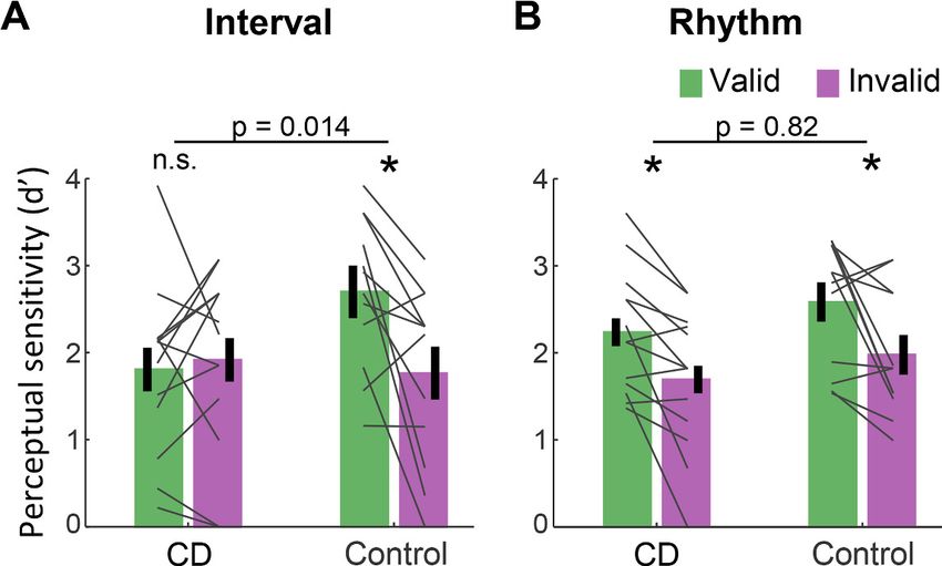
Figure 2 presents d’ values for short interval targets. A three-way ANOVA revealed a significant
validity effect across tasks and groups (F(1,23)=14.75, p=0.001, h2p =0.39). Across tasks, the validity
effect was larger in the control compared to the CD group (cue validity group interaction, F(1,23)
Figure 2. Absence of validity effect in individuals with cerebellar degeneration following interval-based, but not
rhythm-based temporal cues. (A) Interval task. Mean d’ for temporally expected (valid) and unexpected (invalid)
short interval targets (600 ms after the WS) for the CD and control groups. Unlike the controls, the CD group
showed no increase in d’ when the target appeared at the expected time. (B) Rhythm task. Both groups show a
similar increase in d’ on valid trials. *pShort report Neuroscience
=4.83, p=0.038, h2p =0.17), but this effect was qualified by a significant cue validity group task
interaction (F(1,23)=4.76, p=0.04, h2p =0.17). We used a series of planned contrasts to evaluate the
validity effect within each task. For the interval task (Figure 2A), the validity effect was significantly
smaller in the CD group relative to the control group (two-way ANOVA, cue validity group interac-
tion: F(1,23)=7.11, p=0.014, h2p =0.24). The control group showed a higher mean d’ for valid com-
pared to invalid trials (t(11)=3.08, p=0.011, Cohen’s d=0.89), while d’ values in the CD group were
not significantly different, and even numerically higher on invalid trials (t(12) = 0.43, p=0.67,
BF10=0.21, moderate evidence against a directional hypothesis of a validity effect). Thus, the CD
group failed to show an enhancement in perceptual sensitivity from an interval-based temporal cue.
A different pattern was observed on the rhythm task (Figure 2B). Here, the magnitude of validity
effect did not differ between the two groups (F(1,23)=0.053, p=0.82, h2pShort report Neuroscience
minimal, delayed until well after stimulus offset. We note that the current results do not address the
question of whether the cerebellum, in addition to its role in temporal orienting of attention, is also
involved in other spheres of attention.
The dissociation between the impairment in the interval task and the preserved performance in
the rhythm task in the CD group has two important implications. First, a longstanding debate in the
timing literature concerns whether temporal anticipation in a rhythmic context is mediated by
rhythm-specific mechanisms (e.g., entrainment) or by the repeated operation of an interval-based
mechanism (Breska and Deouell, 2017; Haegens and Zion Golumbic, 2018; Drake and Botte,
1993). Our results are at odds with the latter hypothesis given that the CD group failed to benefit
from the interval cues yet showed normal benefits from the rhythm cues. Beat-based timing may rely
on cortico-striatal circuits even in the absence of movement (Cannon and Patel, 2021), consistent
with previous findings of impaired rhythm-based temporal prediction in Parkinson’s disease
(Breska and Ivry, 2018).
Second, the selective contribution of the cerebellum in interval-based but not rhythm-based con-
texts has been observed in multiple timing domains, including duration judgments, timed move-
ment, and timed motor preparation (Breska and Ivry, 2018; Breska and Ivry, 2020; Grube et al.,
2010; Spencer et al., 2003). Our findings extend this functional specificity to the attentional
domain, pointing to a generalized role for the cerebellum in interval timing, at in the sub-second
range. Notably, while inferences from single dissociations such as that observed in the present study
can be limited by concerns about differences in task difficulty, this concern is alleviated by the com-
parable benefits observed in healthy controls from interval- and rhythm-based cues (Breska and
Ivry, 2018; Breska and Ivry, 2020; Breska and Deouell, 2017) (also observed in current dataset:
cue validity task interaction within the control group, F(1,11)=0.86, p=0.37, h2p =0.07).
Computationally, how might the cerebellum contribute to the attentional control of perceptual
sensitivity in time? Given the cerebellar involvement in interval-based timing across timing domains,
an intuitive hypothesis is that the cerebellum is necessary for the temporal processing of isolated
intervals (Ivry and Keele, 1989; Ivry and Schlerf, 2008). This could reflect a central role in one or
both of two putative functional subcomponents in interval timing models (Addyman et al., 2016;
Gibbon et al., 1984), the formation of a temporal representation or retrieval processes related to
interval memory. By this view, predictive processing in non-cerebellar circuits (e.g., prefrontal cortex)
relies on cerebellar interval timing capacities to parameterize the temporal dimension of the predic-
tion. In rhythm-based orienting, an interval-based mechanism would not be required as the temporal
parameters are contained within ongoing neural dynamics. However, a broader hypothesis is that
the interval-based prediction itself is formed within the cerebellum, part of the cerebellar role in pre-
diction in the motor domain and beyond (Sokolov et al., 2017; Wolpert et al., 1998; Miall et al.,
1993). By this view, these cerebellar temporal predictions guide proactive modulation in non-cere-
bellar circuits according to task goals (e.g., to prepare perceptual or motor systems). In rhythm-
based orienting, dedicated prediction mechanisms are not necessary due to the self-sustaining
limit cycle properties of the putatively entrained oscillatory dynamics. Future work should aim to
explore the separability of timing and prediction, identifying the cerebellar computations that pro-
vide the essential information for temporal orienting.
Materials and methods
Key resources table
Reagent type (species) or resource Designation Source or reference Identifiers Additional information
Software, algorithm MATLAB 2019a Mathworks RRID:SCR_001622
Software, algorithm R 3.6.3 R project for statistical computing RRID:SCR_001905
Participants
Fifteen individuals with CD and 14 age-matched neurotypical control individuals were recruited for
the study. The data from two individuals from each group were discarded: One was unable to per-
form the task, two showed no convergence on the staircase procedure used to determine the per-
ceptual threshold, and one asked to terminate the session prematurely. Thus, the final sample size
Breska and Ivry. eLife 2021;10:e66743. DOI: https://doi.org/10.7554/eLife.66743 5 of 11Short report Neuroscience
was 13 CD and 12 control participants. The sample size was determined using power calculations to
allow 80% power to detect effects with Cohen’s d = 0.8 (a conservative estimate, given the typical
effect size of temporal cuing in prior studies: Cohen’s d = 1–1.5; Rohenkohl et al., 2012;
Breska and Ivry, 2018; Rohenkohl et al., 2014). All participants provided informed consent and
were financially compensated for their participation. The study was approved by the Institutional
Review Board at the University of California, Berkeley.
Participants in the CD group (9 females, 12 right-handed, mean age = 56.2 years, SD = 11.1) had
been diagnosed with spinocerebellar ataxia, a slowly progressive adult-onset degenerative disorder
in which the primary pathology involves atrophy of the cerebellum. We did not test patients who
presented symptoms of multisystem atrophy. Eight individuals in the CD group had a specific
genetic subtype (SCA3 = 2, SCA6 = 3, SCA17 = 1, SCA35 = 1, AOA2 = 1) and the other five individ-
uals had CD of unknown/idiopathic etiology. All of the CD participants provided a medical history to
verify the absence of other neurological conditions, and were evaluated at the time of testing with
the Scale for the Assessment and Rating of Ataxia (SARA) (Schmitz-Hübsch et al., 2006). The mean
SARA score was 13.5 (SD = 6.3). Control participants (8 females, 11 right-handed, mean age = 59.1,
SD = 10.2) were recruited from the same age range as the CD group, and, based on self-reports,
did not have a history of neurological or psychiatric disorders. The CD and control groups did not
differ significantly in age (p=0.52).
All participants were prescreened for normal or corrected-to-normal vision and intact color vision.
We also screened for professional musical training or recent amateur participation in musical activi-
ties (e.g., playing a musical instrument or singing in a choir), with the plan to exclude individuals with
such experience (none did). All of the participants completed the Montreal Cognitive Assessment
(MoCA) (Nasreddine et al., 2005) as a simple assessment of overall cognitive competence.
Although we did not select participants to provide a match on this measure, there was no significant
group difference (CD: mean = 26.7, control: mean = 27.5, p=0.32).
Stimuli and task
For the experimental task, participants discriminated the orientation of a masked visual target,
whose timing was cued on each trial (Figure 1). The target was a grayscale, luminance-defined sinu-
soidal Gabor gratings (size: 400 400 pixels, 11 11 cm, 10˚ visual angle; spatial frequency = 1
cycle/degree; Gaussian standard deviation = 2.5˚) that was either oriented horizontally or vertically.
The target was embedded in a dynamic, white noise mask. This mask was a square (size: 400 400
pixels) in which each pixel was randomly assigned a luminance value between 0.25 and 0.75 (with 0
and 1 being black, RGB: [0,0,0] and white, RGB: [255,255,255], respectively). The luminance value for
each pixel in the mask was updated every 16.6 ms (once per monitor refresh cycle) throughout the
trial. The contrast of the target relative to the background noise was adjusted for each participant
(see below). Temporal cues were provided by black squares (size: 200 200 pixels, 5.5 5.5 cm, 5˚
visual angle). All stimuli were created in MATLAB (MathWorks, Natick, MA) and presented using the
Psychtoolbox v.3.0 package for MATLAB (Pelli, 1997; Brainard, 1997). The stimuli were presented
foveally on a gray background (RGB: [128,128,128]) on a 24-in monitor (resolution: 1920 1080 pix-
els, refresh rate: 60 Hz) at a viewing distance of ~65 cm.
The dynamic noise mask remained visible throughout the duration of the trial. The other stimuli
were superimposed on this mask. The suprathreshold temporal cue involved the serial presentation
of two or three black squares (100 ms duration each), with the first black square always appearing
750 ms after the onset of the dynamic mask. The black squares were followed by a suprathreshold
white square, the WS (also 100 ms duration), which was followed in turn by the near-threshold target
(50 ms duration) after either 600 ms (early target) or 1000 ms (late target). Note that given the
screen refresh rate, the 50 ms target was successively embedded in three different masks.
The dynamic noise mask remained visible for 1700 ms after WS onset regardless of the target tim-
ing (1100 ms after early target onset, 700 ms after late target onset). After the termination of the
mask, the screen was blank for a variable interval of 400–900 ms (randomly selected). At the end of
this interval, a visual instruction appeared, requesting that the participant indicates the perceived
orientation of the target (e.g., vertical (press X) ? horizontal (press M)). Responses were made with
the X and M keyboard keys, assigned randomly for each participant and fixed for the entirety of the
experiment. The participants were instructed that the discrimination would be difficult and, if uncer-
tain, to make their ‘best guess’. Note that the procedure involved a long delay between stimulus
Breska and Ivry. eLife 2021;10:e66743. DOI: https://doi.org/10.7554/eLife.66743 6 of 11Short report Neuroscience
offset and the response cue (1500–2000 ms for short interval targets and 1300–1600 ms for long
interval targets). This long delay, coupled with the instructions, was included to eliminate the
demands on motor preparation around the time of the target presentation and, as such, assure that
observed benefits from the temporal cues arose from processes involved in perceptual
discrimination.
Two types of sequences, tested in separate blocks, were used to provide temporal cues. In the
interval task, the sequence consisted of two black squares, with a stimulus onset asynchrony (SOA)
of either 600 ms (short cue) or 1000 ms (long cue). The SOA between the second black square and
WS was randomly set on each trial to be either 1.5 or 2.5 times the cue interval on that trial (short
cue trials: 900/1500 ms; long cue trials: 1500/2500 ms), strongly reducing any periodicity between
the timing of the cue and target (Breska and Deouell, 2017). In the rhythm task, the sequence con-
sisted of three black squares, presented periodically with an SOA of 600 ms (short cue, equivalent to
1.66 Hz) or 1000 ms (long cue, 1 Hz). The SOA between the third black square and WS was the
same duration as the cue SOA for that trial. Thus, the WS fell on the ‘beat’ established by the tem-
poral cues.
In both tasks, the SOA between the WS and target was either the same SOA as defined by the
temporal cue (valid trial, 62.5% of trials) or the non-cued SOA (invalid trials, 25% of trials). This ratio
was selected to incentivize the participant to use the temporal cues to facilitate performance on this
challenging task. On the remaining 12.5% of the trials, no target was presented and the visual
instruction screen did not appear. No response was required on these catch trials and the inter-trial
interval commenced after the dynamic noise was terminated. We included these (no-response) catch
trials to add uncertainty even when a target was not presented at the early target time, by this fur-
ther increasing the difficulty of the task and exploring a possible validity effect in late targets.
Procedure
Upon arrival, all participants provided consent, demographic information, and completed the
MoCA. The CD participants also provided their clinical history and were evaluated with the SARA.
The experiment was conducted in a quiet, dimly lit room. The session began with a familiarization
stage, in which participants performed four practice trials with 100% target contrast followed by four
with 40% target contrast. The latter were included to demonstrate to the participants how difficult it
could be to make a simple orientation judgment when the contrast of the target was similar to that
of the mask.
Following this familiarization phase, we used an adaptive method to determine, on an individual
basis, the target contrast level expected to produce discrimination accuracy of ~79% (descending
staircase procedure, 3 down 1 up [Levitt, 1971], step size = 2%, 10 reversals). We opted to target
79% accuracy to provide sufficient room to detect improvement (to a ceiling of 100% performance)
or impairment (to a floor of 50% performance). Importantly, for this adaptive procedure, only rhyth-
mic temporal cues were used, and the target always appeared at the expected time (valid). Our rea-
soning here was that if the CD group were able to use the temporal cues to modulate perception, it
is more likely to occur in this task (and have a similar threshold value as controls) given previous
work showing that these individuals are not impaired in utilizing rhythmic temporal cues (Breska and
Ivry, 2018). In this way, we would be positioned to ask if the CD group showed an impaired validity
effect in the rhythm task as well as overall performance (valid and invalid trials) on the interval task.
The contrast level identified from the adaptive procedure for a given individual was used in the main
experiment for both tasks. Consistent with our expectation, the mean contrast level did not differ
between groups (t(23)=0.83, p=0.41).
In the main experiment, participants preformed four blocks of each task, alternating between
rhythm and interval blocks (eight blocks total). Each block consisted of 32 trials, 16 with the short
temporal cue and 16 with the long temporal cue. Of these 16 trials, the target appeared at the cued
time on 10 trials (valid), the uncued time on 4 trials (invalid), and did not appear on 2 trials (catch).
When present, the target was horizontal on 50% of the trials and vertical on the other 50% of the tri-
als. Short breaks were provided between each block.
To ensure that the target contrast fell in a range that would be optimal for detecting a validity
effect, we calculated the averaged performance on valid trials across the two tasks after each pair of
blocks. If the accuracy was higher than 99%, we reduced the target contrast for subsequent blocks
by 4%, and if it was lower than 60%, we increased it by 4%. Block pairs in which performance was
Breska and Ivry. eLife 2021;10:e66743. DOI: https://doi.org/10.7554/eLife.66743 7 of 11Short report Neuroscience
above 99% or below 60% were not included in the d’ analyses (four excluded blocks across all partic-
ipants; exclusion had no impact on the statistical tests).
Prior to the first block for each task, the experimenter demonstrated the trial sequence and then
conducted practice trials until the participant could describe how the cues were predictive of the
onset time of the target. For subsequent blocks, the participant first completed two practice trials as
a reminder of the format for the temporal cues in the forthcoming block. Participants received feed-
back on their performance after these practice trials (but not after any of the staircase or experimen-
tal trials).
Statistical analysis
The data were analyzed using custom MATLAB scripts and R50. Following standard practices for
data analysis in non-speeded, 2-AFC tasks, we quantified discrimination performance by calculating,
on an individual basis, a d-prime (d’) score separately for each combination of task, target interval
and cue validity. These values were calculated by subtracting the z-score of the percentage of hits
from the z-score of the percentage of false alarms (referring to vertical and horizontal categories as
‘stimulus present’ and ‘stimulus absent’, respectively, in classic signal detection terminology). As the
‘hit’ category was arbitrarily assigned to one orientation and the two orientations were equally prob-
able, we did not calculate or analyze the criterion index. An increase in perceptual sensitivity due to
temporal anticipation should be manifest as an increase in d’ when the target appeared at the
expected time compared to when it appeared at the unexpected time (validity effect).
Previous work indicates that validity effects from temporal cues in two-interval designs such as
that used here are usually attenuated for late onset targets, either due to re-orienting of attention in
time or foreperiod effects (Correa et al., 2006). As such, our a priori plan was to focus on trials with
short interval targets to increase sensitivity for detecting attenuation of the validity effect. To confirm
that this pattern was present in our data, we subjected d’ values to an omnibus four-way mixed
ANOVA with a between-subject factor group (CD/control), and within-subject factors task (interval/
rhythm), target interval (early/late), and cue validity (valid/invalid), and tested the directional hypoth-
esis of a larger effect of the cue validity factor for target appearing at early compared to late inter-
vals using a one-tailed test. As expected (see Results), we observed a significant cue validity
target interval interaction in the expected direction, and post hoc comparisons showed that the
validity effect was only significant in the early interval condition.
The d’ values for short interval targets were analyzed using a mixed ANOVA with a between-sub-
ject factor group (CD/control), and within-subject factors task (interval/rhythm) and cue validity
(valid/invalid). To assess the effect of cue validity within each group and task, we used within-subject
t-tests. To compare the cue validity effect between groups within each task, we used a mixed
ANOVA with factors group (CD/control) and cue validity (valid/invalid). Finally, to assess
context specificity within the CD group, we performed an orthogonal contrast, comparing the cue
validity effects between tasks using a repeated-measures ANOVA with factors task (interval/rhythm)
and cue validity. In all analyses, effect sizes were estimated using Cohen’s d and partial eta-squared
(h2p ).
Acknowledgements
We thank Arohi Saxena for assistance in data collection.
Additional information
Competing interests
Richard B Ivry: Senior editor, eLife. The other author declares that no competing interests exist.
Funding
Funder Grant reference number Author
National Institutes of Health NS092079 Richard B Ivry
Breska and Ivry. eLife 2021;10:e66743. DOI: https://doi.org/10.7554/eLife.66743 8 of 11Short report Neuroscience
National Institutes of Health NS116883 Richard B Ivry
The funders had no role in study design, data collection and interpretation, or the
decision to submit the work for publication.
Author contributions
Assaf Breska, Conceptualization, Data curation, Software, Formal analysis, Validation, Investigation,
Visualization, Methodology, Writing - original draft, Project administration, Writing - review and edit-
ing; Richard B Ivry, Conceptualization, Resources, Supervision, Funding acquisition, Project adminis-
tration, Writing - review and editing
Author ORCIDs
Assaf Breska https://orcid.org/0000-0002-6233-073X
Richard B Ivry https://orcid.org/0000-0003-4728-5130
Ethics
Human subjects: All participants provided informed consent to participate in the study and for the
publication of de-identified data. The study was approved by the Institutional Review Board at the
University of California, Berkeley (CPHS# 2016-02-8439).
Decision letter and Author response
Decision letter https://doi.org/10.7554/eLife.66743.sa1
Author response https://doi.org/10.7554/eLife.66743.sa2
Additional files
Supplementary files
. Source data 1. Raw data.
. Source code 1. Preprocessing code (MATLAB).
. Source code 2. Statistical analysis code (R).
. Transparent reporting form
Data availability
De-identified source data files for all figures and analyses in the article have been provided. Addi-
tional demographic information was not uploaded as it was not used in any analysis reported in the
text, and can be provided upon request in personal communication with the corresponding author,
without additional restrictions.
References
Addyman C, French RM, Thomas E. 2016. Computational models of interval timing. Current Opinion in
Behavioral Sciences 8:140–146. DOI: https://doi.org/10.1016/j.cobeha.2016.01.004
Allen G, Buxton RB, Wong EC, Courchesne E. 1997. Attentional Activation of the Cerebellum Independent of
Motor Involvement. Science. 275:1940–1943. DOI: https://doi.org/10.1126/science.275.5308.1940
Bolger D, Coull JT, Schön D. 2014. Metrical Rhythm Implicitly Orients Attention in Time as Indexed by Improved
Target Detection and Left Inferior Parietal Activation. Journal of Cognitive Neuroscience 26:593–605.
DOI: https://doi.org/10.1162/jocn_a_00511
Brainard DH. 1997. The Psychophysics Toolbox. Spatial Vision 10:433–436. DOI: https://doi.org/10.1163/
156856897X00357
Breska A, Deouell LY. 2017. Neural mechanisms of rhythm-based temporal prediction: Delta phase-locking
reflects temporal predictability but not rhythmic entrainment. PLOS Biology 15:e2001665. DOI: https://doi.org/
10.1371/journal.pbio.2001665
Breska A, Ivry RB. 2018. Double dissociation of single-interval and rhythmic temporal prediction in cerebellar
degeneration and Parkinson’s disease. PNAS 115:12283–12288. DOI: https://doi.org/10.1073/pnas.
1810596115, PMID: 30425170
Breska and Ivry. eLife 2021;10:e66743. DOI: https://doi.org/10.7554/eLife.66743 9 of 11Short report Neuroscience
Breska A, Ivry RB. 2020. Context-specific control over the neural dynamics of temporal attention by the human
cerebellum. Science Advances 6:eabb1141. DOI: https://doi.org/10.1126/sciadv.abb1141
Cannon JJ, Patel AD. 2021. How Beat Perception Co-opts Motor Neurophysiology. Trends in Cognitive Sciences
25:137–150. DOI: https://doi.org/10.1016/j.tics.2020.11.002
Corbetta M, Shulman GL. 2002. Control of goal-directed and stimulus-driven attention in the brain. Nature
Reviews Neuroscience 3:201–215. DOI: https://doi.org/10.1038/nrn755
Correa A, Lupiáñez J, Tudela P. 2005. Attentional preparation based on temporal expectancy modulates
processing at the perceptual level. Psychonomic Bulletin & Review 12:328–334. DOI: https://doi.org/10.3758/
BF03196380, PMID: 16082814
Correa A, Lupiáñez J, Tudela P. 2006. The attentional mechanism of temporal orienting: determinants and
attributes. Experimental Brain Research 169:58–68. DOI: https://doi.org/10.1007/s00221-005-0131-x,
PMID: 16273403
Coull JT, Nobre AC. 1998. Where and When to Pay Attention: The Neural Systems for Directing Attention to
Spatial Locations and to Time Intervals as Revealed by Both PET and fMRI. The Journal of Neuroscience 18:
7426–7435. DOI: https://doi.org/10.1523/JNEUROSCI.18-18-07426.1998
Davranche K, Nazarian B, Vidal F, Coull J. 2011. Orienting Attention in Time Activates Left Intraparietal Sulcus
for Both Perceptual and Motor Task Goals. Journal of Cognitive Neuroscience 23:3318–3330. DOI: https://doi.
org/10.1162/jocn_a_00030
Denison RN, Heeger DJ, Carrasco M. 2017. Attention flexibly trades off across points in time. Psychonomic
Bulletin & Review 24:1142–1151. DOI: https://doi.org/10.3758/s13423-016-1216-1
Drake C, Botte M-C. 1993. Tempo sensitivity in auditory sequences: Evidence for a multiple-look model.
Perception & Psychophysics 54:277–286. DOI: https://doi.org/10.3758/BF03205262
Fernández A, Denison RN, Carrasco M. 2019. Temporal attention improves perception similarly at Foveal and
parafoveal locations. Journal of Vision 19:12. DOI: https://doi.org/10.1167/19.1.12, PMID: 30650437
Fiebelkorn IC, Kastner S. 2020. Functional Specialization in the Attention Network. Annual Review of Psychology
71:221–249. DOI: https://doi.org/10.1146/annurev-psych-010418-103429
Gibbon J, Church RM, Meck WH. 1984. Scalar timing in memory. Annals of the New York Academy of Sciences
423:52–77. DOI: https://doi.org/10.1111/j.1749-6632.1984.tb23417.x
Gómez CM, Delinte A, Vaquero E, Cardoso MJ, Vázquez M, Crommelinck M, Roucoux A. 2001. Current source
density analysis of CNV during temporal Gap paradigm. Brain Topography 13:149–159. DOI: https://doi.org/
10.1023/A:1007816201345
Green DM, Swets JA. 1966. Signal detection theory and psychophysics. New York: Wiley.
Grube M, Cooper FE, Chinnery PF, Griffiths TD. 2010. Dissociation of duration-based and beat-based auditory
timing in cerebellar degeneration. PNAS 107:11597–11601. DOI: https://doi.org/10.1073/pnas.0910473107
Haarmeier T, Thier P. 2007. The attentive cerebellum - myth or reality? The Cerebellum 6:177–183. DOI: https://
doi.org/10.1080/14734220701286187
Haegens S, Zion Golumbic E. 2018. Rhythmic facilitation of sensory processing: A critical review. Neuroscience &
Biobehavioral Reviews 86:150–165. DOI: https://doi.org/10.1016/j.neubiorev.2017.12.002
Ivry RB, Keele SW. 1989. Timing Functions of The Cerebellum. Journal of Cognitive Neuroscience 1:136–152.
DOI: https://doi.org/10.1162/jocn.1989.1.2.136
Ivry RB, Schlerf JE. 2008. Dedicated and intrinsic models of time perception. Trends in Cognitive Sciences 12:
273–280. DOI: https://doi.org/10.1016/j.tics.2008.04.002
Levitt H. 1971. Transformed Up-Down Methods in Psychoacoustics. The Journal of the Acoustical Society of
America 49:467–477. DOI: https://doi.org/10.1121/1.1912375
Miall RC, Weir DJ, Wolpert DM, Stein JF. 1993. Is the Cerebellum a Smith Predictor? Journal of Motor Behavior
25:203–216. DOI: https://doi.org/10.1080/00222895.1993.9942050
Miniussi C, Wilding EL, Coull JT, Nobre AC. 1999. Orienting attention in time. Modulation of brain potentials.
Brain: A Journal of Neurology 122:1507–1518. DOI: https://doi.org/10.1093/brain/122.8.1507, PMID: 10430
834
Morillon B, Schroeder CE, Wyart V, Arnal LH. 2016. Temporal Prediction in lieu of Periodic Stimulation. The
Journal of Neuroscience 36:2342–2347. DOI: https://doi.org/10.1523/JNEUROSCI.0836-15.2016
Nasreddine ZS, Phillips NA, Bédirian V, Charbonneau S, Whitehead V, Collin I, Cummings JL, Chertkow H. 2005.
The Montreal cognitive assessment, MoCA: a brief screening tool for mild cognitive impairment. Journal of the
American Geriatrics Society 53:695–699. DOI: https://doi.org/10.1111/j.1532-5415.2005.53221.x, PMID: 15
817019
Nobre AC, van Ede F. 2018. Anticipated moments: temporal structure in attention. Nature Reviews
Neuroscience 19:34–48. DOI: https://doi.org/10.1038/nrn.2017.141
O’Reilly JX, Mesulam MM, Nobre AC. 2008. The cerebellum predicts the timing of perceptual events. Journal of
Neuroscience 28:2252–2260. DOI: https://doi.org/10.1523/JNEUROSCI.2742-07.2008
Pelli DG. 1997. The VideoToolbox software for visual psychophysics: transforming numbers into movies. Spatial
Vision 10:437–442. DOI: https://doi.org/10.1163/156856897X00366
Perrett SP, Ruiz BP, Mauk MD. 1993. Cerebellar cortex lesions disrupt learning-dependent timing of conditioned
eyelid responses. The Journal of Neuroscience 13:1708–1718. DOI: https://doi.org/10.1523/JNEUROSCI.13-04-
01708.1993
Posner MI, Petersen SE. 1990. The attention system of the human brain. Annual Review of Neuroscience 13:25–
42. DOI: https://doi.org/10.1146/annurev.ne.13.030190.000325, PMID: 2183676
Breska and Ivry. eLife 2021;10:e66743. DOI: https://doi.org/10.7554/eLife.66743 10 of 11Short report Neuroscience
Praamstra P. 2006. Neurophysiology of Implicit Timing in Serial Choice Reaction-Time Performance. Journal of
Neuroscience 26:5448–5455. DOI: https://doi.org/10.1523/JNEUROSCI.0440-06.2006
Ravizza SM, Ivry RB. 2001. Comparison of the Basal Ganglia and Cerebellum in Shifting Attention. Journal of
Cognitive Neuroscience 13:285–297. DOI: https://doi.org/10.1162/08989290151137340
Raymond JE, Shapiro KL, Arnell KM. 1992. Temporary suppression of visual processing in an RSVP task: an
attentional blink? Journal of Experimental Psychology: Human Perception and Performance 18:849–860.
DOI: https://doi.org/10.1037/0096-1523.18.3.849
Rohenkohl G, Cravo AM, Wyart V, Nobre AC. 2012. Temporal expectation improves the quality of sensory
information. Journal of Neuroscience 32:8424–8428. DOI: https://doi.org/10.1523/JNEUROSCI.0804-12.2012,
PMID: 22699922
Rohenkohl G, Gould IC, Pessoa J, Nobre AC. 2014. Combining spatial and temporal expectations to improve
visual perception. Journal of Vision 14:8. DOI: https://doi.org/10.1167/14.4.8, PMID: 24722562
Samaha J, Bauer P, Cimaroli S, Postle BR. 2015. Top-down control of the phase of alpha-band oscillations as a
mechanism for temporal prediction. PNAS 112:8439–8444. DOI: https://doi.org/10.1073/pnas.1503686112
Sanabria D, Capizzi M, Correa Á, Correa Á. 2011. Rhythms that speed you up. Journal of Experimental
Psychology: Human Perception and Performance 37:236–244. DOI: https://doi.org/10.1037/a0019956
Schmitz-Hübsch T, du Montcel ST, Baliko L, Berciano J, Boesch S, Depondt C, Giunti P, Globas C, Infante J,
Kang JS, Kremer B, Mariotti C, Melegh B, Pandolfo M, Rakowicz M, Ribai P, Rola R, Schöls L, Szymanski S, van
de Warrenburg BP, et al. 2006. Scale for the assessment and rating of ataxia: development of a new clinical
scale. Neurology 66:1717–1720. DOI: https://doi.org/10.1212/01.wnl.0000219042.60538.92, PMID: 16769946
Shalev N, Nobre AC, van Ede F. 2019. Time for What? Breaking Down Temporal Anticipation. Trends in
Neurosciences 42:373–374. DOI: https://doi.org/10.1016/j.tins.2019.03.002
Sokolov AA, Miall RC, Ivry RB. 2017. The Cerebellum: Adaptive Prediction for Movement and Cognition. Trends
in Cognitive Sciences 21:313–332. DOI: https://doi.org/10.1016/j.tics.2017.02.005
Spencer RMC, Zelaznik HN, Diedrichsen J, Ivry RB. 2003. Disrupted Timing of Discontinuous But Not Continuous
Movements by Cerebellar Lesions. Science. 300:1437–1439. DOI: https://doi.org/10.1126/science.1083661
Townsend J, Courchesne E, Covington J, Westerfield M, Harris NS, Lyden P, Lowry TP, Press GA. 1999. Spatial
Attention Deficits in Patients with Acquired or Developmental Cerebellar Abnormality. The Journal of
Neuroscience 19:5632–5643. DOI: https://doi.org/10.1523/JNEUROSCI.19-13-05632.1999
van Ede F, Rohenkohl G, Gould I, Nobre AC. 2020. Purpose-Dependent consequences of temporal expectations
serving perception and action. The Journal of Neuroscience 40:7877–7886. DOI: https://doi.org/10.1523/
JNEUROSCI.1134-20.2020, PMID: 32900836
Wolpert DM, Miall RC, Kawato M. 1998. Internal models in the cerebellum. Trends in Cognitive Sciences 2:338–
347. DOI: https://doi.org/10.1016/S1364-6613(98)01221-2
Breska and Ivry. eLife 2021;10:e66743. DOI: https://doi.org/10.7554/eLife.66743 11 of 11You can also read