The Prevalence of Gastrointestinal Parasites and Seroprevalence of Toxoplasma gondii in Captive Ocelots (Leopardus pardalis) in Trinidad, West ...
←
→
Page content transcription
If your browser does not render page correctly, please read the page content below
Hindawi
Veterinary Medicine International
Volume 2021, Article ID 8820548, 5 pages
https://doi.org/10.1155/2021/8820548

Research Article
The Prevalence of Gastrointestinal Parasites and
Seroprevalence of Toxoplasma gondii in Captive
Ocelots (Leopardus pardalis) in Trinidad, West Indies

          Alissa Bally ,1 Stacy Francis-Charles ,2 Tariq Ackbar,3 Yadel Beharrylal,3
          Roxanne Charles ,1 Asoke Basu ,1 and Rod Suepaul 1
          1
            Department of Basic Sciences, School of Veterinary Medicine, University of the West Indies, St. Augustine, Trinidad and Tobago
          2
            Small Animal Clinic, School of Veterinary Medicine, St. Georges University, True Blue, Grenada
          3
            Veterinarian in Private Practice, Caroni, Trinidad and Tobago

          Correspondence should be addressed to Rod Suepaul; rod.suepaul@sta.uwi.edu

          Received 5 June 2020; Revised 21 April 2021; Accepted 5 May 2021; Published 27 May 2021

          Academic Editor: Carlos Gonz lez Rey

          Copyright © 2021 Alissa Bally et al. This is an open access article distributed under the Creative Commons Attribution License,
          which permits unrestricted use, distribution, and reproduction in any medium, provided the original work is properly cited.

          This study was conducted from November 2010 to June 2011 to determine the prevalence of gastrointestinal parasites and the
          seroprevalence of Toxoplasma gondii in captive ocelots (Leopardus pardalis) in Trinidad. Faecal samples were collected and
          analyzed using faecal flotation to identify helminth ova and protozoan cysts and oocysts. Serum samples from captive ocelots were
          screened for T. gondii using a latex agglutination test kit. Of the 19 ocelots examined, the most prevalent parasites noted were ova
          of ascarids, strongyles, and Capillaria spp. The serum of three of the 13 (23.1%) ocelots tested was positive for T. gondii antibodies.
          These ocelots are therefore a potential source of T. gondii infection to both humans and other animals. This is the first documented
          report of endoparasites in local captive ocelots within Trinidad and provides useful data to support further research of the captive
          and wild populations.

1. Introduction                                                           Trinidad, ocelots have been reported to be sighted in both
                                                                          northern and southern regions and are notorious for the
The ocelot, Leopardus pardalis, is the largest felid of the               predation of domestic fowls. These felids are designated as
genus Leopardus. The pelage is short and thick with a                     an environmentally sensitive species providing legislative
variable coloration that depends on the habitat in which it is            protection for the species. A few that were trapped by
found. Distinctive facial patterns allow relatively easy rec-             hunters in the past have been relocated to the state zoo and
ognition of individuals [1]. Ocelots commonly inhabit the                 other wildlife protection facilities.
tropical rainforests of Trinidad and Central and South                        Parasitic infections in captive wildlife can result in death to
America. They can also be found in marshes, mangroves,                    the affected animals, act as a predisposing factor for the de-
thorn scrub regions, and savannah grasslands. They spend                  velopment of secondary infectious diseases and exert a negative
most of their time in elevated regions below 1200 meters                  impact on reproduction. This is especially important in en-
[1–3]. The diet of ocelots consists mainly of small rodents               dangered species [6]. Leopardus pardalis is the host to a di-
with some medium-sized animals, reptiles, birds, and                      versity of endoparasites including Taenia spp., strongyles,
aquatic species including crustaceans [4, 5]. These felids are            Paragonimus spp., Toxocara cati, Capillaria spp., spirurids,
thus very important in neotropical ecosystems, since they are             Aelurostrongylus abstrusus, acanthocephalans (e.g., Oncicola
apex predators in the regulation of prey populations [4].                 spp.), Hammondia pardalis, and Isospora spp. [1]. They are also
Ocelots are generally not a threat to man since they prefer to            known to host Toxoplasma gondii, Ancylostoma tubaeforme,
hide from or evade human encounters naturally. In                         Uncinaria spp., Crenosoma spp., and Spirometra spp. [7].
2 Veterinary Medicine International
Toxoplasma gondii is a zoonotic protozoan parasite that
is found in a wide range of mammals and birds. Toxo-
plasmosis is responsible for abortions in livestock especially
sheep and is a common cause of pathology in marsupials [8]. 9 ocelots 2 ocelots
Toxoplasmosis in humans causes mental retardation, sei-
N
zures, blindness, and death when transmitted congenitally W E
and may be fatal to immunocompromised individuals S
[9, 10]. Felids are the sole definitive hosts in which the 3 ocelots
parasite can complete both sexual and asexual stages. Pre-
vious reports from neighboring Latin American countries 2 ocelots
have documented Toxoplasma gondii in ocelots [7, 11]. 3 ocelots
Oocysts shed in ocelot faeces can become infective to other
mammalian hosts (including cats, dogs, and humans) via the
faecal-oral route [7, 11–13]. Ocelots prey on small mammals,
including agouti and rodents, which can act as intermediate
hosts with bloodborne tachyzoites and tissue bradyzoite North
cysts which are infective to felids and other intermediate Central
hosts through predation and carnivorism [14]. South
This current study therefore aims to identify the en- Figure 1: Map of Trinidad displaying the geographic locations of
doparasite species which may be found within the captive captive ocelots sampled throughout this study.
ocelot population in Trinidad and determine the serological
status of Toxoplasma gondii in these felids. The ultimate goal
is to create and increase the awareness to the public and parasitological analysis. Samples were refrigerated at 4°C for
scientific community of the potential parasitic infections of further processing for a maximum of 72 hours.
veterinary and public health significance that can be
transmitted by ocelots. 2.2. Flotation Technique. Three grams of faeces were
weighed and mixed with 45 ml of 33.3% zinc sulphate so-
2. Materials and Methods lution and then filtered into a clean faecal cup using a tea
strainer and gauze. The supernatant was added to a floa-
Ethical approval was granted by the University of the West tation vial until a positive meniscus was formed. A coverslip
Indies Campus Research Ethics Committee prior to com- was placed on the meniscus and left undisturbed for 10–15
mencement of this study. A listing of persons holding legal minutes. The coverslip was then removed and placed onto a
licenses for captive ocelots was sourced from the Forestry microscopic slide. Each slide was examined microscopically
Division, Ministry of Agriculture, Land, and Forestry of at 10x and 40x objectives for detection of any parasitic ova,
Trinidad and Tobago. This list revealed five legal captive oocysts, cysts, or larvae [15].
locations within North, Central, and South Trinidad from
which all owners were contacted. The objectives of the study
and samples needed were then explained to willing partic- 2.3. Blood Collection. Feline subjects were anesthetized using
ipants. Site visits to these locations showcased ocelots in a combination of 12 mg/kg ketamine and 1 mg/kg xylazine
apparent good health. The cats were kept in cages and intramuscularly [16]. Each ocelot was placed in lateral re-
maintained in clean, sanitary conditions. Based on locality, cumbency, and one forelimb was partially shaved to gain
11 ocelots were obtained from North Trinidad from two visual access to the cephalic vein. This area was swabbed with
sites, three ocelots were obtained from Central Trinidad 70% alcohol, after which 5 ml of blood was removed via
from one site, and five were obtained from two sites on the venipuncture using a 20-gauge hypodermic needle and
southern end of the island (Figure 1). syringe. Blood samples collected from all cats were placed in
Faecal samples were collected from 17 wild-caught and individually labeled red-topped tubes.
two captive-born ocelots at all participating sites. Blood After blood collection, the ocelots were placed in re-
samples were taken from only 13 cats dispersed over three of covery cages or areas prepared with bedding and low noise
the five sites based on the owner’s consent. and light intensity to allow a smooth recovery without
causing injury on awakening. The animals were monitored
during recovery for any complications due to anesthesia.
2.1. Fecal Specimen Collection. Faecal samples were collected Blood samples were transported in a cooler at 4°C to the
from 19 individual animal enclosures and placed in clean haematology laboratory of the UWI-SVM. Sera were sep-
plastic bags or plastic faecal cups. To minimize handling arated using centrifugation and stored at −80°C until further
stress of the cats, freshly voided faecal samples no older than testing. Serum samples were tested by a latex agglutination
12 hours were collected off the ground of enclosures. The test for the presence of antibodies against Toxoplasma gondii
samples were then transported in a cooler with ice packs to
the Parasitology Laboratory at the University of the West
®
using the Toxotest-MT (Eiken Chemical Co., Ltd., Tokyo,
110-8404, Japan) according to the manufacturer’s instruc-
Indies-School of Veterinary Medicine (UWI-SVM) for tions. The test is a modified indirect agglutination testVeterinary Medicine International 3
Table 1: Prevalence of gastrointestinal parasites among the captive ocelot population in Trinidad.
Parasites detected Number positive (n � 19) Prevalence (%)
Nematodes
Ascarids 7 36.8
Strongyles 6 31.6
Trichostrongylus spp. 2 10.5
Capillaria spp. 3 15.8
Trichuris spp. 2 10.5
Trematodes
Schistosoma spp. 3 15.8
Protozoans
Balantidium coli 1 5.3
Unidentified oocyst 2 10.5
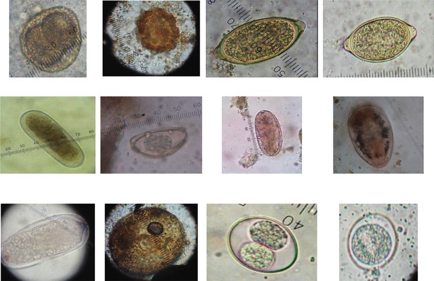
Toxocara spp. Toxascaris spp. Trichuris spp. Capillaria spp.
Trichostrongylus Enterobius Strongyle ovum Schistosoma spp.
spp. vermicularis
Schistosoma Unidentified
Balantidium cyst Unidentified oocyst
spp. oocyst
Figure 2: Images of gastrointestinal parasites identified among the captive ocelot population in Trinidad.
predominantly utilized as a screening test with a sensitivity (Schistosoma spp.) and intestinal protozoa (Balantidium and
of 99% and specificity of 81% with confidence values of Isospora spp.). A total of seven (36.8%) ocelots harboured
94.4% and 92.5%, respectively. Based on the manufacturer’s two or more parasites (nematodes, trematodes, or protozoa),
guidelines, a titer ≥64 was regarded positive. while three (15.8%) exhibited a mixed infection of both
helminths and protozoa. Up to five parasite genera were
observed in one felid. The mean number of parasite genera
3. Results and Discussion hosted by the infected ocelots in this study was 2.4.
3.1. Faecal Analysis. Of the 19 faecal samples tested, gas- Previous reports from Trinidad detected one species of
trointestinal parasites were observed in the faeces of 11 Acanthocephala (Echinopardalis pardalis), one cestode
(57.9%) ocelots (Table 1). A total of eight parasitic genera (Diphyllobothrium sp.), three nematode species (Trichoce-
were detected in gastrointestinal tracts of these felids by phalus serratus, Molineus pardalis, and trichostrongyle spp.),
faecal flotation (Table 1). The most prevalent parasites were and an enteric arthropod (Parrocephalus stilesi) in non-
nematodes (ascarids and strongyles) followed by trematodes captive ocelots [13, 17–19]. However, in this current study,4 Veterinary Medicine International
Table 2: Serological results for Toxoplasma gondii in 13 individual ocelots based on the percent of positivity regarding the gender,
geographical location in Trinidad, adult, and immature ocelots.
Male (n � 7) Female (n � 6) North (n � 8) Central (n � 3) South (n � 2) Adult (n � 11) Immature (n � 2)
Positive (%) 0 23.1 7.69 7.69 7.69 23.1 0
Negative (%) 53.8 23.1 53.8 15.4 7.69 61.5 15.4
Toxocara spp., strongyle spp., Trichostrongylus spp., Capil- due to the small number of ocelots tested in this study. If this
laria spp., coccidian oocysts, Balantidium spp. cyst, and study is to be repeated using a larger number of captive
Schistosoma spp. have been identified in the faeces of ocelots, the data attained can be more reliable allowing more
infested ocelots (Figure 2). Since these cats are carnivorous valid and direct comparisons to be made between both
and feed on a range of invertebrates, there is the possibility countries since Trinidad is geographically similar to the
that some parasites were spurious [6]. It can also be sus- mainland of South America. A higher frequency of ocelots,
pected that the captive living conditions such as husbandry 15/22 (68.18%), was found to be seropositive for T. gondii
practices and prolonged confinement may also play a role in antibodies in a survey at a single location in Brazil [21]. In
the rate of infestation of these parasites. Environmental Mexico, two of three ocelots had antibodies to T. gondii at a
contamination as a result of humans serving as fomited may zoo and 18 of 26 (69%) free ranging ocelots were found to be
also provide additional avenues for infestation particularly seropositive [22].
for those kept within a zoo environment where there are Although the sample size in this study was too small to
multiple visitors and zoo workers [20]. detect any statistically significant differences within the
ocelot population, the parasite diversity will aid in assessing
the health status and possible parasite transmission by these
3.2. Serology. The sera of the thirteen [13] ocelots subjected cats. Another shortcoming of sample collection was the use
to the latex agglutination test revealed three (23.1%) sero- of faeces that was not freshly voided. This sampling tech-
positive animals. nique can potentially minimize parasite detection due to
Seropositive animals were found in North, Central, and degradation of trophozoites or hatching of thin-shelled ova
South Trinidad resulting in a total percent of positivity of such as hookworms before analysis [23]. To detect these
23.1%. This indicates that these animals have been exposed parasites effectively, it would be prudent to collect rectal
to T. gondii and are thus possible sources of infection to faecal samples with analysis within 30–60 minutes of col-
other intermediate hosts including humans. Based on lo- lection. This study thus serves as a pilot for future studies for
cation, the prevalence of T. gondii infection was approxi- the screening of parasites and other pathogens in the wild
mately 7.69% with a higher presentation of adult females at ocelot population in Trinidad and Tobago.
23.1% as opposed to males and immature animals at 0% in
this sample population (Table 2).
The Fischer two-tailed exact test revealed a significant 4. Conclusions
(P < 0.05) relationship between those from the North against
the Central and South. There was no significant relationship More than 50% of the captive ocelot population in Trinidad
with sex or age (adult vs. juvenile). harbour endoparasites and approximately 25% are sero-
In recent times, a few of these animals have been utilized positive for T. gondii. Care must be taken when handling the
in the petting zoo and wildlife awareness programmes and faeces of captive ocelots, and proper waste disposal must be
interacted closely with the public. This could be a cause for implemented to prevent environmental contamination and
concern, though minimal, for handlers and other persons in infection of other animals and humans with these parasites.
contact with infected ocelot faeces. An immunodeficient or
pregnant animal may increase the risk of infection from the
environment where actively infected ocelots defecate. The Data Availability
results of this survey indicate that these animals in particular
should be regularly tested to determine the possible risk to The parasitology and serology results data used to support
their handlers and the wider population. the findings of this study are included within the article.
The small numbers tested were largely due to the small
captive population which is a reflection of the small size of
the island and local ocelot population. Some owners did not Conflicts of Interest
volunteer their animals for serological testing due to the risks
The authors declare that there are no conflicts of interest.
associated with tranquilization. Therefore, statistical rigor
was affected. However, it was found that finding a sero-
positive ocelot was higher for females (23.1%; n � 6) than for
males (0%; n � 7).
Acknowledgments
The seroprevalence of T. gondii found in this study The authors would like to thank the University of the West
(23.1%; n � 13) was lower as compared to a national survey of Indies for granting approval and provision of the necessary
captive ocelots in Brazil (57.7%; n � 168) [11]. This may be funding to carry out this study.Veterinary Medicine International 5
References [20] A. Q. Mir, K. Dua, L. D. Singla, S. Sharma, and M. P. Singh,
“Prevalence of parasitic infection in captive wild animals in
[1] J. Kittel and P. Myers, Leopardus Pardalis. Animal Diversity Bir Moti Bagh mini zoo (Deer Park), Patiala, Punjab,” Vet-
Web, 2001, https://animaldiversity.ummz.umich.edu/site/acc erinary World, vol. 9, no. 6, pp. 540–543, 2016.
ounts/information/Leopardus_pardalis.html. [21] L. S. Ullmann, R. C. da Silva, and W. de Moraes, “Serological
[2] C. A. Gonzales, D. E. Brown, and J. P. Gallo-Reyonoso, survey of Toxoplasma gondii in captive Neotropical felids
“Ecology, distribution and conservation status of Leopardus from Southern Brazil,” Veterinary Parasitology, vol. 102,
pardalis in Mexico. Oryx,” The International Journal of pp. 217–224, 2001.
Conservation, vol. 37, no. 3, pp. 358–364, 2003. [22] C. Alvarado-Esquivel, E. A. Gayosso-Dominguez, I. Villena,
[3] O. E. Ramı́rez-Bravo, E. Bravo-Carrete, C. Hernández-Santı́n, and J. P. Dubey, “Seroprevalence of Toxoplasma gondii in-
S. Schinkel-Brault, and K. Chris, “Ocelot (Leopardus pardalis) fection in captive mammals in three zoos in Mexico city,
distribution in the state of Puebla, Central Mexico,” Therya, Mexico,” The Journal of Zoo and Wildlife Medicine, vol. 44,
vol. 1, no. 2, pp. 111–120, 2010. pp. 803–806, 2013.
[4] K. H. Redford and J. F. Eisenberg, Mammals of the Neotropics: [23] Centers for Disease Control and Prevention, Stool Specimens -
The Southern Cone, The University of Chicago, Chicago, IL, Specimen Processing, CDC, Atlanta, GA, USA, 2016, https://
USA, 1992. www.cdc.gov/dpdx/diagnosticprocedures/stool/specimenproc.
[5] K. C. Abreu, R. F. Moro-Rios, J. E. Silva-Pereira, html.
J. M. D. Miranda, and E. F. Jablonski, “Feeding habits of ocelot
(Leopardus pardalis) in Southern Brazil,” Mammalian Biol-
ogy, vol. 73, no. 5, pp. 407–411, 2008.
[6] M. S. Panayotova-Pencheva, “Parasites in captive animals: a
review of studies in some European zoos,” Der Zoologische
Garten, vol. 82, no. 1, pp. 60–71, 2013.
[7] C. V. Fiorello, R. G. Robbins, L. Maffei, and S. E. Wade,
“Parasites of free-ranging small canids and felids in the
Bolivian chaco,” Journal of Zoo and Wildlife Medicine, vol. 37,
no. 2, pp. 130–134, 2006.
[8] P. J. Canfield, W. J. Hartley, and J. P. Dubey, “Lesions of
toxoplasmosis in Australian marsupials,” Journal of Com-
parative Pathology, vol. 103, no. 2, pp. 159–167, 1990.
[9] J. L. Jones, A. Lopez, M. Wilson, J. Schulkin, and R. Gibbs,
“Congenital toxoplasmosis: a review,” Obstetrical & Gyne-
cological Survey, vol. 56, pp. 296–305, 2001.
[10] G. Saadatnia and M. Golkar, “A review on human toxo-
plasmosis,” Scandinavian Journal of Infectious Diseases,
vol. 44, pp. 805–814, 2012.
[11] J. C. Silva, S. Ogassawara, C. H. Adania et al., “Seroprevalence
of Toxoplasma gondii in captive neotropical felids from
Brazil,” Veterinary Parasitology, vol. 102, pp. 217–224, 2001.
[12] C. V. Fiorello, S. L. Deem, M. E. Gompper, and E. J. Dubovi,
“Seroprevalence of pathogens in domestic carnivores on the
border of Madidi National Park, Bolivia,” Animal Conser-
vation, vol. 7, pp. 45–54, 2004.
[13] S. Patton, A. Rabinowitz, S. Randolph, and S. S. Johnson, “A
coprological survey of parasites of wild neotropical felidae,”
The Journal of Parasitology, vol. 72, no. 4, pp. 517–520, 1986.
[14] E. Rendón-Franco, L. Xicoténcatl-Garcı́a, C. P. Rico-Torres
et al., “Toxoplasmosis seroprevalence in wild small rodents,
potentially preys of ocelots in north-eastern Mexico,” Para-
site, vol. 21, p. 57, 2014.
[15] J. D. Broussard, “Optimal fecal assessment,” Clinical Tech-
niques in Small Animal Practice, vol. 18, no. 4, pp. 218–230,
2003.
[16] M. Fowler, S. Zalmir, and Cubas, Biology, Medicine, and
Surgery of South American Wild Animals, John Wiley & Sons,
Hoboken, NJ, USA, 2008.
[17] K. Sookdeo, “Ocelot-king of our jungle,” Trinidad Newsday,
2014.
[18] T. W. M. Cameron, “Studies on the endoparasitic fauna of
Trinidad mammals: V. Further parasites from the ocelot,”
Canadian Journal of Research, vol. 15, no. 1, pp. 24–27, 1937.
[19] T. W. M. Cameron, “Studies on the endoparasitic fauna of
Trinidad: III. Some parasites of Trinidad carnivores,” Cana-
dian Journal of Research, vol. 14, no. 4, pp. 25–38, 1936.You can also read