Biomechanical evaluation of self-cinching stitch techniques in rotator cuff repair: The single-loop and double-loop knot stitches
←
→
Page content transcription
If your browser does not render page correctly, please read the page content below
Open Medicine 2021; 16: 293–298
Research Article
Stephan Frosch*, Gottfried Buchhorn, Fabian Kück, Tim Alexander Walde,
Wolfgang Lehmann, Christopher Spering
Biomechanical evaluation of self-cinching stitch
techniques in rotator cuff repair: The single-loop
and double-loop knot stitches
https://doi.org/10.1515/med-2021-0211
received September 21, 2020; accepted December 22, 2020
1 Introduction
Abstract: In rotator cuff repair, strong and reliable suturing The rotator cuff tear is one of the most common shoulder
is necessary to decrease failure rates. The biomechanics injuries causing pain and shoulder dysfunction [1–3].
of two self-cinching stitches – the single-loop knot stitch Restoration of full rotator cuff integrity is the aim of sur-
(SLKS) and the double-loop knot stitch (DLKS) – and the gical repair in order to reduce pain and improve shoulder
modified Mason-Allen stitch (mMAS) were compared. function [4]. Early failure after rotator cuff repair is the
Twenty-seven porcine infraspinatus tendons were rando- most common complication and rerupture rates of 15–94%
mized among the three stitches. Each was cyclically loaded of chronic, massive rotator cuff tears are reported [1,3–7].
(10–80–200 N for 50 cycles each) while the gap formation The risk of a rerupture is multifactorial, depending on tear
was measured. Next, ultimate load to failure was tested. The size and thickness peculiarity, age of the patient, and repair
gap widths after cyclic loading were 8.72 ± 0.93 mm for the technique [3].
DLKS, 8.65 ± 1.33 mm for the mMAS, and 9.14 ± 0.89 mm for Arthroscopic as well as mini-open procedures are
the SLKS, without significant differences. The DLKS showed common in rotator cuff repair. Arthroscopic repair tech-
the highest ultimate load (350.52 ± 38.54 N) compared niques have become popular in recent years, with pos-
with the mMAS (320.88 ± 53.29 N; p = 0.304) and the sible advantages in visualization of tears and additional
SLKS (290.54 ± 60.51 N; p < 0.05). The DLKS showed similar intra-articular lesions, less scar formation, and shorter
reliability and better strength compared with the mMAS,
postoperative recovery [4]. On the other hand, arthro-
while the SLKS showed a slight but not significant decrease
scopic repair can be technically demanding and time-
in performance. In our experience, the DLKS and SLKS have
consuming in comparison with mini-open procedures
clinical advantages, as they are easy to perform and the self-
[8]. The modified Mason-Allen stitch (mMAS) technique
cinching loop knot allows the surgeon to grasp degenerative
is common in mini-open procedures and considered to be
tendon tissue. Initial intraoperative tightening of the suture
superior to the simple or mattress stitch with respect to
complex (preloading) before locking is important in order to
initial fixation strength [9,10]. Furthermore, the mMAS
decrease postoperative elongation.
shows similar biomechanical and clinical results when
Keywords: rotator cuff repair, suture techniques, tendon compared with double-row fixation [9–11]. Rotator cuff
repair, cyclic loading, ultimate load failures often occur during the early postoperative stage,
while the integrity of the suture mostly depends on the
fixation of the suture–tendon interface [12,13]. Therefore,
techniques that create strong and reliable sutures are
* Corresponding author: Stephan Frosch, Department of Trauma
Surgery, Orthopaedics and Plastic Surgery, University Medical
required. In a previous biomechanical cadaver study,
Center Göttingen, Robert-Koch-Strasse 40, 37075, Goettingen, the double-loop knot stitch (DLKS) showed superior ulti-
Germany, e-mail: Stephan.Frosch@med.uni-goettingen.de mate-load-to-failure strength when compared with the
Gottfried Buchhorn, Tim Alexander Walde, Wolfgang Lehmann, mMAS (382.2 vs 309.3 N; p < 0.05) [14]. Especially in
Christopher Spering: Department of Trauma Surgery, Orthopaedics
mini-open procedures, where space for the use of a round
and Plastic Surgery, University Medical Center Göttingen,
Robert-Koch-Strasse 40, 37075, Goettingen, Germany
needle under the acromion is limited, the horizontal
Fabian Kück: Department of Medical Statistics, University Medical stitch configuration of the loop in the single-loop knot
Center Göttingen, Humboldtallee 32, 37073 Goettingen, Germany stitch (SLKS) and DLKS makes repairs relatively easy to
Open Access. © 2021 Stephan Frosch et al., published by De Gruyter. This work is licensed under the Creative Commons Attribution 4.0
International License.
294 Stephan Frosch et al.
perform compared with the bulky vertical stitch of the solitary needle. The sutures were placed at a distance of
mMAS. The self-cinching loop knot of the SLKS and 15 mm from the end of a tendon. Care was taken to always
DLKS enhances transverse compression of the tendon use comparably placed and sized portions of the tendon.
tissue as axial strain increases. This effect allows a more The width and thickness of the tendon in the plane of the
effective grasping of frayed tendon tissue and enables the sutures were measured. To avoid the need for an addi-
surgeon to grab smaller parts of the tendon without losing tional knot to anchor the ends of the threads, a custom-
the slipping resistance of the suture. made compensator device was designed. By means of
Cyclic loading, rather than ultimate-load-to-failure adjustable deflection rollers, the branches of the thread
testing, simulates repetitive loading of the tendon in were oriented parallel to the direction of tension. The
the early postoperative stage. In order to examine the thread ends were then each clamped to a branch of the
repetitive load resistance of the SLKS and DLKS in com- axially centred compensator to allow for length compen-
parison with the mMAS, we performed a cyclic loading sation in the case of single-sided suture lengthening. This
program using harvested porcine infraspinatus tendons. allowed both ends of the suture to be equally loaded.
It was hypothesized that the DLKS and the SLKS would Three suture techniques were tested as follows:
yield better or at least equal results in cyclic loading 1. The mMAS technique [15].
compared with the mMAS. 2. The SLKS, requiring two horizontal passes through the
tissue to form a self-cinching sling with a knot that
tightens continuously as tension on the thread increases
(Figure 1) [14]. Care was taken to not completely pene-
trate the tendon but rather to only encompass the upper,
2 Materials and methods bursal portion of the cross-section.
3. The DLKS, made of two consecutive single-loop knot
2.1 Sample preparation stitches, is created using a single thread (Figures 2 and 3)
[14]. This stitch is performed using the same techniques
Twenty-seven porcine shoulders were harvested from as the SLKS, but with a mirrored second stitch.
corpses of Göttingen minipigs (female adult pigs of similar
weight and age) and stored at about −38°C. The animals
had been sacrificed for a previous unrelated experiment,
and the research related to animals use has been complied
with all the relevant national regulations and institutional 2.3 Biomechanical testing
policies for the care and use of animals. The shoulders
were thawed at room temperature 10 h before preparation. The specimens were subjected to cyclic loading and ulti-
The infraspinatus muscle and tendon were exposed with mate load to failure using a Zwick 1446 universal testing
care and dissected from the protruding scapula crista. The machine (UTM) (Zwick-Roell AG, Ulm, Germany). The
tendon was then cut sharply, directly from its bony inser- fixation protocol described by Baums et al. was used [16].
tion at the tuberculum of the humerus. The latter was
inspected for regular anatomy and discarded. All tendons
were roughly 25–30 mm long and had a cross-section
of approximately 15 × 6 mm. The preparations were then
randomly allocated to three groups of nine samples each.
In each group, one of the three suture configurations
was tested. The testing began immediately after tendon
preparation.
2.2 Suture
A high-strength, multistrand polyethylene suture, Fiber- Figure 1: Single-loop knot stitch (SLKS). Schematic illustration on
Wire No. 2 (Arthrex, Karlsfeld/München, Germany), was the left. The photo on the right shows that only a smaller part of the
taken from a reel and combined with a round, sharpened tendon is grasped with the SLKS.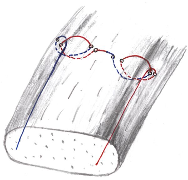
Self-cinching stitches in rotator cuff repair 295
(Figure 3). Each bracket had three transverse recesses,
5 mm deep, to be filled with muscle tissue under com-
pression. To achieve reliable fixation and to prevent slip-
page of the muscle, the metal bags of the brackets were
filled with pellets of dry ice to freezing the protuberances
and prevent slippage. Care was taken to freeze only the
clamped part of the muscle, while the downward-pro-
truding tendon and suture remained unaffected. The
cryo-jaw was attached to the load cell and crossbar of
the UTM with a cardan joint. The compensator device
was mounted on the UTM base in order to load the two
threads equally (Figure 3). The data were recorded using
Figure 2: Double-loop knot stitch (DLKS), made of two consecu-
tive SLKS.
testing software (textX-pert V 112.1, Zwick-Roell AG, Ulm,
Germany). The elongation (precision = 0.5 mm) and load
(precision = 0.1 N) were measured and displayed as a
load/elongation curve. The maximum possible error of
transverse movement was 0.05%. The calibrated force
transducer (maximum load 500 N) had an accuracy of
1% with values above 200 N.
After pre-tension to 40 N, the prepared specimens
were cyclically loaded at a displacement rate of 1 mm/s.
The cyclic loading started at 10–80 N for 50 cycles and
was gradually increased by 20 N every 50 cycles (10–100 N,
10–120 N, etc.) until it reached 10–200 N. After 50 cycles at
10–200 N, the ultimate load to failure was tested. The failure
of the ultimate load (Fmax [N]) testing was defined as 20%
loss of the ultimate tensile strength independent of failure
mode (suture thread cutting through the tendon or breaking
of the suture thread).
2.4 Statistical analysis
The distribution of gap formation (mm) and Fmax (N) were
described by their mean ± standard deviation. The mean
was first calculated per animal in order to have just one
representative value per animal within each category.
Gap formation was visualized separately for each method
and force level.
In order to account for the dependencies within the
same animal, linear mixed effects models were used with
the method, force level, and their interaction as fixed
effects for the gap formation and method as fixed effect
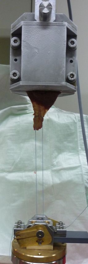
Figure 3: Part of the Zwick UTM test setup. DLKS is shown. The for the ultimate load. General linear hypothesis testing
infraspinatus muscle is clamped using two metal brackets of a cryo-
was carried out for the method comparisons within each
jaw (upper part of the picture). The compensator device is mounted
force level.
on the UTM base (lower part of the picture).
The significance level was set to α = 5% for all statis-
tical tests. All analyses were performed with the statis-
In short, the infraspinatus muscle was clamped in com- tical programming environment R (version 3.4.0, www.
pression using the two metal brackets of a cryo-jaw r-project.org).
296 Stephan Frosch et al.
3 Results Table 2: p value of pairwise comparison of the gap formation in
cyclic loading of the DLKS, SLKS, and mMAS
In cyclic loading, the DLKS and mMAS showed compar-
able gap formation results (8.72 ± 0.93 mm vs 8.65 ± Comparison (N) p value
1.33 mm, p = 1) after 350 cycles (Tables 1 and 2; Figure 4). DLKS.80 – mMA.80 0.9316
The gap formation of the SLKS was somewhat higher DLKS.80 – SLKS.80 0.9843
(9.14 ± 0.89 mm) but not significantly different than the mMA.80 – SLKS.80 1
DLKS.100 – mMA.100 0.9697
DLKS ( p = 0.26) or the mMAS ( p = 0.32) (Table 1;
DLKS.100 – SLKS.100 0.9884
Figure 4). mMA.100 – SLKS.100 1
Each DLKS and mMAS suture–tendon complex sur- DLKS.120 – mMA.120 0.9575
vived 350 cycles. One SLKS suture–tendon complex did DLKS.120 – SLKS.120 1
not survive the 10–160 N loading and one did not survive mMA.120 – SLKS.120 0.9938
the 10–180 N loading due to suture cutting-out. DLKS.140 – mMA.140 0.961
DLKS.140 – SLKS.140 1
The DLKS showed the highest ultimate load with a
mMA.140 – SLKS.140 0.9979
mean value of 350 N (±38.54), compared with the mMAS DLKS.160 – mMA.160 0.7872
(320.88 ± 53.29 N; p = 0.304) and the SLKS (290.54 ± DLKS.160 – SLKS.160 1
60.51 N; p < 0.05), while the only significant difference mMA.160 – SLKS.160 0.6429
was between the DLKS and SLKS (p < 0.05) (Table 1). DLKS.180 – mMA.180 1
DLKS.180 – SLKS.180 0.5395
mMA.180 – SLKS.180 0.242
DLKS.200 – mMA.200 1
DLKS.200 – SLKS.200 0.2644
4 Discussion mMA.200 – SLKS.200 0.3225
The most important finding of this study is that the DLKS
and to a lesser extent the SLKS showed excellent bio- massive tears with degenerative and/or frayed tendon
mechanical behaviour under cyclic loading conditions tissue. The self-cinching property of the loop knot enhances
when compared with the mMAS as the gold standard in tissue grip as axial strain increases and enables the surgeon
(mini) open rotator cuff repair. to grasp frayed tendon tissue more effectively. The loop
The arthroscopic treatment of chronic, massive rotator knot enhances transverse compaction of the tendon fibres
cuff tears can be technically demanding, with longer and thereby increases resistance against axial cutting of the
operative times, higher costs, and possible increased rerup- thread through parallel-running fibre sheaths of the tendon.
ture rates compared with open treatment [8,17]. Therefore, The findings of Ponce et al. in their biomechanical
open and mini-open procedures are still common and fre- study support our understanding of the beneficial effect
quently used in rotator cuff repair [8]. Both the SLKS and induced by transverse compaction of the tendon [18].
DLKS are applicable to arthroscopic technique, but less They compared (among other stitch techniques) the bio-
technically demanding in open repair. The advantages of mechanical properties of three self-cinching stitches.
the loop–knot technique are most effective in chronic, The configuration of the lasso-mattress stitch induces
Table 1: Mean ± standard deviation of gap formation in cyclic loading (10 to 80–200 N) and Fmax (N) in ultimate load testing within each
stitch technique
Force (N) DLKS mMAS SLKS
Gap formation (mm) 80 1.68 ± 0.62 1.15 ± 0.32 1.19 ± 0.3
100 2.88 ± 0.62 2.45 ± 0.83 2.46 ± 0.36
120 4.19 ± 0.99 3.71 ± 1 4 ± 0.7
140 5.73 ± 0.92 5.19 ± 1.17 5.46 ± 0.79
160 7.09 ± 0.83 6.44 ± 1.03 6.96 ± 1.03
180 7.83 ± 0.91 7.56 ± 0.99 8.42 ± 0.89
200 8.72 ± 0.93 8.65 ± 1.33 9.38 ± 1.14
Fmax (N) (ultimate load to failure) 350.52 ± 38.54 320.88 ± 53.29 290.54 ± 60.51Self-cinching stitches in rotator cuff repair 297
forces acting on the supraspinatus from 60 N during basic
elevation of the arm, up to 117 N with maximal isometric
abduction, and of 175–353 N with maximal concentric ele-
vation of the arm [19–22]. However, the results of the pre-
sent biomechanical study cannot be directly applied to
clinical treatment. From our results, we conclude that
the DLKS, SLKS, and mMAS are suitable for passive
mobilization in the early postoperative phase, but active
mobilization could overstrain the suture–tendon com-
plex over time.
Lorbach et al. examined the single-row modified
Mason-Allen stitch in a biomechanical laboratory study
Figure 4: Gap formation in cyclic loading (10 to 80–200 N) of the using porcine infraspinatus tendons [23]. The cyclic loading
three types of stitches: DLKS (brown), SLKS (blue), and mMAS
of the specimens started at 20 N for 50 cycles, increasing
(green).
stepwise by 20 N until it reached 200 N for 50 cycles. Only
the results for 100, 160, and 200 N were reported. The mean
considerable transverse compaction of the tendon tissue elongation of the construct was 6.4 mm after 100 N, 9.7 mm
as axial strain is applied. In contrast, the configuration after 160 N, and 12.3 mm after 200 N of 50 cycles at each
of the self-cinching lasso-loop and the double-cinch force level. The reported values are somewhat high com-
stitch induces more axial compression of the threads, pared with our results, but the differences between them
parallel to the fibre sheaths of the tendon, as axial strain and our values of about 3–4 mm are consistent throughout
increases. Consequently, the lasso-mattress stitch bore different loadings. This might be due to that study’s dif-
superior ultimate loads compared with the lasso-loop ferent preloading of the suture–tendon construct of 10 N
stitch (148.1 vs 64.7 N) and double-cinch stitch (148.1 compared with 40 N in our study. In progressive cyclic
vs 97.1 N). Furthermore, the lasso-loop stitch showed loading, the cinching loop tightens up to a certain extent,
superior results in ultimate loading conditions when which increases the thread length between knot and
compared with the mMAS (148.1 vs 128.3 N) and simple anchor, leading to additional elongation. Ponce et al. con-
stitches such as the mattress stitch (148.1 vs 67.1 N) and firmed additional elongation of self-cinching sutures in
the simple stitch (148.1 vs 47.1 N). The authors concluded loading configurations [18]. Therefore, initial intraoperative
that self-cinching stitches lead to superior tissue-holding tightening of the knot (preloading) before locking the stitch
strength in comparison with other comparable noncinching is important to decrease postoperative elongation.
simple stitches. These findings are consistent with our It is possible to place two separate DLKSs at the
results, as the DLKS showed superior results compared proximal and distal ends of the rupture and tie the
with the mMAS (345.56 vs 320.88 N in ultimate load), and opposing threads on each side of the tendon (two knots).
with a previous study, where the DLKS showed significantly Alternatively, both DLKSs can be placed with one contin-
superior values compared with the mMAS (382.2 vs 309.3 N; uous thread and one final locking knot. It should be noted
p = 0.038) [14]. The forces survived by the SLKS in ultimate that tightening two consecutive DLKSs with one thread is
load testing were insignificantly lower than those survived more difficult because of the self-cinching mechanism.
by the mMAS. However, it is notable that the amount of One limitation of this study is that the results of an
tendon tissue grasped by the SLKS was considerably less in vitro animal study cannot be directly translated to
than that grasped by the mMAS, which might explain these suture techniques for the rotator cuff in human patients.
findings. However, the mechanical properties of pig infraspinatus
Cyclic loading, rather than ultimate load-to-failure tendons are considered comparable with those of human
configurations, simulates repetitive loading of the tendon tendons and are similar to human rotator cuff tendons
during the early stages of rehabilitation. The results from in size, shape, histological parameters, and mechanical
cyclic loading did not significantly differ among the properties [24,25]. Furthermore, the present in vitro animal
DLKS, SLKS, and mMAS. All three suture configurations model is frequently used in the literature and allows for
reached 120 N in cyclic loading before gap formation easy comparison of results. The enhanced transverse com-
exceeded 5 mm. Notably, gap formation exceeding 5 mm pression force on the tendon encompassed by the suture
is considered a clinically relevant failure of the suture– raises concerns of local tendon necrosis. Theoretically,
tendon complex. Force analysis of the rotator cuff predicts larger tendon cross-sections better withstand compression298 Stephan Frosch et al.
forces of constriction. Gerber et al. demonstrated for the double-row suture bridge reconstruction for supraspinatus
mMAS that these forces do not cause long-term histological tendon tears: a matched-pair analysis. Am J Sports Med.
changes within the tendon and that they are biologically 2012;40(12):2777–85.
[12] Anderl W, Heuberer PR, Laky B, Kriegleder B, Reihsner R,
tolerated [26]. Further histologic investigations regarding
Eberhardsteiner J. Superiority of bridging techniques with
self-cinching stitches are necessary. medial fixation on initial strength. Knee Surg Sports Traumatol
Arthrosc Off J ESSKA. 2012;20(12):2559–66.
Conflict of interest: Authors state no conflict of interest. [13] Lorbach O, Tompkins M. Rotator cuff: biology and current
arthroscopic techniques. Knee Surg Sports Traumatol Arthrosc
Off J ESSKA. 2012;20(6):1003–11.
Data availability statement: The datasets generated
[14] Frosch S, Buchhorn G, Hoffmann A, Balcarek P, Schuttrumpf JP,
during and/or analysed during the current study are August F, et al. Novel single-loop and double-loop knot stitch
available from the corresponding author on reasonable in comparison with the modified Mason-Allen stitch for rotator
request. cuff repair. Knee Surg Sports Traumatol Arthrosc Off J ESSKA.
2015;23(5):1552–8.
[15] Schneeberger AG, von Roll A, Kalberer F, Jacob HA, Gerber C.
Mechanical strength of arthroscopic rotator cuff repair tech-
niques: an in vitro study. J Bone Jt Surg Am.
References 2002;84(12):2152–60.
[16] Baums MH, Buchhorn GH, Spahn G, Poppendieck B,
[1] Cummins CA, Murrell GA. Mode of failure for rotator cuff repair Schultz W, Klinger HM. Biomechanical characteristics of
with suture anchors identified at revision surgery. J Shoulder single-row repair in comparison to double-row repair with
Elb Surg Am Shoulder Elb Surg. 2003;12(2):128–33. consideration of the future configuration and future
[2] Fehringer EV, Sun J, VanOeveren LS, Keller BK, Matsen FA3rd. material. Knee Surg Sports Traumatol Arthrosc.
Full-thickness rotator cuff tear prevalence and correlation with 2008;16(11):1052–60.
function and co-morbidities in patients sixty-five years and older. [17] Walton JR, Murrell GA. A two-year clinical outcomes study of
J Shoulder Elb Surg Am Shoulder Elb Surg. 2008;17(6):881–5. 400 patients, comparing open surgery and arthroscopy for
[3] Le BT, Wu XL, Lam PH, Murrell GA. Factors predicting rotator rotator cuff repair. Bone Jt Res. 2012;1(9):210–7.
cuff retears: an analysis of 1000 consecutive rotator cuff [18] Ponce BA, Hosemann CD, Raghava P, Tate JP, Eberhardt AW,
repairs. Am J Sports Med. 2014;42(5):1134–42. Lafosse L. Biomechanical evaluation of 3 arthroscopic self-
[4] Millar NL, Wu X, Tantau R, Silverstone E, Murrell GA. Open cinching stitches for shoulder arthroscopy: the lasso-loop,
versus two forms of arthroscopic rotator cuff repair. Clin lasso-mattress, and double-cinch stitches. Am J Sports Med.
Orthop Relat Res. 2009;467(4):966–78. 2011;39(1):188–94.
[5] Gazielly DF, Gleyze P, Montagnon C. Functional and anatomical [19] Chang YW, Hughes RE, Su FC, Itoi E, An KN. Prediction of
results after rotator cuff repair. Clin Orthop Relat Res. muscle force involved in shoulder internal rotation. J Shoulder
1994;304:43–53. Elb Surg Am Shoulder Elb Surg. 2000;9(3):188–95.
[6] Chillemi C, Petrozza V, Garro L, Sardella B, Diotallevi R, [20] Hughes RE, An KN. Force analysis of rotator cuff muscles. Clin
Ferrara A, et al. Rotator cuff re-tear or non-healing: histo- Orthop Relat Res. 1996;330:75–83.
pathological aspects and predictive factors. Knee Surg Sports [21] Juul-Kristensen B, Bojsen-Moller F, Finsen L, Eriksson J,
Traumatol Arthrosc Off J ESSKA. 2011;19(9):1588–96. Johansson G, Stahlberg F, et al. Muscle sizes and moment
[7] Galatz LM, Ball CM, Teefey SA, Middleton WD, Yamaguchi K. arms of rotator cuff muscles determined by magnetic
The outcome and repair integrity of completely arthroscopi- resonance imaging. Cells Tissues Organs.
cally repaired large and massive rotator cuff tears. J Bone Jt 2000;167(2–3):214–22.
Surg Am. 2004;86-A(2):219–24. [22] Wuelker N, Plitz W, Roetman B, Wirth CJ. Function of the
[8] Elkins A, Lam PH, Murrell GAC. A novel, fast, safe, and effective supraspinatus muscle Abduction humerus studied cadavers.
all-inside arthroscopic rotator cuff repair technique: results of Acta Orthop Scand. 1994;65(4):442–6.
1000 consecutive cases. Orthop J Sports Med. [23] Lorbach O, Bachelier F, Vees J, Kohn D, Pape D. Cyclic loading
2019;7(8):2325967119864088. of rotator cuff reconstructions: single-row repair with modified
[9] Ma CB, MacGillivray JD, Clabeaux J, Lee S, Otis JC. suture configurations versus double-row repair. Am J Sports
Biomechanical evaluation of arthroscopic rotator cuff stitches. Med. 2008;36(8):1504–10.
J Bone Jt Surg Am. 2004;86-A(6):1211–6. [24] Gerber C, Schneeberger AG, Beck M, Schlegel U. Mechanical
[10] Nelson CO, Sileo MJ, Grossman MG, Serra-Hsu F. Single-row strength of repairs of the rotator cuff. J Bone Jt Surg Br.
modified mason-allen versus double-row arthroscopic rotator 1994;76(3):371–80.
cuff repair: a biomechanical and surface area comparison. [25] Yamada H, Evans FG. Strength of biological materials.
Arthrosc J Arthrosc Relat Surg Off Publ Arthrosc Assoc N Am Int Baltimore: Williams & Wilkins; 1970. p. 297.
Arthrosc Assoc. 2008;24(8):941–8. [26] Gerber C, Schneeberger AG, Perren SM, Nyffeler RW.
[11] Gerhardt C, Hug K, Pauly S, Marnitz T, Scheibel M. Experimental rotator cuff repair A preliminary study. J Bone Jt
Arthroscopic single-row modified mason-allen repair versus Surg Am. 1999;81(9):1281–90.You can also read