Intrapleural Migration of a Breast Prosthesis after Redo Thoracotomy for Recurrent Lung Cancer
←
→
Page content transcription
If your browser does not render page correctly, please read the page content below
American Journal of Surgery Case Reports

Case Report

    Intrapleural Migration of a Breast Prosthesis after Redo
           Thoracotomy for Recurrent Lung Cancer
Gregory Fishberger4, Jobelle JAR Baldonado1,2,3, Joseph R. Garrett1, Carla C Moodie1 and Eric M.Toloza1,2,3*
Department of Thoracic Oncology, Moffitt Cancer Center, Tampa, FL, USA
1

Department of Surgery, University of South Florida Health Morsani College of Medicine, Tampa, FL, USA
2

Department of Oncologic Sciences, University of South Florida Health Morsani College of Medicine, Tampa, FL, USA
3

Morsani College of Medicine, University of South Florida Health, Tampa, FL, USA
4

Abstract
Introduction: Breast prostheses have the potential to migrate into the intrapleural cavity and occasionally rupture during thoracic surgery, particularly when the
fibrous capsule around the prosthesis is disrupted. Here, we present a case of breast prosthesis intrapleural migration after re-do thoracotomy for lung cancer.
Case: A 60-year-old woman presented with history of left thoracotomy and left upper lobectomy for stage-1 adenocarcinoma 9 years previously and who
underwent re-do left thoracotomy, pleurolysis, and Left Lower Lobar (LLL) superior segmentectomy for metachronous stage-1 adenocarcinoma 7 months prior to
presentation, both at an outside facility. Her second thoracotomy was complicated by hypoxia refractory to steroids and antibiotics for a right upper lobar infiltrate
noted on chest X-ray on postoperative day (POD)#3. Computerized Tomography (CT)-angiogram on POD#5 revealed no pulmonary embolus and showed her
bilateral breast prostheses to be intact, but the left pleural chest tube was noted to pass through the left breast prosthesis capsule, with an adjacent Intercostal Space
(ICS) defect. Her left chest tube was removed on POD#7, and she was discharged to home on POD#11. Four months after the 2nd thoracotomy, she reported falling
in the shower, which resulted in 10% compression fracture of the T12 vertebra. She was instructed to wear a Thoracic-Lumbar-Sacral Orthosis (TLSO) brace with
bed rest until she underwent T11 and T12 kyphoplasty. Subsequent lung cancer surveillance CT scan revealed that the left breast prosthesis had ruptured and
migrated into the left hemithorax. She underwent left Video-Assisted Thoracoscopic (VATS) surgery using a port incision along the left 8th ICS at the posterior
axillary line and another along the left 6th ICS at the anterior axillary line. The intrapleurally displaced left breast prosthesis was retrieved from within a fibrous
capsule between the LLL basilar segments and the left diaphragm. Skin-to-skin operative time was 51 min, and intraoperative estimated blood loss was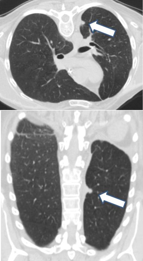
American Journal of Surgery Case Reports

                                                                                         Final pathology revealed a 1.5 cm moderately-differentiated
                                                                                     adenocarcinoma, with visceral pleural invasion, but with uninvolved
                                                                                     resection margins and uninvolved level 10 L (hilar) and 11 L
                                                                                     (interlobar) Lymph Nodes (LNs) (pT2N0M0). One month after the
                                                                                     second thoracotomy, a recurrent left pleural effusion prompted a
                                                                                     repeat left thoracentesis of 250 mL of serous pleural fluid that was
                                                                                     again negative for malignancy on cytology.
                                                                                         Four months after the 2nd thoracotomy (3 months prior to this
                                                                                     presentation), she reported slipping and falling onto her buttock
                                                                                     while leaving the shower, which resulted in a 10% compression
                                                                                     fracture of the T12 vertebra noted on Magnetic Resonance Imaging
                                                                                     (MRI). She was then instructed to wear a Thoracic-Lumbar-Sacral
                                                                                     Orthosis (TLSO) brace with bed rest for the next 3 months. During
                                                                                     the following 3 weeks, she complained that the TLSO brace pressed
                                                                                     against her central venous catheter port on her left anterior chest and
                                                                                     that she experienced disuse weakness of her muscles, which prompted
                                                                                     her subsequently undergoing T11 and T12 kyphoplasty, which
                                                                                     incidentally resulted in pulmonary cement embolization.
                                                                                          Lung cancer surveillance with CT scan 2 months after the
                                                                                     kyphoplasty (one month prior to this presentation) noted intact
                                                                                     subpectoral right breast prosthesis, but revealed that the left breast
                                                                                     prosthesis had ruptured and was now displaced into the inferior
                                                                                     left hemithorax just superior to the left hemidiaphragm, which was
                                                                                     confirmed by MRI of the thorax.
                                                                                         She was referred by her local medical oncologist to our thoracic
                                                                                     surgery clinic for evaluation. She underwent fiberoptic bronchoscopy
                                                                                     and left Video-Assisted Thoracoscopic (VATS) surgery through a 3
                                                                                     cm port incision along the left 8th ICS at the posterior axillary line and
                                                                                     another 3 cm port incision along the left 6th ICS at the anterior axillary
                                                                                     line. She then underwent left VATS lysis of pleural adhesions between
 Figure 1: Coronal (top panel) and axial (bottom panel) images from Com-             the LLL basilar segments and the left hemidiaphragm and between
 puterized Tomography (CT) scan taken 4 months prior to the re-do left tho-
 racotomy and showing a 1 cm paravertebral left lung nodule (arrows) that is         the LLL basilar segments and the pericardium.
 suggestive of recurrent left lung cancer.
                                                                                         The intrapleurally-displaced left breast prosthesis was identified
                                                                                     encapsulated beneath the left 8th ICS port incision between the LLL
desaturation and hypoxia, which required Intensive Care Unit (ICU)                   basilar segments and the left hemidiaphragm and was grasped and
admission, but which were refractory to steroids and antibiotics                     retrieved with a ring clamp, after the fibrous capsule was incised with
for suspected aspiration pneumonia based on Right Upper Lobar                        cautery. Left VATS pulmonary decortication was then performed to
(RUL) infiltrate noted on chest x-ray on postoperative day (POD)#3.                  dissect the fibrous capsule free from the LLL visceral pleura and from
Modified barium swallow study was performed, and her diet was                        the left hemidiaphragm, with no evidence of diaphragmatic hernia
modified; however, she continued to have difficulty.                                 and with subsequent insertion of 32-French angled left pleural chest
                                                                                     tube along the lateral costophrenic recess.
    Computerized Tomography (CT)-angiogram on POD#5 revealed
no evidence of pulmonary embolus, but did reveal pulmonary edema,                        Total skin-to-skin operative time was 51 min, and the total
which responded to furosemide diuresis. Although the patient                         intraoperative Estimated Blood Loss (EBL) was
American Journal of Surgery Case Reports
Over the past year, she developed worsening back pain. Over the
past 4 months, she also developed a psoriatic flare throughout her
extremities, which was treated with prednisone and methotrexate.
Serial chest CT scans and subsequent thoracic and lumbar MRI
revealed abnormal enhancement in the paraspinous soft tissue
extending from T7 through T12 and abutting the lateral surfaces of
multiple vertebral bodies, but without abnormal signal within the
vertebrae. The MRI also demonstrated a chronic 25% loss of height
of the anterior T12 vertebra that appeared unchanged compared to
imaging studies over the preceding 2-1/2 years.
Neurosurgical consultation was obtained and CT-guided needle
biopsy of the left paraspinous muscle abnormality and the LLL
lung nodule each revealed adenocarcinoma. She was subsequently
treated with 70-Gray External-beam Radiation Therapy (XRT) in 35
fractions to the LLL lung cancer and the left paraspinal metastatic
Adenocarcinoma (Figure 2 and 3).
Figure 3: Coronal (upper left panel), axial (upper right panel), and sagittal
(lower left panel) from Computerized Tomography (CT) scan taken 6 months
after re-do thoracotomy for recurrent lung cancer and showing the left breast
prosthesis (arrows) now located within the lower left hemithorax just above
the left hemidiaphragm; and chest radiograph Posterior-Anterior (PA; lower
middle panel) and lateral (lower right panel) views taken 5 days prior to Vid-
eo-Assisted Thoracoscopic (VATS) retrieval of the intrapleurally-migrated left
breast prosthesis (arrows).
surgical intervention [7]. Excess thickening of the capsule and
capsular contracture can lead to compression of the breast implant,
producing a visible deformation of the breast or prosthesis rupture
[8]. Subpectoral placement results in reduced risk of capsule
contracture, even though this method produces more discomfort after
surgery [9,10]. Treatment following rupture of saline breast implants
is fairly uncomplicated, in that the saline is absorbed by the body, and
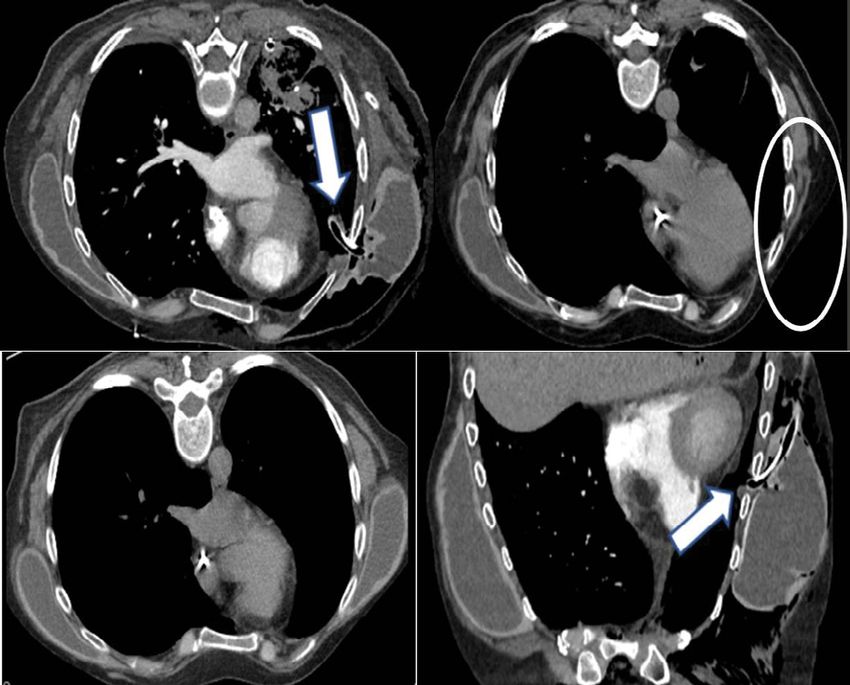
Figure 2: Axial image from Computerized Tomography (CT) scan taken 4
the implant is then explanted [11]. Rupture of silicone prosthetics
months prior to re-do thoracotomy for recurrent left lung cancer and show-
ing presence of bilateral breast prostheses (upper left panel); coronal (upper requires removal of the silicone at the rupture site and explantation
right panel) and axial (bottom left panel) images from another CT scan taken of the remaining prosthetic [12]. If silicone thorax occurs, the silicone
5 days after the re-do thoracotomy showing continued presence of bilateral must be removed from the pleural cavity [13].
breast prostheses and the left pleural chest tube passing through left breast
prosthesis fibrous capsule and into the left pleural cavity through an underly- Intrathoracic migration of breast prosthesis is an exceedingly rare
ing chest wall defect along the left 4th intercostal space (arrows); and axial
event. Instances of such migration typically occur after access to the
image from new CT scan taken 6 months after the re-do thoracotomy now
showing the absence of the left breast prosthesis (white oval). thoracic cavity has been required and a chest wall defect has been
created [1]. Other risk factors involve intraoperative laceration of
the pleura or accidental needle puncture [14]. Migration risk is likely
Discussion impacted by the prothesis placement. Subglandular placement carries
The primary techniques of breast prosthesis implantation include the benefit of protection posteriorly by the pectoralis major muscle
subglandular and subpectoral, and both methods may be utilized for [2]. Subpectoral placement leaves the implant in contact with the chest
cosmetic breast augmentation [2]. Subglandular placement involves wall and intercostal muscles that are then traversed by chest tubes or
implantation within the retromammary space between breast tissue during minimally invasive VATS and thoracotomy interventions [15].
and the pectoralis major muscle [3]. Subpectoral implantation Subpectorally-implanted breast prostheses are, thus, at risk of being
involves placing the breast prosthesis below the pectoralis major injured and of migrating into the pleural cavity through defects in
muscle following the dissection and release of the inferior muscular the intercostal muscles created during thoracotomy, especially if the
attachments [2,4]. The upper pole is located in the submuscular plane, prosthetic fibrous capsule has been disrupted and if the prosthesis
while the lower pole is located in the subglandular plane [5]. has ruptured [16]. Therefore, the fibrous capsule plays an essential
role in maintaining the position of the implant, particularly after
Following implantation of the prosthesis, a collagen scar tissue
the aforementioned intrathoracic interventions when subpectoral
capsule forms around the implant, thus isolating the implant [6].
prosthesis placement has been utilized. Migration through an ICS
The integrity of this fibrous capsule ultimately proves beneficial
defect may be further promoted by extrathoracic pressure, such as
in instances in which the chest wall is traversed for intrathoracic
that experienced with a TLSO brace [17].
© 2021 - Medtext Publications. All Rights Reserved. 060 2021 | Volume 1 | Article 1016American Journal of Surgery Case Reports
Previous case reports have described intrathoracic migration of a bilateral mastectomy for ductal carcinoma, followed by silicone breast
breast prosthesis following relatively similar clinical courses to that in prosthesis 23 years previously. Five months prior to presentation, the
our case: 1) A breast prosthesis is placed for cosmetic purposes or for patient underwent VATS right middle lobectomy with mediastinal
reconstruction post-mastectomy; 2) The prosthetic fibrous capsule is LN dissection, during which a defect was created in the anterior chest
disrupted during chest tube placement [13], VATS surgery [18-20], wall between the right 3rd and 4th ribs. At presentation, the patient
minimally invasive cardiac surgery [7,21], or thoracotomy [1]; 3) A complained of being unable to feel her right breast implant. During
communication remains between the implant capsule and pleural implant retrieval, the posterior aspect of the implant capsule was
space that facilitates the migration of the prosthesis through a defect found to have been disrupted, and a pool of free silicone was found
in the chest wall into the pleural space. within the pleural space, indicating implant rupture.
Mehta et al. [1] described the first reported case of intrathoracic Intrathoracic migration of a breast prosthesis has also been
migration of breast prosthesis post-thoracotomy (Table 1). In that described after minimally invasive cardiac surgery for mitral valve
case, a 52-year-old woman, with history of breast augmentation 14 repair [7,21]. During the procedure, the fibrous capsule was disrupted,
years previously, underwent right thoracotomy for lobectomy and and the breast implant was explanted during surgery [7]. At the end
mediastinal LN dissection for NSCLC, during which the posterior of the operation, the implant was placed back into the implant pocket.
aspect of the prosthetic capsule had been partially opened. On However, the disruption of the prosthesis capsule provided the
POD#12, she complained of progressive dyspnea and breast volume opportunity for subsequent intrathoracic migration.
asymmetry. Chest CT scan revealed intrathoracic migration of the
Herein we report a case of a 60-year-old woman with a cosmetic
right silicone breast prosthesis through a defect in the right thoracic
implant and history of left thoracotomy and left upper lobectomy 9
wall, and re-do thoracotomy was performed to extract the intact
years previously. Seven months prior to the patient’s presentation, she
prosthesis from above the right hemidiaphragm.
underwent repeat left thoracotomy and LLL superior segmentectomy.
Intrathoracic migration of breast prosthesis has been reported to On POD#5 of the second thoracotomy, a CT angiogram revealed the
occur in cases of multiple revision surgeries for breast augmentation. left pleural chest tube was passing through the left breast prosthesis
Kim et al. [14] reported the case of a 34-year-old woman with history of capsule, although the prosthesis appeared intact and not punctured,
two revisions over three years (Table 1). Dissection of dense adhesions thus creating a communication between the prosthesis capsule and
between the prosthesis and the chest wall during the second revision the left pleural cavity. Four months after the second thoracotomy, the
surgery resulted in a chest wall defect along the 4th ICS, which allowed patient slipped in the shower and required a TLSO brace for three
migration of the prosthesis through the defect into the pleural cavity months. Two months after subsequent kyphoplasty, a lung cancer
when the patient massaged her breasts after the second revision. surveillance CT scan revealed that the left breast prosthesis was
ruptured and was now located within the inferior left hemithorax
Sykes et al. [18] described the first reported case of intrathoracic
just above the left hemidiaphragm. The breast prosthesis was
breast prosthesis migration following VATS surgery (Table 1). The
found encapsulated between the LLL visceral pleural and the left
authors report the case of a 72-year-old woman, with history of
hemidiaphragm. The left breast prosthesis and fibrous capsule were
Table 1: Case Reports of Intrathoracic Migration of a Breast Prosthesis.
Most Recent Most Recent
Time from
Surgery Surgical Time from
Reason for Initial
Patient Implant Implant Implant Completed Approach Most Recent
Case Report Initial Breast Implant
Age Type Laterality Position Prior to Prior to Surgery to
Prosthesis Placement
Implant Implant Migration
to Migration
Migration Migration
Breast
Chen et al. [16] 29 Cosmetic Silicone Left Unknown N/A 2 months 2 months
augmentation
Lobectomy;
Mehta et al. [1] 52 Cosmetic Silicone Right Subpectoral mediastinal LN Thoracotomy 14 years 12 days
dissection
Breast
Kim et al. [14] 34 Cosmetic N/A Right Subpectoral augmentation N/A 3 years 1 month
revision
Lobectomy;
Sykes and Rosella [18] 72 Reconstructive Silicone Right Subpectoral mediastinal LN VATS 23 years 7 months
dissection
Lehoux et al. [19] 71 Reconstructive Silicone Right Subpectoral Lobectomy VATS 22 years 6 months
Lobectomy;
Roussel et al. [20] 72 Reconstructive Silicone Right Subpectoral mediastinal LN VATS 24 years 5 months
dissection
Mitral valve
Fong and Hoffman [21] 59 Reconstructive N/A Right Subpectoral MIS N/A N/A
repair
Mitral
Songcharoen et al. [7] 61 Reconstructive Saline Right Subpectoral MIS 5 years 5 months
valvuloplasty
Re-do
Our Case 60 Cosmetic N/A Left Subpectoral Thoracotomy N/A 6 months
lobectomy
N/A: Not Available; VATS: Video-Assisted Thoracoscopy; LN: Lymph Node; MIS: Minimally Invasive Surgery
© 2021 - Medtext Publications. All Rights Reserved. 061 2021 | Volume 1 | Article 1016American Journal of Surgery Case Reports
removed by VATS without complications. be experienced with a TLSO brace [17]. Thus, thoracic procedures
in patients with existing breast prostheses, especially subpectorally-
Several factors likely contributed to intrathoracic migration
implanted breast prostheses, require care to avoid injury to the
of the left breast prosthesis. The patient had undergone two left
prosthesis and to avoid disrupting the prosthetic fibrous capsule in
thoracotomies, each requiring the division of the intercostal
order to minimize the risk of migration of the breast prosthesis into
musculature and which disrupted the muscular integrity of the chest
the pleural cavity.
wall. Further, the left chest tube placed during the second thoracotomy
had passed through the left breast prosthesis capsule. While the Acknowledgements
prosthesis had not yet migrated, the risk of intrathoracic migration Funding
was greatly increased due to the formation of the chest wall defect. This report was supported in part by a 2021 Summer Scholarly
Prior case reports note that the strong negative pressure differential Award presented to G.F. from the Office of Research, Innovation, and
between the pleural cavity and the prosthesis capsule during Scholarly Endeavors (RISE) at the University of South Florida (USF)
inspiration draws the breast prosthesis through the chest wall defect Health, Morsani College of Medicine in Tampa, FL, USA. None of the
into the pleural cavity [14,18,20]. In our case, the pressure differential other authors had any funding source to report.
was exacerbated by the TLSO brace. The TLSO brace compressed the
patient’s anterior chest and resulted in persistently elevated external Informed consent
pressure on the chest, which likely promoted gradual migration of Permission was obtained from the patient for publication of this
the left breast prosthesis into the pleural cavity while the patient was case report and any accompanying images for education purposes
wearing the brace. as part of our institutional surgical informed consent. A copy of the
written consent is available for review by the Editor-in-Chief of this
Compared to minimally invasive VATS surgery, thoracotomy
journal.
involves more substantial division of the intercostal musculature to
access the intrapleural space [22]. In this context, there would be greater References
risk of a post-surgical chest wall defect facilitating communication 1. Mehta AM, Bard MP, van Straten A, van Beijeren I, Rijna H. Intrathoracic migration
between the implant capsule and the intrapleural cavity. Interestingly, of a breast prosthesis after thoracotomy. J Thorac Cardiovasc Surg. 2008;135(1):206-7,
207.e1.
a literature search of ‘"intrathoracic"& "thoracotomy" & "migration"
& "breast"’ on PubMed reveals the case by Mehta et al. [1] as the only 2. Coombs DM, Grover R, Prassinos A, Gurunluoglu R. Breast augmentation surgery:
report of a breast prosthesis undergoing intrathoracic migration after Clinical considerations. Cleve Clin J Med. 2019;86(2):111-22.
thoracotomy. The remaining case reports occurred in the context 3. Adams WP, Mallucci P. Breast augmentation. Plast Reconstr Surg.
of breast revision surgery, VATS surgery, or minimally invasive 2012;130(4):597e-611e.
cardiac surgery, which suggests that disruption of the prosthesis
4. Spear SL, Jespersen MR. Breast implants: saline or silicone? Aesthet Surg J.
fibrous capsule more directly impacts the likelihood of prosthesis 2010;30(4):557-70.
intrathoracic migration rather than the surgical approach.
5. Tebbetts JB. A system for breast implant selection based on patient tissue
While intrathoracic migration of breast prosthesis is an characteristics and implant-soft tissue dynamics. Plast Reconstr Surg.
uncommon occurrence, measures can be taken to minimize the risk 2002;109(4):1396-409;discussion 1410-5.
of this postoperative complication. The fibrous capsule that forms 6. Bucky LP, Ehrlich HP, Sohoni S, May JW Jr. The capsule quality of saline-filled smooth
around the breast prosthesis isolates and keeps the implant in place silicone, textured silicone, and polyurethane implants in rabbits: a long-term study.
[2]. By ensuring that the fibrous capsule is avoided and kept intact Plast Reconstr Surg. 1994;93(6):1123-31; discussion 1132-3.
during thoracic procedures, the breast prosthesis will remain isolated
7. Songcharoen SJ, McClure M, Aru RG, Songcharoen S. Intrathoracic migration
from any defect in the chest wall created during chest tube placement, of a breast implant after minimally invasive cardiac surgery. Ann Plast Surg.
minimally invasive approaches, or thoracotomy. In order to avoid 2015;74(3):274-6.
impacting the breast prosthesis capsule, image-guided chest tube
8. Hillard C, Fowler JD, Barta R, Cunningham B. Silicone breast implant rupture: a
placement could prove highly beneficial. For procedures involving
review. Gland Surg. 2017;6(2):163-8.
thoracotomies or VATS, preoperative imaging of patients with existing
breast protheses would aid in surgical planning to avoid the capsule if 9. Henriksen TF, Fryzek JP, Holmich LR, McLaughlin JK, Kjoller K, Hoyer AP, et
al. Surgical intervention and capsular contracture after breast augmentation: a
possible. Furthermore, the breast prosthesis should not be explanted
prospective study of risk factors. Ann Plast Surg. 2005;54(4):343-51.
unless necessary. If the prosthesis capsule must be disrupted, the
capsule should be prophylactically sutured closed or reinforced, and 10. Stevens WG, Nahabedian MY, Calobrace MB, Harrington JL, Capizzi PJ, Cohen R, et
the chest wall defect meticulously repaired. al. Risk factor analysis for capsular contracture: a 5-year Sientra study analysis using
round, smooth, and textured implants for breast augmentation. Plast Reconstr Surg.
Conclusion 2013;132(5):1115-23.
Breast prostheses, especially those implanted subpectorally, are at 11. Swezey E, Shikhman R, Moufarrege R. Breast Implant Rupture. StatPearls. 2021.
risk of being injured and of migrating into the pleural cavity through
12. Seiler SJ, Sharma PB, Hayes JC, Ganti R, Mootz AR, Eads ED, et al. Multimodality
defects in the intercostal muscles created during thoracic surgery,
Imaging-based Evaluation of Single-Lumen Silicone Breast Implants for Rupture.
particularly if the prosthesis fibrous capsule has been disrupted
Radiographics. 2017;37(2):366-82.
and if the prosthesis has ruptured [16]. We report a case of breast
prosthesis migration into the pleura cavity after re-do thoracotomy 13. Rice DC, Agasthian T, Clay RP, Deschamps C. Silicone thorax: a complication
of tube thoracostomy in the presence of mammary implants. Ann Thorac Surg.
for recurrent lung cancer, in which both the intercostal muscles and
1995;60(5):1417-9.
the prosthesis capsule were disrupted. Migration through an ICS
defect may be facilitated by extrathoracic pressure, such as what might 14. Kim H, Heo C, Baek R, Minn K, Kim S, Chun S. Breast implant migration into pleural
© 2021 - Medtext Publications. All Rights Reserved. 062 2021 | Volume 1 | Article 1016American Journal of Surgery Case Reports
cavity. J Plast Reconstr Aesthet Surg. 2009;62(5):e89-90. 19. Lehoux JM, Tchantchaleishvili V, Jones CE. Intrathoracic migration of a silicone breast
implant after video-assisted thoracoscopic surgery. Ann Thorac Surg. 2013;96(1):326.
15. Lesavoy MA, Trussler AP, Dickinson BP. Difficulties with subpectoral augmentation
mammaplasty and its correction: the role of subglandular site change in revision 20. Roussel LO, Koltz PF, Langstein HN. Intrathoracic Breast Implant Migration following
aesthetic breast surgery. Plast Reconstr Surg. 2010;125(1):363-71. Video-Assisted Thorascopic Surgery. Plast Reconstr Surg. 2015;135(6):1075e-6e.
16. Chen ZY, Wang ZG, Kuang RX, Wang BT, Su YP. Implant found in thoracic cavity 21. Fong TC, Hoffmann B. Images in clinical medicine. Disappearance of a breast
after breast augmentation. Plast Reconstr Surg. 2005;116(6):1826-7. prosthesis during Pilates. N Engl J Med. 2011;365(24):2305.
17. Pham VM, Houilliez A, Schill A, Carpentier A, Herbaux B, Thevenon A. Study of the 22. Kirby TJ, Mack MJ, Landreneau RJ, Rice TW. Lobectomy--video-assisted thoracic
pressures applied by a Cheneau brace for correction of adolescent idiopathic scoliosis. surgery versus muscle-sparing thoracotomy. A randomized trial. J Thorac Cardiovasc
Prosthet Orthot Int. 2008;32(3):345-55. Surg. 1995;109(5):997-1001; discussion 1001-2.
18. Sykes JB, Rosella PA. Intrathoracic migration of a silicone breast implant 5 months
after video-assisted thoracoscopic surgery. J Comput Assist Tomogr. 2012;36(3):306-
7.
© 2021 - Medtext Publications. All Rights Reserved. 063 2021 | Volume 1 | Article 1016You can also read