The Role of Ultrasonography in The Diagnosis of Oral and Maxillofacial Disease - Open Journal Systems
←
→
Page content transcription
If your browser does not render page correctly, please read the page content below
Indian Journal of Forensic Medicine & Toxicology, January-March 2021, Vol. 15, No. 1   1853

       The Role of Ultrasonography in The Diagnosis of Oral and
                         Maxillofacial Disease

          Ni Putu Mira Sumarta1, David Buntoro Kamdjaja2, Roberto Manahan Yantie Simandjuntak3
   1
    Researchers staff in Department of Oral and Maxillofacial Surgery, Faculty of Dental Medicine, Universitas
                                         Airlangga, Surabaya-Indonesia

                                                          Abstract
       Objective: Ultrasonography, as a diagnostic tool, constitutes a non-invasive, cost-effective, readily-available
       and repeatable imaging technique. Ultrasonography has been used as a means of diagnosing various medical
       conditions for many years. However, in the field of maxillofacial surgery it represents a relatively new aid
       in the diagnosis of various diseases affecting the oral and maxillofacial regions. These include: infection,
       soft-tissue related diseases and vascular anomalies which can be detected using Doppler ultrasonography.
       This article presents four cases, in which ultrasonography was employed to confirm diagnoses and act as a
       guide to treatment. Methods: Four cases of soft tissue swelling and enlargement were diagnosed with the
       aid of ultrasonography, namely: a submasseteric abscess, a nasolabial cyst, a dermoid cyst and a left buccal
       space abscess caused by a foreign body (i.e. a fish bone). Result: In the case of a submasseteric abscess,
       ultrasonography was used in confirming the diagnosis and therapy, while determining the maximal point of
       the abscess. In the cases of both cysts, ultrasonography highlighted well-defined cystic lesions with internal
       echo showing fluid accumulation, while in the buccal space abscess, an ultrasonogram confirmed the exact
       location of the fish bone. Conclusion: Ultrasonography is a quick, widely-available, relatively inexpensive,
       painless procedure which can be repeated as often as necessary without risk to the patient. Thus, ultrasonography
       is a valuable diagnostic aid to the oral and maxillofacial surgeon in achieving early and accurate diagnosis.

       Keywords: Abscess, Cyst, Maxillofacial abnormalities, Ultrasonography.

                       Introduction                                reflected waves is digitalized and thousands of such
                                                                   measurements generate an ultrasound cross-sectional
     Ultrasonography (USG), as a diagnostic tool, is               image which is then recorded on the monitor in order
a non-invasive, cost-effective, readily-available and              to enable its interpretation. Ultrasonography images
repeatable imaging technique. Although used as a                   comprise: hypoechoic (low reflection of sound waves)
diagnostic tool in the treatment of various medical                that appear black, isoechoic (intermediate reflection of
conditions since 1940, in the field of maxillofacial               sound waves) that appear heterogeneously grey, and
surgery it represents a relatively new diagnostic aid.1,2          hyperechoic (high reflection of sound waves) that appear
Medical ultrasound devices use ultrasound waves of 2-20            white. Bone tissue, empty space and water are generally
MHz. USG technique is based on the variable acoustic               hypoechoic, while bone margin is hyperechoic and
impedance produced at tissue interphases as sound waves            muscular tissue is isoechoic. 4
reflected at various organ surfaces to produce images.3
The reflected sound beam produces diagnostic anatomic                   USG is used as an aid in the diagnosis of various
information relating to the size, shape and internal               diseases in the oral and maxillofacial regions such as
structure of normal tissues and also pathologic processes.         infection, soft-tissue related diseases such as those
The time interval between the ultrasound wave’s being              afflicting the salivary gland, lymphnode reactions,
emitted from the transducer and the registering of the             cysts and neoplasm. Vascular anomalies can also be
reflected wave produces a measurement of the distance              detected using Doppler ultrasonography.1,3,5 Recently,
between the skin and the organ and also the location               USG became more popular in dentomaxillofacial region
of the pathology. The resulting information from the               because of increasing radiation dose concerns and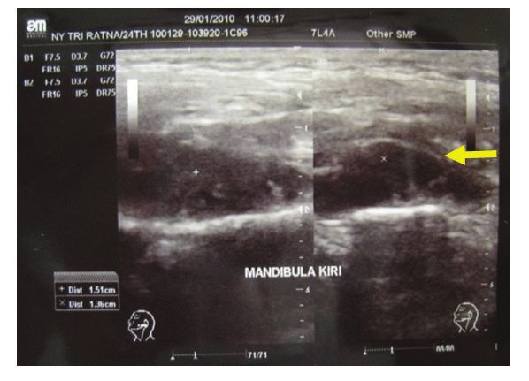
1854     Indian Journal of Forensic Medicine & Toxicology, January-March 2021, Vol. 15, No. 1

economic limitations.6                                                cheek and unability to open her mouth after having her
                                                                      lower left first molar extracted ten days prior to admission.
     The purpose of this clinical study is to present four
                                                                      Clinical examination findings included: patient looking
cases of soft tissue swelling where USG was used as
                                                                      unwell, presence of a diffuse, hard and painful swelling
an aid in confirming diagnosis and supporting surgical
                                                                      in the left masseter region and limited ability to open
treatment.
                                                                      the mouth (i,e, less than 1 cm wide). No fluctuation was
                                                                      encountered, while intraoral examination confirmed
                 Material and Methods
                                                                      no signs of post-extraction infection of the socket.
    Clinical study was conducted through retrospective                Clinical diagnosis of a submasseteric space abscess
medical record study of four cases of soft tissue swelling            was conducted and surgical drainage was planned. A
and enlargement in patients of the Oral and Maxillofacial             USG examination completed for confirmation revealed
Department, Universitas Airlangga Dental Hospital                     a hypoechoic lesion in the submasseteric region with
where diagnosis was confirmed using USG (GE), as well                 fluid echo intensity at a maximal point of 1.57 cm. The
as an aid in surgical theraphy.                                       presence of a submasseteric abscess was confirmed by
                                                                      means of USG examination (figure 1).
       Case 1

       A 24-year old female presented swelling of the left

            Figure 1. Ultrasonogram showed hypoechoic lesion (x sign) in submasseteric space abscess.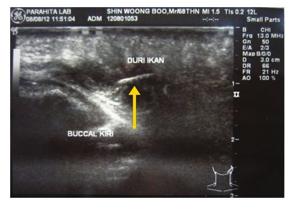
Indian Journal of Forensic Medicine & Toxicology, January-March 2021, Vol. 15, No. 1 1855
An intraoral incision with Swann Morton surgical measuring ± 2 cm in diameter with a soft consistency
blade no.11 was performed to evacuate pus from the and painless on palpation. Intraoral examination
submasseteric space abscess, 1 cc pus mixed with revealed a flattened left upper anterior vestibulum due
blood being drained. On evaluation, pain was found to to enlargement with defined borders, measuring ± 2
have diminished, mouth opening had widened and the cm in diameter, with a soft consistency and painless on
swelling had gradually subsided. palpation. Periapical radiograph examination was within
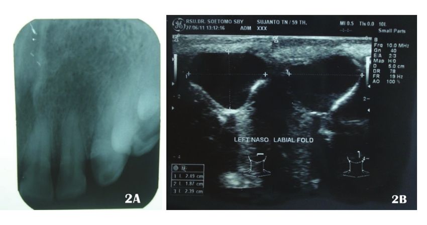
normal limits (Figure 2A). Diagnosis of a nasolabial
Case 2
cyst was made and confirmed by means of USG which
A 59-year old male attended with painless enlargement showed a thin walled cystic lesion with defined border
of the left nasal base which had been developing during and internal echo, measuring 2,49 x 1,87 x 2,39 cm in
the ten years prior to admission. Clinical examination the left nasolabial fold (Figure 2B).
revealed soft tissue enlargement with defined borders in
Extirpation of the cyst was performed through
the left nasolabial region resulting in a narrowing of the
an intra oral approach, healing was effective and no
left nasolabial sulcus when compared to the right side,
recurrence was found upon evaluation.
Figure 2. Periapical radiograph showed normal appearance in nasolabial cyst (2A). Ultrsonogram showed
cystic lesion in the left nasolabial fold (2B).
Case 3 and backwards. The consistency was firm and tender
on palpation. Submandibular node enlargement was
A 25-year old female had complained for the evident.
previous 12 years of a recurrent neck swelling that
forced her tongue upwards and backwards, thereby Clinical diagnosis of the infected dermoid cyst
impairing both her breathing and ability to swallow. She was conducted and, after aspiration made from the
also complained of pain and fever, but was generally submental region for decompression revealed an
in good condition. The submental swelling was extremely viscous, yellowish fluid, the patient was
erythematous, well-defined, measured 4 cm in diameter sent for USG examination. The USG revealed a well-
and extended sublingually, pushing the tongue upwards defined heterogeneous echoic lesion, measuring 3.34 cm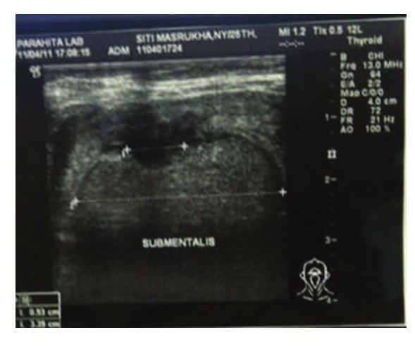
1856     Indian Journal of Forensic Medicine & Toxicology, January-March 2021, Vol. 15, No. 1

in diameter, with mixed content and debris. There was                     The treatment consisted of extirpation of the cyst
also post-aspiration defect with adjacent fluid collection            through an extra oral approach under general anesthesia,
and no intralesion vascularization was found. The                     healing was effective and no recurrence was found upon
submandibular glands were enlarged and diagnosis of an                evaluation.
infected dermoid cyst was confirmed (figure 3).

 Figure 3. Ultrasonogram of Dermoid cyst showed a well defined heterogenous echoic lesion measuring 3,34
  cm in diameter, with mixed content and debris. There was also post aspiration defect with adjacent fluid
                           collection, and no intralesion vascularization found.

       Case 4                                                         palpation. An initial, provisional diagnosis was one of a
                                                                      left buccal space abscess caused by a foreign body (fish
     A 68 year old male presented with swelling on his
                                                                      bone). This was subsequently confirmed by USG which
left buccal region that had been present with intermittent
                                                                      revealed a foreign body (fish bone) in the left buccal
pain for one week prior to admission,. The patient had
                                                                      region measuring 7 mm and 8.4 mm in depth from the
suffered a fish bone puncture to the left buccal region upon
                                                                      skin surface. There was also adjacent fluid collection
eating three months previously. Clinical examination
                                                                      around the fish bone, edematous surrounding tissue and
confirmed well-defined erythematous swelling in the
                                                                      increased vascularization (figure 4).
left buccal region measuring 2 cm in diameter, with firm
consistency and little pain on palpation. Well-defined                    Surgical drainage and exploration of the fish bone
intraoral swelling in left buccal mucosa measuring 2                  using an intraoral approach was performed. Upon
cm in diameter, with a soft consistency and tender on
Indian Journal of Forensic Medicine & Toxicology, January-March 2021, Vol. 15, No. 1 1857
exploration, a 7 mm long fish bone was found and evacuated. The healing process was successful and no recurrence
found.
Figure 4. Ultrasonogram showed foreign body measuring 7 mm and 8,4 mm in depth from skin surface
in the left buccal region (yellow arrow). There was also adjacent fluid collection arround the fish bone,
oedematous sorrunding tissue, and increased vascularization in buccal space abscess caused by foreign body.
Result conducted by Akinbami, et al. (2006) confirmed the
reliability of ultrasonography in the diagnosis of
All cases presented showed that USG 100%
pleomorphic adenoma as being 80% and 100% for
accuracy in diagnosis of soft tissue enlargement in oral
adenocarcinoma and hemangioma. It was also 100%
and maxillofacial region, as well as guiding surgical
in the majority of cyst and salivary gland swellings.
drainage and approach.
Ultrasonography was also 100% specific in the diagnosis
of monomorphic adenoma and hemangioma.7 Research
Discussion
conducted by Chandak et al., (2011) to evaluate USG in
USG has been traditionally employed in the the diagnosis of head and neck swelling, showed that this
assessment of soft tissues in the abdomen and pelvis. form of diagnosis provided a sensitivity and accuracy
Its role in oral and maxillofacial surgery is less widely rate of 98,5% compared to that of clinical diagnosis at
recognized. Recently, a considerable body of literature 85,7%.8
and research reports about the reliable use of USG in
diagnostic processes relating to oral and maxillofacial All of the four cases reported in this paper
lesions. Research into the accuracy, sensitivity, showed congruence between clinical, ultrasonographic
specificity and predictive value of ultrasound as a and histopathological diagnosis. This is consistent with
means of diagnosis of cervico-facial soft tissue swelling the findings in the research reports referred to above.1858 Indian Journal of Forensic Medicine & Toxicology, January-March 2021, Vol. 15, No. 1
In aiding therapy such as abscess drainage, Craniomand Pract. 2005;23(2): 100-12.
ultrasonography can delineate the location and extent 3. Mohan KR, Rao NK, Krishna GL, Kumar VS,
of abscess formation. USG is capable of measuring Ranganath N, Lakshmi UV. Role of Ultrasonography
the distance from skin to oral mucosa, denoted a third in Oral dan Maxillofacial Surgery: A Review of
dimension of the swelling and quantification of pus Literature. J Maxillofac Oral Surg. 2015;14(2):
through its anechoic pattern and inflammatory zone.9 162-70.
Ultrasonography can also be used intraoperatively to 4. Gold L, Nazarian LN, Johar AS, Rao VM.
aid in the aspiration, incision and drainage of pus.10 Characterization of maxillofacial soft tissue
As in the case of submasseteric abscesses and buccal vascular anomalies by ultrasound and color doppler
space abscesses discussed in this report, the distance imaging: an adjuvant to computed tomography and
of the maximal point of the abscess, the location and magnetic resonance imaging. J Oral Maxillofac
distant of fish bone from the skin can be detected using Surg. 2003; 61:19-31.
ultrasonography. 5. Joshi PS, Pol J, Sudesh AS. Ultrasonography – a
diagnostic modality for oral and maxillofacial
Conclusion diseases. Contemp Clin Dent. 2014; 5(3): 345-51.
Ultrasonography is a quick, widely available, 6. Eviergen Ş, Kamburoǧlu K. Review on
relatively inexpensive, painless procedure which can be the applications of ultrasonography in
dentomaxillofacial region. World J Radiol.
repeated as often as necessary without risk to the patient.
2016;8(1): 50-8.
Thus, ultrasonography is a valuable diagnostic aid to the
oral and maxillofacial surgeons for early and accurate 7. Akinbami BO, Ugboko VI, Owotade FJ, Obiechina
diagnosis, as well as in surgical treatment of oral and AE, Adetiloye VO, Ayoola A. Aplications of
ultrasonography in the diagnosis of soft tissue
maxillofacial soft tissue enlargement.
swellings of the cervicofacial region. WAJM.
Ethical Clearance: Nil. 2006; 25(2): 110-8.
8. Chandak R, Degwekar, Bhowte RR, Motwani M,
Sources of Funding: Self-funded. Banode P, Chandak M, et al. An evaluation of
Conflict of Interest: There are no conflict of efficacy of ultrasonography in the diagnosis of head
and neck swellings. Dentomaxillofac Radiol. 2011;
interest.
40: 213-21.
References 9. Srinivas K, Sumanth KN, Chopra SS.
Utrasonographic evaluation of inflammatory
1. Pandey PK, Umarani M, Kotrashetti S, Baliga S.
swellings of buccal space. Indian J Dent Res. 2009;
Evaluation of ultrasonography as a diagnostic tool
20(4): 458-62.
in maxillofacial space infection. J Oral Maxillofac
Res. 2011; 2(4):1-6. 10. Thiruchelvam JK, Songra AK, Ng SY.
Intraoperative ultrasound imaging to aid abscess
2. Imbeau J. Introduction of through-transmission
drainage-a technical note. Int J Oral Maxillofac
alveolar ultrasonography in dental medicine. J
Surg. 2002; 31: 442-3.You can also read