CTU findings of duplex kidney in kidney: A rare duplicated renal malformation
←
→
Page content transcription
If your browser does not render page correctly, please read the page content below
Open Medicine 2021; 16: 651–654
Case Report
Nanai Xie#, Xu Huang#, Jie Zhou, Heng Zhang, Wanling Ma*
CTU findings of duplex kidney in kidney:
A rare duplicated renal malformation
https://doi.org/10.1515/med-2021-0271 incomplete fusion of upper and lower kidney moieties
received January 2, 2021; accepted March 11, 2021 accompanied by complete or incomplete duplications of
Abstract: Duplex kidney is a common congenital malfor- pyeloureteral [4].
mation appeared as duplication of pelvis and ureter. However, duplicated renal malformation located in
However, renal duplication within sinus renalis is an sinus renalis is an extremely rare anatomical variation.
extremely rare variation of the renal collecting system. To the best of our knowledge, there is rare report con-
In this study, we report a case of an asymptomatic kidney cerning this malformation. In this study, we reported an
disease in a 33-year-old man, who demonstrates abnormal extremely rare renal duplication: another little duplex
echo of renal sinus anomaly discovered incidentally in kidney located in renal sinus. It is generally asympto-
ultrasound examination. Computed tomography urography matic or presents with non-specific symptoms, and thus
(CTU) exhibited the other small duplex kidney located in missed easily by clinician. On the plain computed tomo-
renal sinus. In the excretory phase images, the contrast graphy (CT) images, the abnormal renal shaped mass
medium within its small renal pelvis could be seen to in the renal sinus is very easily misdiagnosed as the renal
flow into the right major renal calices. This case exhibited pelvis mass. Moreover, distinguishing it from renal
a very rare anatomical variation of duplicated renal columnar hypertrophy is very difficult during arterial
malformation. and venous phases of the enhancement because of its
similar enhancement to kidney. However, the CT pyelo-
Keywords: kidney, urinary tract, computed tomography graphic phase images demonstrate clearly that tubular
urography, duplex kidney, intrarenal kidney contrast medium from the renal shaped mass merges
into the renal pelvis, which verifies the existence of the
duplicated renal pelvis.
1 Introduction
The renal duplication is a common renal malformation 2 Case report
with an incidence of approximately 0.8% [1,2]. It usually
appears as a renal unit comprised of two renal pelvis and Incidentally, a 33-year-old man was diagnosed as hyper-
kidney systems [3]. This malformation is characterized by trophied column of Bertin by ultrasound, which demon-
strated abnormal echo mass in renal sinus (Figure 1). The
patient was asymptomatic. All laboratory results and
# Nanai Xie and Xu Huang contributed equally to this work. physical examinations were normal. No palpable mass
Nanai Xie and Xu Huang should be listed as co-first author.
and percussion pain were found in bilateral renal areas.
CT urography (CTU) was performed to evaluate the
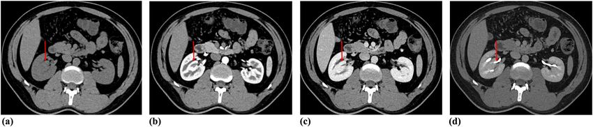
renal lesion more accurately. Plain CT demonstrated an
* Corresponding author: Wanling Ma, Department of Radiology,
Longgang District People’s Hospital of Shenzhen & The Third isodensity mass to normal renal parenchyma with unclear
Affiliated Hospital (Provisional) of The Chinese University of Hong boundary in his right renal sinus without hydronephrosis
Kong, Shenzhen, Guangdong Province, 518172, China, (Figure 2a). The size of lesion was 14 × 5 × 3 mm. Renal
e-mail: mawanling321@163.com, tel: +86-0755-28932321, contrast-enhanced CT (CECT) showed the intrarenal sinus
fax: +86-0755-28932321
mass that exhibited the enhancement mode similar to
Nanai Xie, Xu Huang, Jie Zhou, Heng Zhang: Department of
Radiology, Longgang District People’s Hospital of Shenzhen & The
kidney: wheel-spoke shaped enhancement of renal cortex
Third Affiliated Hospital (Provisional) of The Chinese University of and no enhancement of renal medulla in corticomedullary
Hong Kong, Shenzhen, Guangdong Province, 518172, China phase, and homogeneous enhancement in parenchymal
Open Access. © 2021 Nanai Xie et al., published by De Gruyter. This work is licensed under the Creative Commons Attribution 4.0
International License.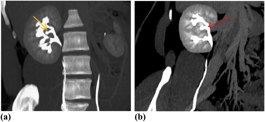
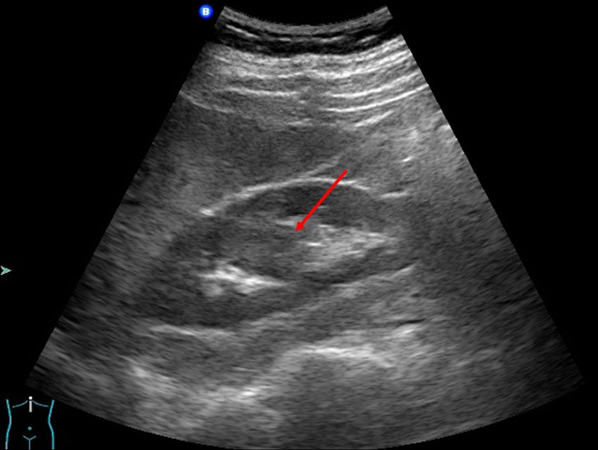
652 Nanai Xie et al. Figure 1: Ultrasound showed a patchy hypoechoic area (red arrow) within the right renal sinus, with slightly irregular shape, clear boundary, the similar echo of renal cortex, and enhanced echo in the rear. phase (Figure 2b and c). Pyelographic phase images demon- The curved planar reconstruction (CPR) image could dis- strated that a renal pelvis shaped structure from mass play the aberrant duplex kidney and renal pelvis in renal merged into the right major renal calyces (Figure 2d). Cor- sinuses and the confluence into the right upper renal onal maximum intensity projection (MIP) CT image in pye- calices in one plane (Figure 3b). lographic phase directly revealed that the mass in renal sinus sent out a renal pelvis shaped structure and Ethics statement: This study protocol was approved by merged into the right upper major calyces (Figure 3a). the Institutional Ethics Committee Board of our hospital. Figure 2: Axial precontrast (a) and contrast-enhanced (b and c) CT showed oval solid mass (red arrows) in right renal sinus with identical density and enhancement pattern to adjacent normal renal cortex. In pyelographic phase image (d) exhibited the small renal pelvis (red arrow) from the pseudo-mass merging into the right renal pelvis.
CT findings of intrarenal kidney 653
Figure 3: Pyelographic phase coronary MIP (a) and CPR (b) images demonstrated that the little renal pelvis (yellow arrow) from renal shaped
mass in the renal sinus drained into the calyces of the upper pole of right kidney (red arrow). MIP = maximum intensity projection; CPR =
curved planar reconstruction.
Written informed consent was obtained from the patient may be a rare and special type of embedded type duplex
for publication of this manuscript and any accompanying kidney [6].
images. Intrarenal duplex kidney is usually discovered by
accident and closely similar to renal column hypertrophy
on ultrasonography. It is usually located in the interior
middle portion of the kidney. The key to diagnose intrarenal
3 Discussion duplex kidney correctly is the presence of normal renal
parenchyma. The key point to diagnose it on imaging is its
Duplex kidney in kidney is very rare. As far as we know, identical density/signal intensity to renal parenchyma on
such cases have rarely been reported previously. The both precontrast and contrast sequences [5]. To distinguish
duplex kidney in renal sinus and the ipsilateral kidney from renal column hypertrophy, it is critical to identify the
were integrated without an apparent borderline between duplicated renal pelvis derived from mass within renal sinus
them and were supplied by the same ipsilateral renal draining into the collection system of kidney on pyelo-
artery. However, there were two renal pelves that finally graphic phase images. Unfortunately, our results lack the
drained into the single ureter. No vascular malformation pathological diagnosis. Notwithstanding its limitation, our
and accessory renal arteries were found in this kind of case did demonstrate that the enhancement of mass within
renal duplication. This disease is very easy to be misdiag- renal sinus was similar to background renal parenchyma
nosed because it is asymptomatic or with non-specific in arterial and venous phase. The pyelographic phase
symptoms and clinically normal renal function. images and CTU confirmed the diagnosis of intrarenal
The intrarenal duplex kidney is comprised of two duplex kidney in our case by showing the duplicated renal
separate kidneys and pelvicalyceal systems just like pelvis from mass in renal sinus merging into the ipsilateral
conventional duplex kidney. However, the conventional renal collecting system.
duplex kidney is frequently associated with duplication Duplex kidneys are common with the incidence of
of ureter. Moreover, the duplicated renal pelves of con- around 0.8% [1,2]. The majority of duplex kidneys
ventional duplex kidney generally locate in the upper are often of no clinical significance and do not become
and lower positions [5]. However, duplex kidney and diseased [4,7,8]. However, almost 50% of the pediatric
renal pelvis locates in renal sinus between normal upper patients with duplex kidneys have complications re-
and lower major renal calices in this report. Therefore, quiring treatment [4]. If the duplication of kidney is
the intrarenal duplex kidney described in this report complicated with renal hypoplasia, atrophy of renal654 Nanai Xie et al.
parenchyma, loss of function, ureteral calculi, severe analyzed during the current study. The authors pre-
infection or leakage due to ectopic ureteral orifice, ure- sented, in the manuscript, all the necessary information
terocele, hydronephrosis, or vesicoureteral reflux, sur- about their case report.
gical treatment is needed [9,10]. Heminephrectomy is
the most commonly used operation for the management
of duplex kidney [11–13]. Intrarenal duplex kidneys are a
very rare congenital anomaly and take up about 15% of References
all duplex kidneys [6]. It is important for urologist to get a
correct preoperative diagnosis of this type duplex kidney [1] Davda S, Vohra A. Adult duplex kidneys: an important differ-
ential diagnosis in patients with abdominal cysts. JRSM Short
to be more cautious during the heminephrectomy. Con-
Rep. 2013;4(2):13. doi: 10.1177/2042533312472126.
servative treatment remains a challenge due to its very [2] Hunziker M, Kutasy B, D’Asta F, Puri P. Urinary tract anomalies
rare occurrence and lack of long-term follow-up data. associated with high grade primary vesicoureteral reflux.
In conclusion, intrarenal duplex kidney is prone to Pediatr Surg Int. 2012;28(2):201–4. doi: 10.1007/s00383-011-
misdiagnosed as a renal mass on ultrasonography and 2986-1.
[3] Horst M, Smith GH. Pelvi-ureteric junction obstruction in
plain CT. CECT and CTU are essential for diagnosing it
duplex kidneys. BJU Int. 2008;101(12):1580–4. doi: 10.1111/
accurately. Prolonged CTU could be helpful in evaluating j.1464-410X.2007.07386.x.
the duplex kidney with poor function and demonstrating [4] Doery AJ, Ang E, Ditchfield MR. Duplex kidney: not just a
the urinary tract malformations more clearly [8]. If the drooping lily. J Med Imaging Radiat Oncol. 2015;59(2):149–53.
renal function of the duplex kidney is well, timely diag- doi: 10.1111/1754-9485.12285.
[5] Ramanathan S, Kumar D, Khanna M, Al Heidous M, Sheikh A,
nosis before operation is crucial to avoid unnecessary
Virmani V, et al. Multi-modality imaging review of congenital
radical surgery.
abnormalities of kidney and upper urinary tract. World J
Radiol. 2016;8:132–41. doi: 10.4329/wjr.v8.i2.132.
[6] Ma R, Wu RD, Liu W, Wang G, Wang T, Xu ZD, et al. A new
classification of duplex kidney based on kidney morphology
Abbreviations and management. Chin Med J (Engl). 2013;126(4):615–9.
PMID: 23422177.
[7] Privett JT, Jeans WD, Roylance J. The incidence and importance
CTU computed tomography urography of renal duplication. Clin Radiol. 1976;27(4):521–30.
CECT contrast-enhanced CT doi: 10.1016/s0009-9260(76)80121-3.
MIP maximum intensity projection [8] Gong H, Gao L, Dai XJ, Zhou F, Zhang N, Zeng X, et al.
CPR curved planar reconstruction Prolonged CT urography in duplex kidney. BMC Urol.
2016;16(1):21. doi: 10.1186/s12894-016-0139-5.
[9] Abel C, Lendon M, Gough DC. Histology of the upper pole in
Funding information: The authors declare that no fund- complete urinary duplication–does it affect surgical manage-
ment? Br J Urol. 1997;80(4):663–5. doi: 10.1046/j.1464-
ings have been received.
410x.1997.00413.x. PMID: 9352710.
[10] Rickwood AM, Reiner I, Jones M, Pournaras C. Current man-
Author contributions: Conceptualization: Wanling Ma. agement of duplex-system ureteroceles: experience with 41
Data curation: Jie Zhou, Xu Huang. Formal analysis: patients. Br J Urol. 1992;70(2):196–200. doi: 10.1111/j.1464-
Nanai Xie. Investigation: Heng Zhang. Resources: Nanai 410x.1992.tb15703.x. PMID: 1393443.
Xie. Writing – original draft: Nanai Xie. Writing – review [11] Gao Z, Wu J, Lin C, Men C. Transperitoneal laparoscopic
heminephrectomy in duplex kidney: our initial experience.
and editing: Wanling Ma.
Urology. 2011;77(1):231–6. doi: 10.1016/
j.urology.2010.02.002.
Conflict of interest: The authors declare that the research [12] Leclair MD, Vidal I, Suply E, Podevin G, Heloury Y.
has been conducted in the absence of any commercial Retroperitoneal laparoscopic heminephrectomy in duplex
or financial relationships that could be construed as a kidney in infants and children: a 15-year experience.
Eur Urol. 2009;56(2):385–9. doi: 10.1016/
potential conflict of interest.
j.eururo.2008.07.015.
[13] Zumsteg J, Roberts WW, Wolf JS. Laparoscopic hemine-
Data availability statement: Data sharing is not applic- phrectomy for benign renal anomalies. J Endourol.
able to this article as no datasets were generated or 2010;24(1):41–7. doi: 10.1089/end.2009.0346.You can also read