Salvage of Lower Limb in Delay-Diagnosed Popliteal Artery Transection Caused by Blunt Trauma
←
→
Page content transcription
If your browser does not render page correctly, please read the page content below
Open Access

Austin Journal of Surgery

Special Article – Surgery Case Reports

Salvage of Lower Limb in Delay-Diagnosed Popliteal
Artery Transection Caused by Blunt Trauma
Chin-Choon Yeh*
                                                                  Abstract
Division of Plastic Surgery, Department of Surgery, Chi-
Mei Medical Center, Taiwan                                            Background: Transection of poplieal artery could block majority of lower
*Corresponding author: Chin-Choon Yeh, Division of                limb perfusion, results in ganrene and amputation finally.
Plastic Surgery, Department of Surgery, Chi-Mei Medical
                                                                       Aim and Objectives: This article presents a case of successful salvage
Center, No.901, Jhonghua Rd., Yongkang Distinct, Tainan
                                                                  of lower limb in delay-diagnosed popliteal artery transection and review of the
County 71004, Taiwan
                                                                  literature. Total transection of popliteal artery could block nearly all perfusion to
Received: January 01, 2020; Accepted: January 22,                 the lower limb and subsequently causes gangrene that need to be amputated.
2020; Published: January 29, 2020
                                                                       Materials and Methods: The patient was a 31-year-old male suffered
                                                                  from left lower limb blunt trauma in a traffic accident. The mechanism is falling
                                                                  down from ridding motorcycle and hitting a telephone pole. Initially vessel injury
                                                                  was not identified and he was discharged from ER after 7-hour observation.
                                                                  However, he felt severe pain and swelling with large area ecchymosis of his left
                                                                  leg after working. So he came back to our ER on post-trauma day #3. Emergent
                                                                  fasciotomy was done but distal perfusion did not recover. Cardio-vascular
                                                                  surgeon was consulted for arterial bypass. However, distal limb did not become
                                                                  warm and pink immediately. We kept medication for vessel patency and anti-
                                                                  coagulation. Fortunately, distal perfusion recovered gradually after several
                                                                  days. But partial muscle and fascia necrosis resulted in bone exposure. Serial
                                                                  debridement and sequestrectomy were done. Finally, we applied Negative
                                                                  Pressure Wound Therapy (NPWT) then STSG eventually closed the wound.
                                                                      Result: We followed up his wound condition for one year, his wound healed
                                                                  well and there was no recurrence of infection or necrosis. Ambulation is re-
                                                                  gained without aid of prosthesis or cruches despite of mild foot-drop.
                                                                      Conclusion: Clinicians should be cautious to unusual manifestations of
                                                                  blunt trauma, which may signalize a concomitant vascular injury. Early detection
                                                                  and re-perfusion as soon as possible are very important and encouraged for
                                                                  successful limb salvage.
                                                                     Keywords: Limb salvage; Artery transection; Limb blunt trauma; Arerial by
                                                                  pass

Introduction                                                                   Case Report
     Popliteal artery injury is an uncommon situation encounterd in                This patient was a 31-year-old male suffered from acute left lower
lower limb trauma. Delay in diagnosis and treatment could increase             limb blunt trauma in a traffic accident which mechenism was falling
morbidity and eventually result in a diaster. Popliteal artery was             down from ridding motorcycle and hitting a telephone pole. He was
anatomically surrounded by the politeal ligamentous, femur bone,               sent to our ER (emergency room) by EMT (Emergency Medical
tibial plateau, and knee joint capsule thus susceptible to high-               Technician). The Glasgow Coma Score was 15 at the scene, no
energy blunt and penetrating trauma [1]. The association between               requiring of intubation. The systolic blood pressure was 122mmHg
popliteal artery injury and tibiofemoral dislocations as well as               while the diastolic blood pressure was 68 mmHg, and the heart rate
femur fractures has been widely discussed in literatures [2,3]. Initial        was 120 beats per minute. CT (Computed Tomography) of brain
physical exams characteristically show marked knee joint instability,          and focused assessment with sonography for trauma yieled negative
and roentgenogram revealed fractures, subluxations and soft tissue             results.
swelling [4].
                                                                                   On physical examination, the left lower extremity was swollen and
    Reviewed literatures about popliteal artery injury caused by               tense, but distal pulses were ausculated by sonogram. Radiographs of
trauma have described intimal tears, dissection, ruptures, or even             the left femur and left knee demonstrated no evidence of fracture or
transection of the popliteal artery upon surgical exploration [5]. We          dislocation. Due to left lower limb pain complained by the patient,
present a unique case of a delay-diagnosed complete left popliteal             he stayed in our ER for 7 hours observation then was discharged.
artery transection following a motorcycle collision, without obvious           Symptoms and signs of vessel injury were not detected at that
associated fracture or evidence of a tibiofemoral dislocation.                 moment.

 Austin J Surg - Volume 7 Issue 1 - 2020                   Citation: Chin-Choon Yeh. Salvage of Lower Limb in Delay-Diagnosed Popliteal Artery Transection Caused by
 ISSN : 2381-9030 | www.austinpublishinggroup.com          Blunt Trauma. Austin J Surg. 2020; 7(1): 1240.
 Yeh. © All rights are reserved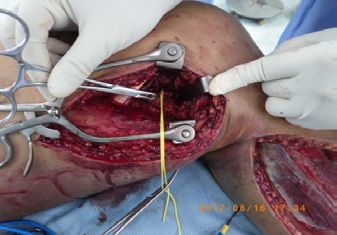
Chin-Choon Yeh Austin Publishing Group
Figure 4: Partail muscle (mainly lateral compartment and anterior
compartment) necrosis with small area of tibia and fibula bone exposure
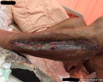
Figure 1: The patient felt severe pain and swelling with large area ecchymosis were noted.
of his left leg after working. So he came back to our ER on post-trauma day
#3.
Figure 2: Emergent fasciotomy was done the compartment pressure was Figure 5: We applied Negative Pressure Wound Therapy (NPWT) then
released, but distal perfusion didn’t recover. Immediately, a vascular surgeon STSG eventually closed the wound.
was consulted for arterial bypass. Popliteal artery transection was observed
at that scene.
popliteal artery with reverse greater sapheneous vein) was done
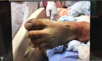
after Angio-graphy examination (Figure 2). Unfortunately, distal
limb did not become warm and pink immediately (Figure 3). We
kept medication for vessel dilatation (Prostaglandin E1) and anti-
coagulation (heparin) infusion for 7 days. Distal perfusion rocovered
and skin color became pink at post-bypass day 3. Partail muscle
(mainly lateral compartment and anterior compartment) necrosis
with small area of tibia and fibula bone exposure were noted (Figures
4-6).
Serial debridement and sequestrectomy with VAC (vacumm
Figure 3: However, distal limb did not become warm and pink at that moment. assited closure) system apply then subsequent skin grafting were done
We kept medication for vessel patency and anti-coagulation. from post-bypass day 12 to day 54. The wound closure was achieved
eventually at post-bypass day 60 and the patient was discharged from
However, he came back to our ER 3 days later because of our ward.
progressive swelling and pain of his left lower limb with large area At OPD (outpatient department) examination and follow-up of
of ecchymosis occured after his working (Figure 1). At this moment, a 3-month period, the skin graft condition is good without infection
labratory data showed Myoglobin was 5437 ng/mL whereas CK nor necrosis. Ambulation without crutch or wheelchair is achieved
(creatine kinase) -total was 31903 U/L. Distal perfusion of left lower except mild frop footnoted.
leg and foot was absent. Due to clinical evidence of left lower extremity
compartment syndrome with rhabdomyolysis, the patient was taken Discussion
to the operating room immediately for emergent fasciotomy to
Although tibiofemoral knee dislocation is a rare and serious
release pressure. However, after we released all compartment of left
injury that occurs in ~0.02–0.1% of all musculoskeletal injuries [6],
lower leg, distal perfusion did not recover at this moment.
it could result in a diasater. These knee dislocations occur in both
A cardio-vascular surgeon was consulted. Operation of arterial high- and low-energy contusional or penetrating traumas and often
bypass (left supra-genicular popliteal artery bypass to infra-genicular spontaneously reduce, thus severe injury could be ignored at first
Submit your Manuscript | www.austinpublishinggroup.com Austin J Surg 7(1): id1240 (2020) - Page - 02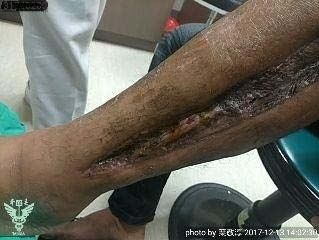
Chin-Choon Yeh                                                                                                                         Austin Publishing Group

                                                                                 high clinical suspicion for popliteal vessel injury must still be kept in
                                                                                 mind. In fact, even in the absence of clinical or radiographic evidence
                                                                                 of significant injury, careful evaluation for vascular injury is still
                                                                                 needed, as tibiofemoral dislocation often spontaneously reduces by
                                                                                 itself, leaving little or no detectable deformity at the time the patient
                                                                                 is seen at first time. In addition, in cases where no trauma history
                                                                                 information is available and the mechanism of injury is unknown, a
                                                                                 severe vascular injury such as arterial transection or total throbosis
                                                                                 should be considered as a potential cause of refractory hypotension
                                                                                 [11].
                                                                                 References
                                                                                 1. Witz M, Witz S, Tobi E, Shnaker A, Lehmann J. Isolated complete popliteal
 Figure 6: We followed up his wound condition for one year, his wound healed        artery rupture associated with knee dislocation. Knee Surg Sports Traumatol
 well and there was nor recurrence of infection or necrosis. Ambulation is re-      Arthrosc 2004; 12: 3-6.
 gained despite of mild foot-drop.
                                                                                 2. Perron AD, Brady WJ, Sing RF. Orthopedic pitfalls in the ED: vascular injury
                                                                                    associated with knee dislocation. Am J Emerg Med. 2001; 19: 583-588.
aid. Physical examination of the knee for ligamentous instability can
                                                                                 3. Seroyer S, Musahl V, Harner C. Management of the acute knee dislocation:
alert the physician to the possibility of an occult knee dislocation. It
                                                                                    the Pittsburgh experience. Injury. 2008; 39: 710-718.
is imperative to recognize the clinical signs and symptoms of these
                                                                                 4. Yahya MM, Mwipatayi BP, Abbas M, Rao S, Sieunarine K. Popliteal artery
dislocations, as they are associated with a significant incidence (25–
                                                                                    injury: Royal Perth experience and literature review. ANZ J Surg. 2005; 75:
40%) of concomitant popliteal vessel injury, with 1.6–13% of cases                  882-886.
requiring acute popliteal vessel repair or re-perfusion [7]. Delayed
                                                                                 5. Gupta R, Quinn P, Rao S, Sleunarine K. Popliteal artery trauma: a critical
diagnosis and treatment of a popliteal vessel injury significantly                  appraisal of an uncommon injury. Injury. 2001; 32: 357-361.
increases patient morbidity.
                                                                                 6. Sillanpaa PJ, Kannus P, Niemi ST, Rolf C, Felländer-Tsai L, Mattila VM.
    Popliteal artery injuries have been traditionally treated with                  Incidence of knee dislocation and concomitant vascular injury requiring
                                                                                    surgery: a nationwide study. J Trauma Acute Care Surg. 2014; 76: 715-719.
open surgical repair or bypass and fasciotomy. Recent advances
in facilities have allowed for endovascular repair in selected                   7. Steele HL, Singh A. Vascular injury after occult knee dislocation presenting
patients. Endovascular techniques are generally introduced in                       as compartment syndrome. J Emerg Med. 2012; 42: 271-274.

hemodynamically stable patients to treat vascular injuries such                  8. Piffaretti G, Tozzi M, Lomazzi C, Rivolta N, Caronno R, Laganà D, et al.
as thrombosis, pseudoaneurysm, dissection, hematoma and                             Endovascular treatment for traumatic injuries of the peripheral arteries
                                                                                    following blunt trauma. Injury. 2007; 38: 1091-1097.
arteriovenous fistula, and have been demonstrated to be successful
specifically in instances of popliteal artery trauma [8-10]. In severe           9. Hutto JD, Reed AB. Endovascular repair of an acute blunt popliteal artery
                                                                                    injury. J Vasc Surg. 2007; 45: 188-190.
lower extremity trauma, clinical or radiographic evidence of distal
femoral fracture or tibiofemoral dislocation should prompt dedicate              10. Megalopoulos A, Siminas S, Trelopoulos G. Traumatic pseudoaneurysm
vascular evaluation, which will often reveal the concomitant                         of the popliteal artery after blunt trauma: case report and a review of the
                                                                                     literature. Vasc Endovascular Surg. 2007; 40: 499-504.
popliteal vessels injury. In milder injuries, physical examination may
demonstrate evidence of ligamentous instability, and radiographs may             11. Weiner EJ, Ditchek JJ, Solomon RJ, Eyerly-Webb S, Kiffin C, Carrillo EH, et
                                                                                     al. A unique case of popliteal artery transection after a motorcycle collision. J
reveal only subtle findings such as nondisplaced fracture, subluxation,              Surg Case Rep. 2017: rjx222.
joint effusion/hematoma and soft tissue swelling. In these cases, a

Submit your Manuscript | www.austinpublishinggroup.com                                                               Austin J Surg 7(1): id1240 (2020) - Page - 03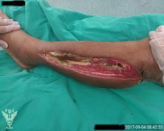
You can also read