Tuberculous Sacroiliitis with Secondary Psoas Abscess: A Case Report
←
→
Page content transcription
If your browser does not render page correctly, please read the page content below
Open Journal of Rheumatology and Autoimmune Diseases, 2021, 11, 48-52
                                                                                                https://www.scirp.org/journal/ojra
                                                                                                          ISSN Online: 2164-005X
                                                                                                            ISSN Print: 2163-9914

Tuberculous Sacroiliitis with Secondary Psoas
Abscess: A Case Report

Kaba Condé1,2, Carlos Othon Guelngar1, Igué Kadidjatou1, D. D. Granga 3, Mamadou Ciré Barry4,
Mamadou Hady1, Karinka Diawara1, Konaté Mamady2, Touré Moriba2, Awada Mohamed2,
Fodé Abass Cissé1
1
 Department of Neurology, Academic Hospital, University of Conakry, Conakry, Guinea
2
 Department of Rheumatology, Academic Hospital, University of Conakry, Conakry, Guinea
3
 Department of Pediatric, Mother and child Hospital, Ndjamena, Chad
4
 Department of Pediatric, Academic Hospital, University of Conakry, Conakry, Guinea

How to cite this paper: Condé, K., Gueln-       Abstract
gar, C.O., Kadidjatou, I., Granga, D.D.,
Barry, M.C., Hady, M., Diawara, K., Ma-         Tuberculous sacroiliitis secondary to a psoas abscess is rare, only a few spo-
mady, K., Moriba, T., Mohamed, A. and           radic cases were reported in the literature. Tuberculous sacroiliitis is rare,
Cissé, F.A. (2021) Tuberculous Sacroiliitis
                                                usually unilateral, its symptomatology is misleading, its diagnosis is often de-
with Secondary Psoas Abscess: A Case
Report. Open Journal of Rheumatology and        layed or even confused with damage to the hip or lumbosacral hinge, most
Autoimmune Diseases, 11, 48-52.                 often related to difficulties exploration of the sacroiliac joint. We report the
https://doi.org/10.4236/ojra.2021.112006        case of a 66-year-old diabetic patient with low back pain, unilateral right with
Received: December 8, 2020
                                                inflammatory appearance, insidious installation, evolving for about 8 months.
Accepted: April 24, 2021                        The diagnosis of tuberculous sacroiliitis was made after biopsy of the sacroi-
Published: April 27, 2021                       liac joint. CT and MRI are necessary for lesion diagnosis. Tuberculosis treat-
                                                ment was started and the abscess was surgically drained. The aim of this work
Copyright © 2021 by author(s) and
Scientific Research Publishing Inc.             was to describe the diagnostic pathway of a patient with tuberculous sacroilii-
This work is licensed under the Creative        tis in a tropical environment. Conclusion: Tuberculous sacroiliitis, secondary
Commons Attribution International               to an abscess of the psoas muscle is an unusual cause of hip pain and is likely
License (CC BY 4.0).
                                                to be overlooked due to its atypical presentation.
http://creativecommons.org/licenses/by/4.0/
                Open Access
                                                Keywords
                                                Tuberculous Sacroiliitis, Psoas Abscess, Guinea

                                              1. Introduction
                                              Osteoarticular tuberculosis is uncommon, estimated at 1% to 3% of extra-pul-
                                              monary tuberculosis [1]. Tuberculous sacroiliitis is rare, usually unilateral, its

DOI: 10.4236/ojra.2021.112006 Apr. 27, 2021                        48      Open Journal of Rheumatology and Autoimmune Diseases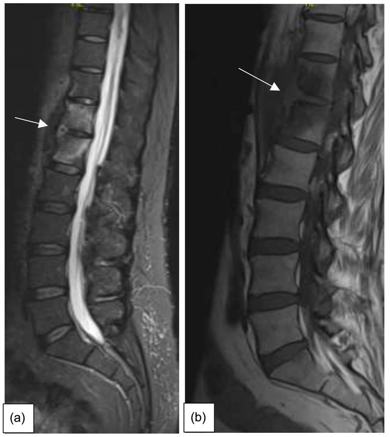
K. Condé et al.
symptomatology is misleading [2], its diagnosis is often delayed or even confused
with damage to the hip or lumbosacral hinge, most often related to difficulties
exploration of the sacroiliac joint [3] [4]. We report a case of tuberculous sacroi-
liitis secondary to an abscess of the psoas muscle.
2. Observation
Patient’s Consent
A 66-year-old patient with a history of blood hypertension and diabetes, hospi-
talized for lumbar-fessalgia, right unilateral inflammatory appearance, insidious
installation, evolving for about 8 months. He had been treated with Coversyl,
Metformin and Tramadol. He describes neither fever, nor the concept of conta-
gion, nor respiratory symptoms, or weight loss. On physical examination, the
patient was in good general condition. There was a pain on the pressure of the
spinous processes. The Schöber index and Lasegue’s sign were impossible due to
the pain. The sacroiliac pressure was painful, Ericksen’s and Volkmann’s ma-
noeuvres were positive. There was no lymphadenopathy. The remainder of the
physical examination was unremarkable.
The biological assessment found a normal blood count, a non-specific biolog-
ical inflammatory syndrome with an erythrocyte sedimentation rate (ESR) acce-
lerated to 86 mm at the 1st hour, a positive C-reactive protein (CRP) at 59.3
mg/l. The hepatic and renal function was normal. The HLA-B27 antigen was
immunologically negative. Blood cultures were positive on two occasions. The
human immunodeficiency virus (HIV) serology was negative.
The standard X-ray of the lumbar spine and pelvis was unremarkable; howev-
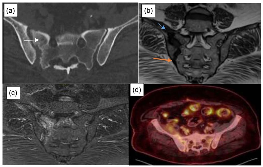
er, CT scan of the pelvis (Figure 1(a)) showed severe right unilateral sacroiliitis
with intra-auricular effusion (Forest stage III). Lumbar (Figure 2) and pelvis
Figure 1. (a) CT scan of the pelvis showing right sacroiliitis. Sacroiliac MRI ((b) sequence
T1 and (c) sequence T2) showing right sacroiliitis (white arrow) with intra-articular effu-
sion and infiltration of the psoas muscle (blue arrow); (d) PET scan showing heterogene-
ous hypermetabolism of the psoas muscle and the right sacroiliac joint (blue arrow).
DOI: 10.4236/ojra.2021.112006 49 Open Journal of Rheumatology and Autoimmune DiseasesK. Condé et al.
Figure 2. Lumbar MRI scan ((a) sequence T1 and (b) sequence T2) showing a D12-L1
spondylodiscitis with infiltration of the pre-vertebral space.
(Figure 1(b) and Figure 1(c)) MRI scar further revealed D12-L1 spondylodisci-
tis with infiltration of the pre-vertebral space and the right psoas muscle. The
PET scan noted heterogeneous hypermetabolism of the psoas muscle and the
right sacroiliac joint (Figure 1(d)). Drainage of the abscess and a fluoroscopic
biopsy of the right sacroiliac joint were positive for mycobacterium tuberculosis.
The chest X-ray was normal while the tuberculin intradermal reaction was posi-
tive at 15 mm. The diagnosis of tuberculous sacroiliitis secondary to a psoas ab-
scess was retained. Quadruple therapy combining isoniazid, rifampicin, pyrazi-
namide and ethambutol was started for 12 months and the abscess was surgically
drained. After a follow-up of 6 months, all the clinical symptoms regressed and
the laboratory parameters returned to normal.
3. Discussion
The incidence rate of tuberculosis continues to decline worldwide [3], but it re-
mains a major public health concern in developing countries due to the AIDS
pandemic [5]. Osteoarticular tuberculosis accounts for 3% of extrapulmonary
tuberculosis, of which 5% - 8% concerns the sacroiliac joint [1] [2] [6]. Tuber-
culous sacroiliitis secondary to psoas abscess is rare, only a few sporadic cases
have been reported in the literature [3] [5] [6]. A predisposing risk factor is
sometimes found HIV infection, postpartum, transplantation, ulcerative colitis,
prolonged corticosteroid therapy [7]. In our case, the only risk factor found was
diabetes.
DOI: 10.4236/ojra.2021.112006 50 Open Journal of Rheumatology and Autoimmune DiseasesK. Condé et al.
In Africa these are short series and occasional observations that have been re-
ported as in the Maghreb especially in Tunisia or Bouajina et al. [8] reported 22
cases and tuberculous sacroiliitis as well as in Sub-Saharan Africa, in Burkina
Fasso [5] have reported 2 cases of tuberculous sacroiliitis. However, there was no
associated psoas abscess as reported in observation.
Tuberculous sacroiliitis is insidious, the time between the onset of clinical
signs and hospitalization is long (approximatively 2 weeks to 6 months) [9]. It is
characterized by the predominance of local symptoms as demonstrated in this
case. The patient had no systemic symptoms and pain is the most common
complaint. The diagnosis of osteoarticular tuberculosis is based on the culture of
biopsy material, sputum is negative except in the very rare cases with concomi-
tant pulmonary tuberculosis were reported [3] [9]. The main species identified is
mycobacterium tuberculosis [3].
The radiological lesions of sacroiliitis are very progressive onset. Tuberculous
arthritis is very destructive, most often giving images of erosion and geodes [9].
CT scan appears to be the test of choice for the detection of sequesters, while
MRI is more sensitive for the detection of early cancellous bone abnormalities as
well as abscesses regardless of their size [10].
The introduction of anti-tuberculosis chemotherapy resulted in a dramatic
improvement in the prognosis. Psoas abscess is treated with antibiotic therapy
combined with drainage [11]. However, tuberculosis abscesses often heal with
anti-tuberculosis treatments without drainage [2] [11]. The duration of treat-
ment for osteoarticular tuberculosis should not be less than 12 months or even
extended to 18 months [12].
4. Conclusion
Tuberculous sacroiliitis is rare and its clinical presentation is atypical. The diag-
nosis must be made in the event of low back pain and/or pain in the hip. Our
case confirms the rarity and the non-specific nature of the clinical manifesta-
tions of tuberculous sacroiliitis, generally leading to a delay in diagnosis. The CT
scan, MRI and biopsy are necessary for diagnosis.
Conflicts of Interest
The authors declare that they have no conflicts of interest.
References
[1] Sahu, R. (2011) Turner Syndrome with Tubercular Osteomyelitis of Iliac Bone: An
Unusual Presentation. Indian Health Journal.
https://www.researchgate.net/publication/261216229_TUBERCULOSE_DE_L%27
OS_ILIAQUE_UNE_LOCALISATION_INHABITUELLE
[2] Prakash, J. (2014) Sacroiliac Tuberculosis—A Neglected Differential in Refractory
Low Back Pain—Our Series of 35 Patients. Journal of Clinical Orthopaedics and
Trauma, 5, 146-153. https://doi.org/10.1016/j.jcot.2014.07.008
[3] Kramer, L., Geib, V., Evison, J., Altpeter, E., Basedow, J. and Brügger, J. (2018) Tu-
DOI: 10.4236/ojra.2021.112006 51 Open Journal of Rheumatology and Autoimmune DiseasesK. Condé et al.
berculous Sacroiliitis with Secondary Psoas Abscess in an Older Patient: A Case
Report. Journal of Medical Case Reports, 18, 237.
https://doi.org/10.1186/s13256-018-1754-4
[4] Özdemir, Z.M., Kahraman, A.S., Görmeli, C.A., Sevimli, R. and Akpolat, N. (2016)
Langerhans Cell Histiocytosis with Atypical Intervertebral Disc and Sacroiliac Joint
Involvement Mimicking Osteoarticular Tuberculosis in an Adult. Balkan Medical
Journal, 33, 573-577. https://doi.org/10.5152/balkanmedj.2016.160492
[5] Diallo, I., Zabsonré, J.T., Tiemtoré Kambou, B.M.A., Sondo, A.K., Sagna, Y. and
Ouédraogo, D.D. (2016) Sacroillite Tuberculeuse: A Propos de deux cas. Pan
African Medical Journal, 25, 69.
[6] Shields, D.W. and Robinson, P.G. (2012) Iliopsoas Abscess Masquerading as ‘Scia-
tica’. BMJ Case Reports, 2012. https://doi.org/10.1136/bcr-2012-007419
[7] Nair, K.R. and Jayachandran, R. (2013) Postpartum Tuberculous Sacroiliitis. Amer-
ican Journal of Orthopedics (Belle Mead, N.J.), 42, 16-17.
[8] Bouajina, E., Harzallah, L., Hachfi, W., Bel Hadj Slama, K., Rammeh, N., Ghan-
nouchi, M., Bahri, M. and Letaief, A. (2005) Sacro-Iliites Tuberculeuses : A propos
de 22 cas. La revue de médecine interne, 26, 690-694.
[9] Ali Chaudhry, L. and Al-Solaiman, S. (2013) Multifocal Tuberculosis: Many Faces
of an Old Menace. The International Journal of Mycobacteriology, 2, 58-69.
https://doi.org/10.1016/j.ijmyco.2013.01.001
[10] Albano, D., Treglia, G., Desenzani, P. and Bertagna, F. (2017) Incidental Unilateral
Tuberculous Sacroiliitis Detected by 18F-FDG PET/CT in a Patient with Abdominal
Tuberculosis. Asia Oceania Journal of Nuclear Medicine and Biology, 5, 144-147.
[11] Navarro López, V. (2015) Worldwide Variations over the Years in Etiology of
Iliopsoas Abscess. Reality or a Selection Bias? Medicina Clínica (Barc), 144,
259-260.
[12] WHO (2010) Treatment of Tuberculosis: Guidelines.
http://www.who.int/tb/publications/2010/9789241547833/en/
DOI: 10.4236/ojra.2021.112006 52 Open Journal of Rheumatology and Autoimmune DiseasesYou can also read