HALF-CUTTING METHOD during Hysterectomy for Large Uterine Cervical Myoma
←
→
Page content transcription
If your browser does not render page correctly, please read the page content below
Open Journal of Obstetrics and Gynecology, 2021, 11, 1196-1201
                                                                                                  https://www.scirp.org/journal/ojog
                                                                                                            ISSN Online: 2160-8806
                                                                                                              ISSN Print: 2160-8792

HALF-CUTTING METHOD during Hysterectomy
for Large Uterine Cervical Myoma

Yosuke Fukui1, Yuki Yamada1, Seiji Mabuchi2

Department of Obstetrics and Gynecology, Nara Medical University, Kashihara, Japan
1

Department of Gynecology, Osaka International Cancer Institute, Osaka, Japan
2

How to cite this paper: Fukui, Y., Yamada,      Abstract
Y. and Mabuchi, S. (2021) HALF-CUTTING
METHOD during Hysterectomy for Large            Hysterectomy for large uterine cervical myoma is a challenging surgical pro-
Uterine Cervical Myoma. Open Journal of         cedure due to the limited operative field for lateral and posterior dissections.
Obstetrics and Gynecology, 11, 1196-1201.       Existing procedures such as performing myomectomy before hysterectomy or
https://doi.org/10.4236/ojog.2021.119113
                                                performing retrograde hysterectomy remain suboptimal in expanding the
Received: August 21, 2021                       operative field, especially in cases with a huge cervical myoma. In this report,
Accepted: September 14, 2021                    we introduce a new procedure, the “HALF-CUTTING METHOD” which can
Published: September 17, 2021                   be used to obtain an adequate surgical field during hysterectomy.

Copyright © 2021 by author(s) and
Scientific Research Publishing Inc.
                                                Keywords
This work is licensed under the Creative        HALF-CUTTING METHOD, Cervical Myoma, Hysterectomy, Surgical Field
Commons Attribution International
License (CC BY 4.0).
http://creativecommons.org/licenses/by/4.0/
               Open Access
                                              1. Introduction
                                              Uterine leiomyomas are the most common pelvic tumors in women, with a pre-
                                              valence of 20% - 40% after the age of 35 years [1]. Of these cases, 95% occur in
                                              the uterine corpus while a frequency of only 0.6% is reported for cervical lei-
                                              omyomas [2].
                                                Cervical myomas differ from their corpus counterparts, where symptoms of
                                              abnormal uterine bleeding predominate. Due to the lack of menstrual symp-
                                              toms, cervical myomas usually grow to large sizes before detection. The size of
                                              cervical myoma in previous reports ranges from 5 - 24 cm [3].
                                                Surgery remains the mainstay of treatment for most leiomyomas. Indications
                                              for surgical intervention include tumor-related bulk symptoms, abnormal ute-
                                              rine bleeding, recurrent pregnancy loss, and infertility. In postmenopausal
                                              women or premenopausal women who are not desirous of preserving their fer-
                                              tility, hysterectomy is the standard treatment as it eliminates both the symptoms
                                              and the possibility of future recurrence.

DOI: 10.4236/ojog.2021.119113 Sep. 17, 2021                       1196                    Open Journal of Obstetrics and Gynecology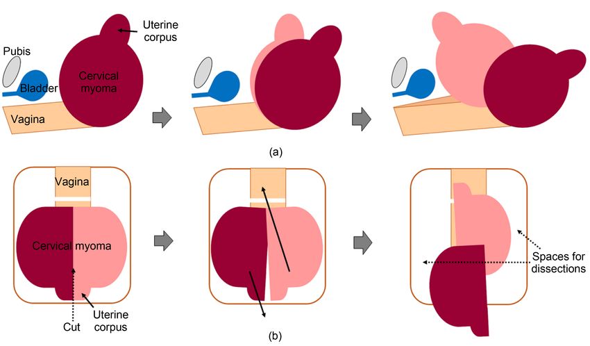
Y. Fukui et al.
Hysterectomy is challenging to perform in cases with a large cervical myoma
due to the limited operative field for the complete incision of the parametrium.
For such cases, previous reports have suggested performing myomectomy before
hysterectomy or performing retrograde hysterectomy to expand the operative
field; however, these procedures are far from optimal. Thus, it is important to
develop new methods to supplant these procedures when they are inefficient or
unsuccessful.
In this report, we introduce a new procedure called the “HALF-CUTTING
METHOD” which can be used to obtain an optimal surgical field for lateral and
posterior dissections during hysterectomy for large cervical myoma.
2. Surgical Procedures
Total abdominal hysterectomy using the HALF-CUTTING METHOD can be
safely performed according to the method described below.
1) Perform the laparotomy, identify the major uterine ligaments, and check
the mobility of the uterus.
2) Incise the round ligaments and the peritoneum of the vesicouterine pouch,
and dissect the bilateral pelvic sidewall triangles, which are surrounded by the
round ligament, infundibulopelvic ligament, and posterior lobe of the broad li-
gament.
3) Incise the ovarian ligaments and fallopian tubes (or infundibulopelvic li-
gaments).
4) After the development of paravesical spaces, identify the uterine artery,
which can be traced from the internal iliac artery, and incise it at its origin.
5) Push the bladder down from the anterior wall of the cervix and vagina as
much as possible, and identify the anterior vaginal fornix by manual examina-
tion in the vaginal canal, then incise the anterior vaginal wall. In cases where the
posterior vaginal fornix was visible, circumferential dissection of the vagina is
done while carefully avoiding rectal injury. By performing stapes 3 - 6, the ute-
rine blood flow can be significantly reduced.
6) The uterus is then cut in half from the fundus to the cervix (cervical myo-
ma). Crossing the half-cut uterus reduces the width of the uterus (Figure 1 and
Figure 2) and allows the surgeon to obtain the optimal surgical field for lateral
and posterior dissections.
7) Identify and isolate the ureters from the posterior leaf of the broad ligament
to the ureteral tunnel. Incise the posterior leaf of the broad ligaments and utero-
sacral ligaments. By changing the position of the half-cut uterus, the parame-
trium can be fully exposed, coagulated and incised.
8) In cases wherein circumferential dissection of the vagina could not be per-
formed in step 6, colpotomy is completed by cutting the posterior vaginal wall.
Then excise the remaining connective tissues surrounding the vagina and re-
move the uterus.
9) Suture the vaginal vault.
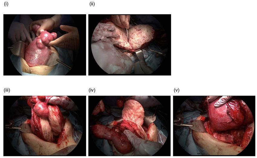
DOI: 10.4236/ojog.2021.119113 1197 Open Journal of Obstetrics and GynecologyY. Fukui et al. Figure 1. Schematic diagram of the HALF-CUTTING METHOD. (a) Longitudinal view, (b) Frontal view. Figure 2. Hysterectomy using the HALF-CUTTING METHOD, shown in representative photos. (i), large cervical myoma occu- pying surgical field. (ii)-(iii), the uterus was cut in half from the fundus to the cervix. (iv)-(v), the uterine width was reduced by crossing the half-cut uterus, and optimal surgical field for lateral and posterior dissections was obtained. DOI: 10.4236/ojog.2021.119113 1198 Open Journal of Obstetrics and Gynecology
Y. Fukui et al.

                                3. Case Presentation
                                A 49-year-old nulliparous (G0P0) Japanese woman presented with abdominal
                                distension. Cytology of the cervix was negative for intraepithelial lesions of ma-
                                lignancy (NILM). MRI revealed an 18 by 15 by 11 cm leiomyoma in the cervix
                                and 4 cm and 5 cm leiomyomas in the uterine corpus. Her past surgical and
                                medical histories were unremarkable. After discussing treatment options, a total
                                abdominal hysterectomy and bilateral salpingectomy were performed. During
                                hysterectomy, an enlarged uterine cervix obstructed the surgical field for lateral
                                and posterior dissections; thus, the HALF-CUTTING METHOD was employed
                                (Figure 2). The procedure lasted for 219 minutes, with intraoperative blood loss
                                of 500 mL. The weight of the removed uterus was 2454 g and pathological analy-
                                sis confirmed leiomyoma. The postoperative course was uneventful, and the pa-
                                tient was discharged on postoperative day 8 without complications.

                                4. Discussion
                                In this report, we introduce a new procedure, termed “HALF-CUTTING
                                METHOD” which can be used during hysterectomy for large cervical myoma.
                                  Large cervical myomas pose challenges during hysterectomy, as the size adds
                                obstruction in the surgical field and complicates lateral or posterior dissections.
                                To our knowledge, two different options have been proposed to safely complete
                                hysterectomy for cervical myoma, namely, performing myomectomy before
                                hysterectomy or performing retrograde hysterectomy [4] [5]. Although these
                                procedures can help expand the operative field in some cases, myomectomy be-
                                fore lateral dissection can cause massive bleeding in cases involving an extremely
                                large cervical myoma. Moreover, performing retrograde hysterectomy often
                                does not sufficiently expand the field for the visualization of ureters, thereby
                                impeding the safe incision of the parametrium, uterosacral ligaments, and the
                                posterior leaf of broad ligaments.
                                  We believe that the use of the HALF-CUTTING METHOD has important
                                clinical implications. Firstly, this method does not require additional surgical
                                devices. Secondly, it can be performed even in cases wherein the ureters are un-
                                identifiable behind the huge cervical myoma. This method requires only the
                                identification of the uterine artery origin from the internal iliac artery and of the
                                anterior vaginal fornix, which are not difficult to achieve. The uterine artery can
                                be identified by tracing the internal iliac artery from paravesical spaces, which
                                easily develop even in cases with large cervical myoma. Additionally, the ante-
                                rior vaginal fornix can easily be identified at the lower end of the cervical
                                myoma, which causes the stretching and upward lift of the vaginal fornixes;
                                hence, cutting the anterior vaginal wall can be safely performed. Thirdly, per-
                                forming this procedure for cervical myoma significantly reduces the width of
                                the uterus by up to 50%, which provides enough space for the identification of
                                ureters and subsequent safe dissection of the posterior and lateral uterine
                                components.

DOI: 10.4236/ojog.2021.119113                       1199                    Open Journal of Obstetrics and Gynecology
Y. Fukui et al.
Although no complications were observed in our cases, we recognize some
potential risks related to the use of the HALF-CUTTING METHOD. Hemorr-
hage may occur from a vaginal wall that was incised before the completion of
lateral and posterior dissections. However, such vaginal wall bleeding is usually
mild, as demonstrated in previous studies on retrograde hysterectomy [6]. There
is another risk for hemorrhage from the half-cut uterine corpus and cervical
myoma. This may be minimized by blocking the blood flow through ligation of
the uterine and ovarian arteries. The blood flow from the vagina can also be sig-
nificantly blocked by cutting the anterior vaginal wall or both the anterior and
posterior vaginal walls; thus, serious hemorrhage from the half-cut uterine cor-
pus and cervical myoma rarely occur.
In conclusion, we developed a new procedure, the “HALF-CUTTING MET-
HOD” which can be employed during hysterectomy for large cervical myoma.
We believe that this procedure enables surgeons to obtain an optimal surgical
field and eliminate the possibility of ureteral injury or massive hemorrhage. This
preliminary but promising result justifies further investigation of this procedure
in larger prospective studies.
Ethical Approval Status
The Ethics Committee of Nara Medical University decided that this case report
does not require IRB approval due to the nature of this study: a report describing
the treatment of a single patient and thus does not meet the definition of human
subjects research.
Conflicts of Interest
The authors declare no conflicts of interest.
References
[1] Laganà, A.S., Alonso Pacheco, L., Tinelli, A., Haimovich, S., Carugno, J., Ghezzi, F.,
Mazzon, I. and Bettocchi, S. (2019) Management of Asymptomatic Submucous
Myomas in Women of Reproductive Age: A Consensus Statement from the Global
Congress on Hysteroscopy Scientific Committee. Journal of Minimally Invasive
Gynecology, 26, 381-383. https://doi.org/10.1016/j.jmig.2018.06.020
[2] Tiltman, A.J. (1998) Leiomyomas of the Uterine Cervix: A Study of Frequency. In-
ternational Journal of Gynecological Pathology, 17, 231-234.
https://doi.org/10.1097/00004347-199807000-00006
[3] Wong, J., Tan, G.H.C., Nadarajah, R. and Teo, M. (2017) Novel Management of
a Giant Cervical Myoma in a Premenopausal Patient. BMJ Case Reports, 2017,
bcr2017221408. https://doi.org/10.1136/bcr-2017-221408
https://casereports.bmj.com/content/casereports/2017/bcr-2017-221408.full.pdf
[4] Hiramatsu, Y. (2019) Hysterectomy for Cervical and Intraligamental Fibroids. Sur-
gery Journal, 6, 2-10. https://doi.org/10.1055/s-0039-1698419
[5] Ishidera, Y., Furugori, M., Hirata, G., Wakabayashi, R., Shigeta, H. and Yoshida, H.
(2021) Total Laparoscopic Hysterectomy for Anterior Cervical Myoma: Possible
Significance of Presurgical Assessment by Magnetic Resonance Imaging. Gynecol-
DOI: 10.4236/ojog.2021.119113 1200 Open Journal of Obstetrics and GynecologyY. Fukui et al.
ogy and Minimally Invasive Therapy, 10, 61-64.
[6] Hiramatsu, Y. (2019) Retrograde Abdominal Hysterectomy. Surgery Journal, 5,
27-32. https://doi.org/10.1055/s-0039-1683919
DOI: 10.4236/ojog.2021.119113 1201 Open Journal of Obstetrics and GynecologyYou can also read