Two Subsequent Metachroneus Solid Tumors: Oncocytic Variant Adrenocortical Carcinoma and Rhabdomyosarcoma of Childhood: Case Report and Literature ...
←
→
Page content transcription
If your browser does not render page correctly, please read the page content below
CASE REPORT DOI: 10.4274/jcrpe.galenos.2020.2020.0060
J Clin Res Pediatr Endocrinol 2021;13(2):225-231
Two Subsequent Metachroneus Solid Tumors: Oncocytic Variant
Adrenocortical Carcinoma and Rhabdomyosarcoma of Childhood:
Case Report and Literature Review
Onur Akın, Erman Ataş, İrem Ayşe Atasoy, Nihal Durmaz, Ömer Kartal
University of Health Sciences Turkey, Gülhane Training and Research Hospital, Clinic of Pediatrics, Ankara, Turkey
What is already known on this topic?
Adrenocortical carcinoma can be related to other cancers.
What this study adds?
Sequence analysis should be performed if fluorescence in situ hybridization analysis is negative. Analysis of Hounsfield unit density of
the tumor on imaging can help to distinguish malign from benign. The Lin-Weiss-Bisceglia system should be used instead of the Weiss
system for the oncocytic variant.
Abstract
Most cases of malignancies appear to be sporadic, but some syndromes are associated with malignancies with germline variants.
Herein, a child with an unusual association of oncocytic variant adrenocortical carcinoma (ACC) and rhabdomyosarcoma (RMS) was
presented. An 18-month-old-boy was admitted with virilization of the genital area, penis enlargement and erection, which had begun six
months earlier. Serum total testosterone (457 ng/dL; NR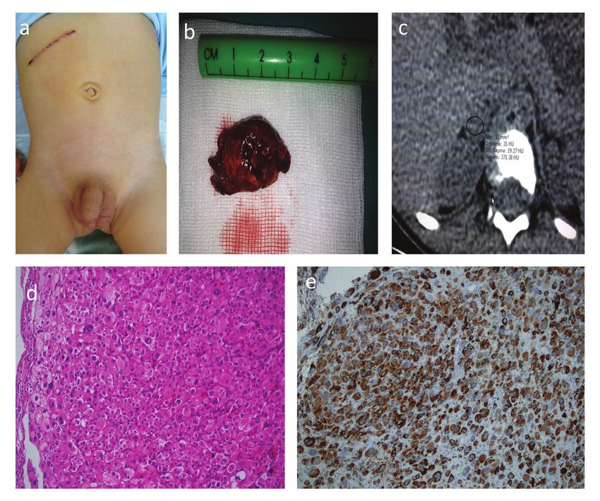
Akın O et al.                                                                                         J Clin Res Pediatr Endocrinol
Two Subsequent Metachroneus Solid Tumors                                                                       2021;13(2):225-231

significant role in the development of cancer (6). Breast         (NR
J Clin Res Pediatr Endocrinol                                                                                             Akın O et al.
2021;13(2):225-231                                                                            Two Subsequent Metachroneus Solid Tumors

Figure 1. a) Enlargement of phallus, stage 2 pubic hair development. b) Abdominal computed tomography showing right adrenocortical
carcinoma. c) The mass after adrenelectomy. d) Oncocytic adrenocortical carcinoma, oxyphilic cell population, hematoxylin-
eosin, immunohistochemical expression, (magnification x200). e) Oncocytic adrenocortical carcinoma, Melan-A (MART-1)
immunohistochemical expression, (magnification x200)

Evaluation After Relapse                                           Table 1. Abnormal hormanal levels changes of patient
                                                                   before and after operation
Three months after adrenalectomy, the child represented
                                                                                       Preoperative Postoperative Postoperative
with swelling of the left leg with a few weeks of history.                                          12th hour     24th hour
A painless, immobile, rough, 6x8 cm swelling between the
                                                                   Total testosteron 457
Akın O et al. J Clin Res Pediatr Endocrinol
Two Subsequent Metachroneus Solid Tumors 2021;13(2):225-231
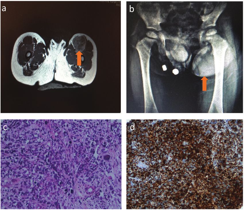
Figure 2. a) Femoral magnetic resonance imaging showing left side rhabdomyosarcoma. B) X-ray graphy showing left femoral mass.
c) Rhabdomyosarcoma, hematoxylin-eosin, (magnification x400). d) Rhabdomyosarcoma, Desmin positivity, (magnification x200)
Treatment with ifosfamide, carboplatin, etoposide (ICE) regimen. After
The patient was treated with chemotherapy consisting of three cycles, he was in remission. Radiotherapy was given
vincristine, cyclophosphamide, and actinomycin-D according for local control of RMS. After the sixth cycle of ICE, the
to the protocol POG D-9602-VAC. Revision of pathological patient was in good condition with no tumor recurrence,
and treatment was stopped. However, he came back six
samples was performed again. Sarcomatous component of
months later with left leg pain due to a mass and the mass-
the oncocytic variant ACC was not found. The patient was
effect on the femoral artery and nerve. Thrombosis and
evaluated ten weeks after starting the VAC regimen, and he
malign mesenchymal tumour protocol and sorafenib were
was in remission at both the ACC and RMS sites. His disease
started. Surgeons recommended amputation due to nerve
was discussed with the radiotherapy department for local
and lymphatic invasion of the tumor. However, the patient
therapy of extremity RMS. Radiation oncology refused to
family refused the amputation. He died, due to progressive
give radiation therapy owing to remission of disease and
disease, 2.5 years after diagnosis.
because the patient was aged under three years. At the 24th
week of treatment, a 2x2 cm sized, inguinal mass in the
left sartorius muscle, which was enhanced after injection Discussion
of contrast agent, and compatible with lymphadenopathy Although most cases of malignancies appear to be sporadic,
was detected with MRI. The mass was excised, and the some syndromes that are associated with malignancy or
pathological result was compatible with RMD metastasis. malignancies can be detected in oncology practice. Even if
The patient was evaluated as relapse and progression under an association is not found, unusual associated malignancies
chemotherapy treatment, and the treatment was changed can be evaluated for germline variants. In this study, we
228J Clin Res Pediatr Endocrinol Akın O et al.
2021;13(2):225-231 Two Subsequent Metachroneus Solid Tumors
aimed to describe an association of cancers with no family to this, a sarcomatous component may be present (1).
history in a child with oncocytic variant ACCs and RMS with RMS as a second cancer developed in the follow-up. RMS
the help of genetic evaluation. represents 6.5% of childhood cancers, and 52.9% of soft
Patients with LFS, BWS, and multiple endocrine neoplasia tissue sarcomas (3). Somatic variants of the TP53 gene can
type 1 (MEN-1) are at risk for developing certain types of be seen in as many as 50% of cases. However, germline
cancers, such as ACC and RMS (9). LFS especially is associated variants are much less common and tend to be associated
with a 40% risk of malignancy before the age of 16 years, with a lower age (average age 22 months) at presentation
high mortality rates, and second primary malignancies, and (18,19). Although this case was hormonally active, the
is an autosomal disorder (10,11). TP53 tumor suppressor sarcomatous component was not found.
gene on chromosome 17p13 in LFS, GNAS 1 variant and There is no definitive pattern on CT scan or MRI (4). On the
abnormalities of 11p15.5 in BWS and variants of the MEN- CT Hounsfield Scale, adenoma/hyperplasia and carcinoma
1 gene on chromosome 11q13 in MEN-1 may be detected are assigned a value of 16.2±13.6 and 36.9±4.1,
in some patients with malignancy (12,13,14,15). Six per respectively (20). Specificity and sensitivity of PET-CT are
cent of patients with second malignancies and no familial >95% (21). The mass was compatible with non-adenoma
features of LFS had a germline TP53 variant in a sample of owing to a Hounsfield Scale value of 35 HU. PET-CT was
59 patients (16). Germline TP53 variants with no familial performed after surgery. Evaluation of disease with PET-CT
features of LFS are identified in 50-80% of children with ACC was normal in staging workup.
and 10% with RMS (17,18). A variant related to malignancy
may not be detected, as was the case in our patient. In Clinicopathological, oncocytic variant ACC differs from
this study, an extensive pedigree was obtained. No family conventional ACC. There is no preference for males or
history of any other malignancies was documented in this females. It is smaller and lighter than conventional ACC and
family. Our patient did not have clinical features of BWS and tends to hold the left side. The oncocytic variant has rare
McCune-Albright syndrome. Thus, we decided to evaluate mitoses including no atypia, low rate of necrosis, fibrosis,
genetically because of sporadic malignancy. and venous, sinusoidal, and capsular invasion (22). These
features were mostly compatible with the pathology and
ACC is an extremely rare tumour. It accounts for 0.2%
clinical features in our case, except the high mitotic rate and
of childhood cancers (12). In our country, ACC accounts
involvement of the right side.
for 6.9% of carcinomas and other malignant epithelial
tumours, and 0.19% of childhood cancer (3). Although Proposed major criteria including a mitotic rate of more
most cases of ACCs appear to be sporadic, some have been than five mitoses per 50 high power fields, any atypical
described as a component of several hereditary cancer mitoses or venous invasion, and minor criteria including
syndromes (9). Virilization can be seen owing to increased large size/weight (>10 cm and >200 g), necrosis, capsular
DHEA and DHEA-SO4 production (13). In this case, DHEA- invasion or sinusoidal invasion, have been investigated for
SO4, androstenedione, and testosterone were all above distinguishing malignant tumors by Bisceglia et al (23).
the reference range contemporary with virilization. The Defining criteria for oncocytic tumors have been outlined
absence of hormonal hyperactivity is associated with poor that include predominantly cells with eosinophilic and
prognosis because of the advanced stage of the tumor at granular cytoplasm, a high nuclear grade, and a diffuse
diagnosis (14). The hormonally active status of this case was architectural pattern. The presence of one major criterion
very significant in early diagnosis together with the evident indicates malignancy, 1-4 minor criteria indicating uncertain
virilization. The levels of total testosterone, androstenedione, malignant potential (borderline) and the absence of all major
DHEA-SO4 were highly elevated at the time of diagnosis. and minor criteria are indicative of a benign mass. In the
After surgery, the levels of these hormones decreased to an case presented the mitotic rate was more than five mitoses
average level in the 24th hour. The survival rates of stage per 50 high power fields. When mitosis was evaluated with
1 are higher than others (15). Our patient was followed- PHH3, 20 mitoses were counted per 50 high power fields.
up without treatment after surgery. The primary site was Venous, capsular, and sinusoidal invasions and necrosis
normal in the follow-up. were not detected. Thus the ACC tumor in our patient met
Oncocytic variant ACC is a rare disease with low incidence. all defining criteria. The massAkın O et al. J Clin Res Pediatr Endocrinol
Two Subsequent Metachroneus Solid Tumors 2021;13(2):225-231
The molecular pathogenesis of sporadic ACCs is less well with the absence of a positive family history of malignancies
understood. Activation of proto-oncogenes and oncogenes strongly suggests a problem in tumor suppressor genes or
on chromosome 4, 5, and 12, and inactivation of tumor new variants. The present case with an initial ACC tumor and
suppressor genes on chromosome arms 1p and 17p may a subsequent RMS tumor had a TP53 variant and inactivated
be related to progression from adenoma to carcinoma (25). TP53 is present in about half of all human cancers. Whole
Loss of heterozygosity (LOH) at 17p13 is common, but genome analyses will provide important information on the
only about one-third of these tumors are associated with development of ACCs and secondary cancer in the future.
a variant of TP53. However, TP53 might not be the only
or major tumor suppressor gene at 17p related to ACCs. Ethics
Another suppressor gene, which is as yet unidentified, may Informed Consent: Informed consent was obtained from
be present and effective in this locus and there is evidence the parents of the patients.
to support this hypothesis (26). ACCs are associated with Peer-review: Externally peer-reviewed.
multiple somatic gene alterations and thus it is difficult to
identify the exact genetic changes (27). Amplification of the Authorship Contributions
steroidogenic factor-1 gene as well as germline TP53 variant Surgical and Medical Practices: Erman Ataş, İrem Ayşe
in Southern Brazil, LOH of 11p15 with overexpression of Atasoy, Nihal Durmaz, Concept: Onur Akın, Erman Ataş,
insulin-like growth factor-2 as well as other growth-related Ömer Kartal, Design: Erman Ataş, Nihal Durmaz, Data
tumor suppressor genes at this locus may explain this Collection or Processing: Erman Ataş, İrem Ayşe Atasoy,
(28,29). Analysis or Interpretation: Onur Akın, Erman Ataş, Nihal
Durmaz, Literature Search: Erman Ataş, İrem Ayşe Atasoy,
Study Limitations Ömer Kartal, Writing: Onur Akın, Erman Ataş, İrem Ayşe
Unfortunately, there was only resource enough to investigate Atasoy.
TP53 in this case, which is the limitation of our study. Financial Disclosure: The authors declared that this study
However, the TP53 variant was positive in sequence analysis. received no financial support.
Use of imaging techniques that use ionizing radiation, such
as PET and CT scans, during the follow-up of this small child References
at risk of other malignancies was inadvisable. Therefore, MRI
1. Thway K, Olmos D, Shah C, Flora R, Shipley J, Fisher C. Oncocytic adrenal
and abdominal USG were used to avoid repeated irradiation cortical carcinosarcoma with pleomorphic rhabdomyosarcomatous
after the second malignancy. External radiotherapy to the metastases. Am J Surg Pathol 2012;36:470-477.
leg after 10-weeks from the beginning of chemotherapy 2. Bilimoria KY, Shen WT, Elaraj D, Bentrem DJ, Winchester DJ, Kebebew
was not given by the radiotherapy department owing to E, Sturgeon C. Adrenocortical carcinoma in the United States: treatment
utilization and prognostic factors. Cancer 2008;113:3130-3136.
remission of the disease. However, it was agreed to provide
3. Kutluk MT, Yeşilipek A. Turkish National Pediatric Cancer Registry
radiotherapy after the recurrence of the disease for local 2002-2008 (Turkish Pediatric Oncology Group and Turkish Pediatric
control. Hematology Society). J Clin Oncol 2013;31(Suppl 15)10067.
The occurrence of ACC as a part of a syndrome is clinically 4. Mearini L, Del Sordo R, Costantini E, Nunzi E, Porena M. Adrenal
oncocytic neoplasm: a systematic review. Urol Int 2013;91:125-133.
significant because of the choice of treatment, caution with Epub 2012 Nov 8
radiotherapy in patients with LFS, individualized screening 5. Lodish MB, Stratakis CA. Rare and unusual endocrine cancer syndromes
for other cancers in these syndromes with mammography, with mutated genes. Semin Oncol 2010;37:680-690.
colonoscopy, and identification family members at risk (30). 6. Figueiredo BC, Stratakis CA, Sandrini R, DeLacerda L, Pianovsky
MA, Giatzakis C, Young HM, Haddad BR. Comparative genomic
hybridization analysis of adrenocortical tumors of childhood. J Clin
Conclusion Endocrinol Metab 1999;84:1116-1121.
7. Sandrini R, Ribeiro RC, DeLacerda L. Childhood adrenocortical tumors.
A multidisciplinary team approach, including oncology, J Clin Endocrinol Metab 1997;7:2027-2031.
surgery, endocrinology, pathology, radiation oncology, and
8. Ferrari A, Bisogno G, De Salvo GL, Indolfi P, Perilongo G, Cecchetto G;
genetic counselling is necessary. The cost-effectiveness Italian Study on Rare Tumours in Paediatric Age (TREP); Associazione
of cancer screening with colonoscopy is not considered Italiana Ematologia Oncologia Pediatrica (AIEOP). The challenge of
very rare tumours in childhood: the Italian TREP project. Eur J Cancer
controversial for well-defined common cancers such as
2007;43:654-659. Epub 2006 Oct 16
colon cancer, but no data are available for ACCs. Genetic
9. Koch CA, Pacak K, Chrousos GP. The molecular pathogenesis of
evaluation should be suggested for patients with a second hereditary and sporadic adrenocortical and adrenomedullary tumors. J
primary cancer. An unusual association of malignancies Clin Endocrinol Metab 2002;87:5367-5384.
230J Clin Res Pediatr Endocrinol Akın O et al.
2021;13(2):225-231 Two Subsequent Metachroneus Solid Tumors
10. Li FP, Fraumeni JF. Prospective study of a family cancer syndrome. 22. Song SY, Park S, Kim SR, Suh YL. Oncocytic adrenocortical carcinomas:
JAMA 1982;247:2692-2694. a pathological and immunohistochemical study of four cases in
11. Eng C, Schneider K, Fraumeni JF, Li FP. Third international workshop comparison with conventional adrenocortical carcinomas. Pathol Int
on collaborative interdisciplinary studies of p53 and other predisposing 2004;54:603-610.
genes in Li-Fraumeni syndrome. Cancer Epidemiol Biomarkers Prev 23. Bisceglia M, Ludovico O, Di Mattia A, Ben-Dor D, Sandbank J, Pasquinelli
1997;6:379-383. G, Lau SK, Weiss LM. Adrenocortical oncocytic tumors: report of 10
12. Young JL, Miller RW. Incidence of malignant tumors in US children. J cases and review of the literature. Int J Surg Pathol 2004;12:231-243.
Pediatr 1975;86:254-258.
24. Michalkiewicz E, Sandrini R, Figueiredo B, Miranda EC, Caran E,
13. Del Gaudio AD, Del Gaudio GA. Virilizing adrenocortical tumors in Oliveira-Filho AG, Marques R, Pianovski MA, Lacerda L, Cristofani
adult women. Report of 10 patients, 2 of whom each had a tumor LM, Jenkins J, Rodriguez-Galindo C, Ribeiro RC. Clinical and outcome
secreting only testosterone. Cancer 1993;72:1997-2003.
characteristics of children with adrenocortical tumors: a report from
14. Neblett WW, Frexes-Steed M, Scott HW Jr. Experience with the International Pediatric Adrenocortical Tumor Registry. J Clin Oncol
adrenocortical neoplasms in childhood. Am Surg 1987;53:117-125. 2004;22:838-845.
15. Dong D, Li H, Yan W, Ji Z, Mao Q. Surgical management and clinical 25. Sidhu S, Marsh DJ, Theodosopoulos G, Philips J, Bambach CP, Campbell
prognosis of adrenocortical carcinoma. 2012;88:400-404. Epub 2012
P, Magarey CJ, Russell CF, Schulte KM, Röher HD, Delbridge L, Robinson
Apr 5
BG. Comparative genomic hybridization analysis of adrenocortical
16. Kakimoto S, Yushita Y, Sanefuji T, Kondo A, Fujishima N, Kishikawa tumors. J Clin Endocrinol Metab 2002;87:3467-3474.
M, Matsumoto K. Non-hormonal adrenocortical adenoma with
oncocytoma-like appearances. Hinyokika Kiyo 1986;32:757-763. 26. Libè R, Groussin L, Tissier F, Elie C, René-Corail F, Fratticci A, Jullian
E, Beck-Peccoz P, Bertagna X, Gicquel C, Bertherat J. Somatic TP53
17. de Krijger RR, Papathomas TG. Adrenocortical neoplasia: evolving
mutations are relatively rare among adrenocortical cancers with the
concepts in tumorigenesis with an emphasis on adrenal cortical
carcinoma variants. Virchows Arch 2012;460:9-18. Epub 2011 Nov 16 frequent 17p13 loss of heterozygosity. Clin Cancer Res 2007;13:844-
850.
18. Felix CA, Kappel CC, Mitusdomi T, Nau NM, Tsokos M, Crouch GD, Nisen
PD, Winick NJ, Helman LJ. Frequency and diversity of p53 mutations 27. Koch CA, Pacak K, Chrousos GP. The molecular pathogenesis of
in childhood rhabdomyosarcoma. Cancer Res 1992;52:2243-2247. hereditary and sopradic adrenocortical and adrenomedullary Tumors.
19. Diller L, Sexsmith E, Gottlieb A, Li FP, Malkin D. Germline p53mutations J Clin Endocrinol Metab 2002;87:5367-5384.
are frequently deleted in young children with rhabdomyosarcoma. J 28. Figueiredo BC, Cavalli LR, Pianovski MA, Lalli E, Sandrini R, Ribeiro RC,
Clin Invest 1995;95:1606-1611. Zambetti G, DeLacerda L, Rodrigues GA, Haddad BR. Amplification of
20. Hamrahian AH, Ioachimescu AG, Remer EM, Motta-Ramirez G, the steriodogenic factor 1 genein childhood adrenocortical tumors. J
Bogabathina H, Levin HS, Reddy S, Gill IS, Siperstein A, Bravo EL. Clin Endocrinol Metab 2005;90:615-619. Epub 2004 Nov 16
Clinical utility of noncontrast computed tomography attenuation value
29. Bourcigaux N, Gaston V, Logie A, Bertagna X, Le Bouc Y, Gicquel C. High
(hounsfield units) to differentiate adrenal adenomas/hyperplasias from
expression of cyclin E and G1 CDK and loss of function of p57KIP2 are
nonadenomas: Cleveland Clinic experience. J Clin Endocrinol Metab
2005;90:871-877. Epub 2004 Nov 30 involved in proliferation of malignant sporadic adrenocortical tumors.
J Clin Endocrinol Metab 2000;85:322-330.
21. Gust L, Taieb D, Beliard A, Barlier A, Morange I, de Micco C, Henry
JF, Sebag F. Preoperative 18F-FDG uptake is strongly correlated with 30. Else T. Association of adrenocortical carcinoma with familial cancer
malignancy, Weiss score, and molecular markers of agressiveness in susceptibility syndromes. Mol Cell Endocrinol 2012;351:66-70. Epub
adrenal cortical tumors. World J Surg 2012;36:1406-1410. 2011 Dec 19
231You can also read