UC Davis Dermatology Online Journal - eScholarship
←
→
Page content transcription
If your browser does not render page correctly, please read the page content below
UC Davis
Dermatology Online Journal

Title
Late-onset pseudoepitheliomatous hyperplasia developing within a red ink tattoo

Permalink
https://escholarship.org/uc/item/7rk415vm

Journal
Dermatology Online Journal, 25(5)

Authors
Badavanis, George
Constantinou, Panayotis
Pasmatzi, Efstathia
et al.

Publication Date
2019

License
CC BY-NC-ND 4.0

Peer reviewed

 eScholarship.org                                Powered by the California Digital Library
                                                                 University of California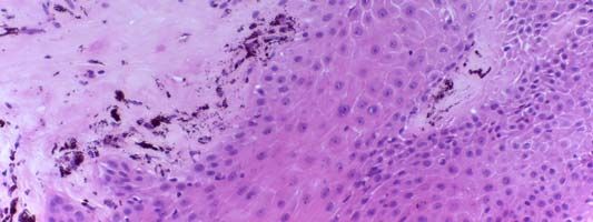
Volume 25 Number 5| May 2019|
Dermatology Online Journal || Case Presentation                                                                           25(5): 10

Late-onset pseudoepitheliomatous hyperplasia developing
within a red ink tattoo
George Badavanis1 MD, Panayotis Constantinou2 MD MSc, Efstathia Pasmatzi3 MD, Alexandra Monastirli1,3
MD, Dionysios Tsambaos1,3 MD PhD
Affiliations: 1Center for Dermatologic Diseases, Limassol, Cyprus, 2Histopathology & Cytology Laboratory, Nicosia, Cyprus,
3
 Department of Dermatology, School of Medicine, University of Patras, Greece
Corresponding Author: Efstathia Pasmatzi MD, Associate Professor, Department of Dermatology, School of Medicine, University of
Patras, P.O. Box 1413, 26504 Rio-Patras, Greece, Tel: 30-261-0 270 577, Fax: 30-261-0 270235, Email: pasmatzi@med.upatras.gr,
pasmatzi@otenet.gr

                                                                      Introduction
  Abstract                                                            Tattooing is defined as the process of implantation
  The popularity of tattoos has increased dramatically
                                                                      of exogenous inerasable pigment into the dermis of
  worldwide particularly in the last three decades,
                                                                      the skin or other parts of the body (e.g., mucosae,
  giving rise to the frequent occurrence of a wide
  spectrum of secondary cutaneous and systemic
                                                                      lips, eyebrows, eyes) of consumers to create a design.
  complications. Pseudoepitheliomatous hyperplasia                    Although tattoos in most cases are decorative, they
  (PEH) is a benign irregular hyperplasia of the                      can also serve religious or medical purposes (e.g.
  epidermis occurring in response to various stimuli,                 breast reconstruction, radiotherapy), or may occur
  that clinically and histopathologically resembles                   accidentally after injuries (traumatic tattoos).
  cutaneous neoplasms such as squamous cell
                                                                      Particularly in the last three decades, the popularity
  carcinoma and keratoacanthoma. In an attempt to
                                                                      of tattoos has impressively increased worldwide and
  improve the awareness of the possible occurrence of
  PEH in tattoos and of its diagnostic and therapeutic                has become mainstream at least for the young
  aspects, we present herein the case of a 30-year-old                generation. It is estimated that more than 100 million
  woman with histologically confirmed PEH related to                  European citizens and about 24% of the US
  a red-ink tattoo. She revealed two important features:              population up to the age of 60 years have one or
  the longest interval reported so far between                        more permanent tattoos on their skin [1]. Already by
  tattooing and onset of PEH (two years) and the lack                 the end of 19th century it was well known that tattoos
  of the otherwise very common lichenoid tissue                       may elicit a variety of mucocutaneous or systemic,
  reaction to red ink. In view of the serious toxicological           early or delayed, acute or chronic complications.
  potential of tattoo inks, implementation of updated                 According to the type of reaction these
  and standardized regulations worldwide regarding                    complications can be classified as hypersensitivity or
  their use in the tattooing process is now urgently
                                                                      inflammatory reactions and as infectious, neoplastic,
  warranted and continuous efforts should be
                                                                      or granulomatous disorders [2].
  undertaken in order to enhance the awareness
  among tattoo artists and the public with regard to                  Pseudoepitheliomatous hyperplasia (PEH) is a
  the possible serious health risks associated with the               benign irregular hyperplasia of the epidermis that
  use of tattoo ink pigments.                                         occurs in response to various stimuli and bears
                                                                      clinical and histopathological similarity to cutaneous
Keywords: red ink, tattoo, pseudoepitheliomatous                      neoplasms, such as squamous cell carcinoma (SCC)
hyperplasia                                                           and keratoacanthoma [3-5]. PEH secondary to tattoo

                                                                -1-
Volume 25 Number 5| May 2019|
Dermatology Online Journal || Case Presentation 25(5): 10
tests and X-ray examination of the leg revealed
normal or negative results. The results of
bacteriological and mycological examination of the
skin lesion were also negative.
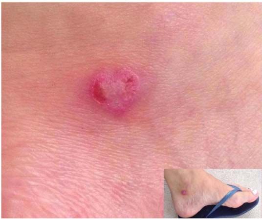
Histopathological examination of a deep biopsy
obtained from the lesional skin revealed epidermal
pseudoepitheliomatous hyperplasia, characterized
by irregular acanthosis and parakeratosis. There
were sharply pointed epithelial strands projecting
into the underlying dermis, whereas significant
cellular atypia was lacking and mitoses were rare.
Prominent extracellular deposits of red exogenous
pigment were found between collagen bundles in
the upper and mid dermis accompanied by a mixed
inflammatory cell infiltrate consisting of
Figure 1. Well demarcated heart-shaped firm nodule with lymphocytes and plasma cells (Figure 2). Thus, the
verrucous and partly erosive surface beneath the medial diagnosis of PEH at the site of the tattoo was
malleolus of the left foot. established. The lesion was totally excised with
excellent cosmetic results.
is a rare complication. In an attempt to improve the
awareness among physicians of the possible
occurrence of PEH in tattoos and of its diagnostic and Case Discussion
therapeutic aspects, we present herein the case of a In view of the millions of tattooed people worldwide,
30-year-old woman with histologically confirmed it is not surprising that tattoo-related complications
late-onset PEH reaction to a red-ink permanent are being increasingly encountered by dermatologists
tattoo.
Case Synopsis
A 30-year-old HIV-negative and otherwise healthy
woman presented to the Center for Dermatologic
Diseases in Limassol, Cyprus, with a one-year history
of a skin lesion progressively arising on the inner
aspect of the ankle of her left foot on a red tattoo
performed by a professional artist in the United
States three years prior. She was not aware of the
origin, the type, or the quantity of the red ink used.
She had no history of malignancy or
immunosuppression and received no medications
on a daily basis. Clinical examination revealed a well-
developed woman in no acute distress that was
remarkable for a well demarcated heart-shaped firm Figure 2. Pseudoepitheliomatous hyperplasia as a reaction in a
nodule (1.3cm in diameter) with a verrucous and red tattoo. The epidermis reveals a distinct hyperkeratosis,
partly erosive surface beneath the medial malleolus parakeratosis and acanthosis with sharply-pointed epithelial
strands projecting into the underlying dermis. In the papillary
of the left foot (Figure 1). There was no evidence of dermis, extracellular deposits of red pigment are seen, that are
lymphadenopathy and/or hepatosplenomegaly. accompanied by a mixed inflammatory cell infiltrate consisting
Routine hematological, biochemical and serological of lymphocytes and plasma cells. H&E, 100×.
-2-Volume 25 Number 5| May 2019|
Dermatology Online Journal || Case Presentation 25(5): 10
Table 1. Mucocutaneous and systemic adverse reactions related to permanent tattoos.
A. Systemic Reactions
[1, 2, 39-41] B. Mucocutaneous Reactions
B1. Hypersensitivity and B4. Granulomatous
inflammatory reactions B2. Infections B3. Neoplasms Reactions
[9, 11, 41-48] [46, 49-54] [12, 13, 29, 46, 55-60] [1, 2, 61-64]
Abdominal compartment Acute GVHD Bacterial Benign Foreign body reaction
syndrome Angioneurotic oedema Abscesses Cutaneous lymphoid Perforating
Bacteremia Bleeding / Hematoma Cellulitis hyperplasia granulomatous reaction
Death Blister formation Cutaneous Dermatofibroma Sarcoidosis
Endocarditis “Blue-foot” discoloration diptheria Epidermal cysts Necrobiotic reaction
Fat necrosis Burning sensation Erysipelas Milia Tuberculoid reaction
Fever Chronic fibrosing vasculitis Gangrene Seborrhoeic keratosis
Gangrene Contact dermatitis Impetigo
Iliopsoas abscess Contact urticaria Leprosy Malignant
Latex allergy Crusting MRSA infections Basal cell carcinoma
Lymphadenopathy Edematous “peau d’ orange” Non-tuberculous Fibrosarcoma
regional or generalized Erythema multiforme mycobacterium protuberans
Lymphoedema Hypertrichosis skin infections Keratoacanthoma
Multiorgan failure Hypo- or Staphylococcal Lymphomas
Necrotizing pneumonia hyperpigmentation scalded skin Melanoma
Pyelonephritis Lichen planus and syndrome Squamous cell
Septic shock lichenoid lesions Syphilis carcinoma
Spinal abscesses Light-induced urticaria Tetanus
Systemic bacterial and Lymphoedema Tuberculosis
viral infections Morphea and
Systemic vasculitis scleroderma-like lesions Viral
Tropical pyomyositis Mucosal ulceration CMV
Uveitis Necrotizing fasciitis or Heparitis B and C
Xanthogranulomatous tissue necrosis HIV
Pain or tenderness HPV
Paradoxical skin darkening HSV
Photodermatitis Molluscum
Pigment diffusion contagiosum
Prurigo nodularis
Pruritus Fungal
Pseudolymphomatous lesion Aspergillosis
Psoriasis Dermatophytosis
Purpura / Petechiae Sporotrichosis
Pyoderma gangrenosum Tinea cutis
Scarring / Keloid Zygomycoses
Subacute or discoid lupus
erythematosus
Sweet syndrome
Swelling
Vasculitis
Wells syndrome
in everyday clinical practice. The prevalence of the numbers of individuals are still lacking. Tattoo-
reported complications varies between 2% and 27% related complications can be acute or have a
[2, 6]. Nevertheless, its accurate estimation is protracted and chronic course [7]. Table 1
presently impossible because a number of cases summarizes the reported mucocutaneous and
remains unreported and controlled studies on large systemic complications of permanent tattoos.
-3-Volume 25 Number 5| May 2019|
Dermatology Online Journal || Case Presentation 25(5): 10
Cutaneous complications of red tattoos are very longest interval reported so far, to our knowledge.
common, with allergic dermatitis, photosensitivity, Additionally, the lichenoid tissue reaction to red ink,
and granulomatous adverse reactions being the that is otherwise very common among PEH patients
most frequent ones [8-11]. Red inks are associated [10], was absent in our case.
with 34% of post-tattoo skin cancers, 50% of SCCs, Upon transfer of the pigment into the dermis using
and 73% of keratoacanthomas [12, 13]. It is believed an electric vibrating device, pigment granules are
that substances contained in red inks (e.g. 2- ingested by skin phagocytes and a transient
anisidine) function as co-carcinogens, especially in inflammatory period of two-week duration is
combination with sunlight exposure [14]. initiated that is characterized by a foreign-body
PEH is a benign proliferative cutaneous disorder, reaction and fibrous tissue formation [4]. Finally, the
occurring in response to various stimuli, which is pigment is encapsulated in dense layers of
regarded as a reactive histopathological pattern connective tissue mainly in the papillary dermis
rather than a distinct nosological entity [15]. either within fibroblasts or between collagen
Clinically PEH occurs as a nodule or plaque of various bundles [11, 29]. Pigment can migrate, however, via
sizes with a verrucoid or vegetative surface, scaling, lymphatics to regional lymph nodes, which in some
and possibly ulceration and crusting [16]. The main cases are filled with pigment [12]. Recently, Sepehri
histopathological feature of PEH is the presence of et al. [30] reported the occurrence of tattoo pigments
follicular infundibulum-derived irregular projections in the blood circulation and in the Kupffer cells of the
of the epidermis extending deep into the reticular mouse liver, as well. These pigments can be
dermis. Orthokeratosis, parakeratosis, degraded under UV irradiation leading to the
hypergranulosis, and keratin pearls are usual formation of toxic or carcinogenic products such as
findings, whereas mitoses are sparse and not- 3, 3-dichlorobenzidine [31-35].
atypical [16]. Owing to their clinical and The exact pathogenetic mechanisms of PEH
histopathological similarities, the distinction secondary to tattoos still remain unknown. Since PEH
between PEH and SCC can be very challenging. A is specifically related to red pigment in permanent
deep biopsy including the base of the lesion and tattoos, it has been hypothesized that early
underlying dermis is usually necessary for a definitive inflammation triggered by the newly introduced
diagnosis. Important histopathological features that exogenous pigment could result in the development
favor the diagnosis of PEH are minimal cytological of PEH [4]. PEH is regarded by some authors as an
atypia, paucity of mitotic activity, absence of atypical autoimmune reaction and epidermal hyperplasia as
mitoses, and absence of necrotic keratinocytes or the result of lymphocyte-derived chemokines
vascular and perineural invasion [16, 17]. inducing keratinocyte proliferation [28].
Since the original description of PEH secondary to a The red tattoo pigmentation was traditionally
tattoo by Sulzberger in 1937, 20 cases have been achieved by the use of cinnabar (mercuric sulfide), a
reported (Table 2), [17-28]. However, owing to well-known allergen that has been implicated in
substantial clinical and histopathological similarity, several cases of delayed hypersensitivity reactions
the possibility that some cases previously reported [1]. Actually, the first described case of tattoo-related
as keratoacanthomas or lichenoid hypersensitivity PEH by Sulzberger [18] was associated with its use.
reactions in tattooed patients were in fact PEH Although cinnabar has been gradually replaced by
cannot be definitely excluded [10]. Interestingly, new mercury-free red pigments such as cadmium
almost all tattoo-related PEH cases reported so far red, iron oxide, ferric sulfate, hematite, cadmium
were associated with the use of either red or purple selenide, sienna, naphthol-AS pigment, azo
dye. The interval between tattooing and the onset of pigments (pigment red 210, 170, 112, 122), and
PEH varies between 4 days and 12 months. In our quinacridones (Violet 19, red 122), [1], adverse
patient PEH occurred two years after tattooing, the reactions in red tattoo areas still continue to occur.
-4-Volume 25 Number 5| May 2019|
Dermatology Online Journal || Case Presentation 25(5): 10
Table 2. Reported cases of PEH after tattoo.
Interval between
tattoo and onset of
Gender Age Tattoo pigment PEH Treatment Authors
1 M 25 Red Unknown Unknown Sulzberger 1937
2 M 23 Red 12 months Surgical excision Goldberg 1959
3 M Unknown Red Unknown Unknown Goldstein 1967
4 M Unknown Red Unknown Unknown Goldstein 1967
5 F 27 Purple 2 months Surgical excision Balfour et al 2003
Red, green,
6 F 59 1 week Surgical excision Cui et al 2007
yellow
7 F 30 Red 1 month Lost to follow up Kluger et al 2008
Topical cosrticosteroids
8 F 32 Red 4 days Kluger et al 2008
(partial response)
Topical cosrticosteroids
9 F 54 Unknown 3 months Kluger et al 2008
(no response)
10 F 24 Red 12 months Surgical excision Then et al 2009
de Freitas
1 month after tattoo Ferreira
11 M 51 Red Unknown
recoloring Hostalácio et al
2011
De Roeck et al
12 F 50 Red 1 month Surgical excision
2012
Topical cosrticosteroids
13 M 31 Red 2 months Breza et al 2013
and surgical excision
7 months after tattoo Topical 5-FU (no Kazlouskaya et al
14 M 47 Red
recoloring response) 2015
Self-resolution 6 weeks Kazlouskaya et al
15 F 44 Red 7 – 9 months
after biopsy 2015
16 F 36 Red Unknown Unknown Kiss et al 2016
Intralesional Tammaro et al
17 F 26 Purple 6 months
corticosteroids 2016
Topical and systemic
2 weeks after tattoo
corticosteroids (initial
18 F 25 Red recoloring with a new Conti et al 2017
clearance, relapse after
red color
10 days)
Recently after injections
Intralesional Tammaro et al
19 F 36 Red of pigment in an 1-year
corticosteroids 2018
tatoo
Broussard-
CO2 laser treatment was
20 F 52 Red 1 year Steinberg et al
programmed
2018
Sunlight exposure of the red tattoo areas prior of the sunlight. It is possible, therefore, that UV irradiation
occurrence of PEH is a common denominator among could have contributed to the development of PEH
the reported cases and is considered as an additional in her tattoo.
pathogenetic factor for this complication [18], Unfortunately, tattoo inks are not regulated by the
possibly through the production of toxic or FDA as they are considered to be cosmetics and
sensitizing substances by photodegradation or additives, whereas in the European Union (EU) they
metabolic activation of the pigment molecules. The are regarded as general consumer products and
tattoo of our patient was located on the ankle of her hence, are regulated under the General Product
left foot and, thus, would likely be heavily exposed to Safety Directive (92/59/EEC). It is obvious, therefore,
-5-Volume 25 Number 5| May 2019|
Dermatology Online Journal || Case Presentation 25(5): 10
that the manufacturers in the US have no obligation properties of red pigment particles associated with
to disclose the chemicals contained in these inks and allergenic or toxic potential [37, 38].
that the ingredients of the latter have never been
tested for safety when injected into the skin [35].
Guidelines in a resolution of the Council of the EU Conclusion
released in 2008 are: The frequency of cutaneous and systemic
complications secondary to tattoos is constantly
1. The maximum permitted concentrations of rising as a result of the increased popularity of
carcinogenic, mutagenic and reproduction-toxic tattooing process. Red tattoos are associated with an
substances in tattoo inks were established, and increased rate of dermatological complications,
2. The necessary conditions for risk evaluation prior including SCCs and keratoacanthomas. PEH is a
to performing tattoos and for the appropriate rarely reported (and possibly underdiagnosed)
information of customers about the health risks of complication of red and purple tattoos that bears
the latter were described [2, 32, 36]. considerable clinical and histopathological similarity
However, the effectiveness of this resolution is to cutaneous neoplasms, and especially SCCs and
practically limited since most inks used for tattooing keratoacanthomas.
are manufactured in, and can be purchased from We present herein the case of a 30-year-old woman
countries outside the EU [2]. with late-onset PEH (two years) to a red-ink tattoo,
The modalities most commonly applied in the histologically characterized by lack of the otherwise
management of PEH secondary to tattoo include very common lichenoid reaction. We review the
surgical excision, topical or intralesional relevant literature, since an extensive knowledge of
corticosteroids, topical 5-FU, or CO2 laser that PEH is mandatory for its early diagnosis and
reportedly reveal varying therapeutic efficacy. There appropriate treatment. Finally, since the
are also some anecdotal reports on the treatment of composition, biokinetics, metabolic activation,
PEH with photochemotherapy (PUVA), phototoxicity, migratory, and carcinogenic potential
phototherapy (narrow band UVB), excimer laser, or of tattoo inks still remains unexplored and a constant
photodynamic therapy, and topical calcineurin reason of serious concern, we feel obliged to
inhibitors based on the clinical and histological emphasize the necessity for urgent implementation
similarity of PEH with hypertrophic lichen planus of updated and standardized regulations worldwide
[10]. Our patient was treated with excellent cosmetic with regard to their use in tattooing. On the other
results by surgical excision, which is the treatment of hand, continuous and concerted efforts should be
choice, particularly for small and well-defined undertaken in order to enhance the awareness
lesions. Carbon dioxide laser treatment is the main among tattoo artists and the public with regard to
alternative, especially for larger lesions, as it the possible serious health risks associated with the
effectively ablates the hypertrophic tissue and leads use in the tattooing process of ink pigments of
to fragmentation of the pigment particles into questionable or completely unknown safety.
smaller sizes, which can then be eliminated by
physiological processes [37, 38]. However, it should
be kept in mind that the latter may result in a Potential conflicts of interest
significant alteration in the structure and chemical The authors declare no conflicts of interests.
References
1. Islam PS, Chang C, Selmi C, et al. Medical complications of tattoos: 2. Kluger N. Cutaneous and systemic complications associated with
A comprehensive review. Clin Rev Allergy Immunol. tattooing. Presse Med. 2016;45:567-76. [PMID:27160631].
2016;50(2):273-86. [PMID:26940693].
-6-Volume 25 Number 5| May 2019|
Dermatology Online Journal || Case Presentation 25(5): 10
3. Fu X, Jiang D, Chen W, Sun BS T, Sheng Z. Pseudoepitheliomatous 24. Kiss F, May K, Piguet V. Image Gallery: Pseudoepitheliomatous
hyperplasia formation after skin injury. Wound Repair Regen. hyperplasia, a rare tattoo reaction. Br J Dermatol.
2007;15:39-46. [PMID:17244318]. 2016;175(3):e112. [PMID:27632970].
4. Kluger N, Durand L, Minier-Thoumin C, et al. 25. Tammaro A, Abruzzese C, Narcisi A et al. Localised
Pseudoepitheliomatous epidermal hyperplasia in tattoos: report pseudoepitheliomatous hyperplasia: unusual cutaneous reaction
of three cases. Am J Clin Dermatol. 2008;9(5):337-40. pattern to tattoo. Int Wound J. 2016 ;13(2):294-5.
[PMID:18717610]. [PMID:24720778].
5. de Roeck A, Joujoux JM, Fournier F, et al. Florid 26. Conti R, Bassi A, Bruscino N, et al. Pseudoepitheliomatous
pseudoepitheliomatous hyperplasia related to tattoo: a case hyperplasia in a tattoo. G Ital Dermatol Venereol. 2017;152(1):71-2.
report. Int Wound J. 2013;10(5):539-41. [PMID:22712583]. [PMID: 27055149].
6. Kazandjieva J, Tsankov N. Tattoos: dermatological complications. 27. Tammaro A, Raffa S, Petrigliano N, et al. Marked
Clin Dermatol 2007;25(4):375-82. [PMID:17697920]. pseudoepitheliomatous hyperplasia secondary to a red-
7. Goldstein N. Mercury-cadmium sensitivity in tattoos: a pigmented tattoo: a case report. J Eur Acad Dermatol Venereol.
photoallergic reaction in red pigment. Ann Intern Med. 1967;67: 2018 Jul;32(7):e272-3. [PMID:29357106].
984-9. [PMID:6050824]. 28. Broussard-Steinberg C, Zemtsov A, Strausburg M, Zemtsov G,
8. Hjerppe A, Hasan T. Short time fun and long time Warren S. Lichenoid reaction pattern with
allergic problems from henna tattoo. Duodecim. 2005; pseudoepitheliomatous hyperplasia – a rare tattoo reaction: a
121(12):1327-9. [PMID:16134512]. case report and review of the literature. Case Rep Dermatol.
9. Tang MM, Beltranimelli I, Perruchoud D. Tattoo complicated by 2018;10(3):268-73.
allergic contact dermatitis and panniculitis. J Eur Acad Dermatol 29. Paprottka FJ, Bountikous S, Lohmeyer JA, Hebebrand D.
Venereol. 2014;28(1):127-8. [PMID: 23495838]. Squamous-cell carcinoma arises in red parts of multicolored
10. Kazlouskaya V, Junkins-Hopkins JM. Pseudoepitheliomatous tattoo within months. Plast Reconstruct Surg Glob Open.
hyperplasia in a red pigment tattoo: a separate entity or 2014;2:e114. [PMID:25289308].
hypertrophic lichen planus-like reaction? J Clin Aesthet Dermatol. 30. Sepehri M, Sejersen T, Qvortrup K, Lerche CM, Serup J. Tattoo
2015;8(12):48-52. [PMID:26705448]. pigments are observed in the Kupffer cells of the liver indicating
11. Shashikumar BM, Harish MR, Shwetha B et al. Hypersensitive blood-borne distribution of tattoo ink. Dermatology.
reaction to tattoos: a growing menace in rural India. Indian J 2017;233(1):86-93. [PMID:28486229].
Dermatol. 2017;62(3):291-6. [PMID:28584372]. 31. Wang L, Yan J, Hardy W et al. Light-induced mutagenicity in
12. Kluger N, Koljonen V. Tattoos, inks and cancer. Lancet Oncol. Salmonella TA102 and genotoxicity/cytotoxicity in human T-cells
2012;13(4):e161-8. [PMID:22469126]. by 3,3'-dichlorobenzidine: a chemical used in the manufacture of
13. Paprottka FJ, Krezdorn N, Narwan M et al. Trendy Tattoos-maybe dyes and pigments and in tattoo inks. Toxicology.
a serious health risk? Aesthetic Plast Surg. 2018;42(1):310-21. 2005;207(3):411-8. [PMID:15664269].
[PMID: 29124377]. 32. Lehner K, Santarelli F, Penning R. et al.The decrease of pigment
14. Lerche CM, Heerfordt IM, Serup J, Poulsen T, Wulf HC. Red tattoos, concentration in red tattooed skin years after tattooing. J Eur Acad
ultraviolet radiation and skin cancer in mice. Exp Dermatol. Dermatol Venereol. 2011;25(11):1340-45. [PMID: 21349116].
2017;26(11):1091-6. [PMID:28500679]. 33. Hauri U, Hohi C. Photostability and breakdown products of
15. Weedon D. Tumor of the epidermis. In: Weedon D, Strutton G. Skin pigments currently used in tattoo inks. Curr Probl Dermatol.
Pathology. New York: Churchill Livingstone, 2002;753-802. 2015;48:164-9. [PMID: 25833639].
16. Zayour M, Lazova R. Pseudoepitheliomatous hyperplasia: a 34. Schreiver I, Hesse B, Seim C et al. Synchrotron-based v-XRF
review. Am J Dermatopathol. 2011;33(2):112-22; quiz 123-6. mapping and μ-FTIR microscopy enable to look into the fate and
[PMID:21399447]. effects of tattoo pigments in human skin. Sci Rep. 2017;7(1):11385.
17. Cui W, McGregor DH, Stark SP, Ulusarac O, Mathur SC. [PMID:28900193].
Pseudoepitheliomaous hyperplasia: an unusual reaction 35. Niederer M, Hauri U, Hohl Ch. Identification of organic pigments
following tattoo: report of a case and review of the litareture. Int J in tattoo inks and permanent make-up using laser desorption
Dermatol. 2007;46:743-5. [PMID:17614808]. ionization mass spectrometry [version 2; referees: 2 approved].
18. Sulzberger MB. Tattoo dermatitis (sensitivity to cinnabar?) Arch F1000 Research. 2018;6:2034(1-14). [PMID:29259773].
Dermatol Syph. 1937; 36:1265. 36. Sepehri M, Lerche CM, Hutton Carlsen K, Serup J. Search for
19. Goldberg HI. Mercurial reaction in a tattoo. Can Med Assoc J. internal cancers in mice tattooed with inks of high contents of
1959;80:203-4. [PMID:13618823]. potential carcinogens: a one-year autopsy study of red and black
20. Balfour E, Olhoffer I, Leffell D, Handerson T. Massive pseudo- tattoo inks banned in the market. Dermatology. 2017;233(1):94-9.
epitheliomaous hyperplasia: an unusual reaction in a tattoo. Am J [PMID:28511186].
Dermatopathol. 2003;25:338-40. [PMID:12876493]. 37. Meesters AA, De Rie MA, Wolkerstorfer A. Generalized eczematous
21. Then M, Mark Boustred A, Clarke LE. Keratoacanthomatous reaction after fractional carbon dioxide laser therapy for tattoo
hyperplasia in response to a tattoo. Dermatol Surg. allergy. J Cosmet Laser Ther. 2016;18(8):456-8. [PMID:27593684].
2009;35(4):685-6. [PMID:19309341]. 38. Bäumler W. Laser treatment of tattoos: basic principles. Curr Probl
22. Bassi A, Campolmi P, Cannarozzo G et al. Tattoo-associated Dermatol. 2017;52:94-104. [PMID:28288450].
skin reaction: the importance of an early diagnosis and proper 39. Duan L, Kim S, Watsky K, Narayan D. Systemic Allergic Reaction to
treatment. Biomed Res Int. 2014;2014:354608. doi: Red Tattoo Ink Requiring Excision. Plast Reconstr Surg Glob Open.
10.1155/2014/354608. [PMID:25147796]. 2016;4(11):e1111. [PMID:27975018].
23. Breza TS Jr, O'Brien AK, Glavin FL. Pseudoepitheliomatous 40. Lemor D, Lazar DB, Emory WB, Nussdorf JD. Sarcoid Uveitis: A look
hyperplasia: an unusual tattoo reaction. JAMA Dermatol. beyond the eyes. Ochsner J. 2016;16(4):551-553. [PMID:27999517].
2013;149(5):630-1. [PMID:23677107].
-7-Volume 25 Number 5| May 2019|
Dermatology Online Journal || Case Presentation 25(5): 10
41. Serup J, Sepehri M, Hutton Carlsen K. Classification of tattoo infection after tattooing. Dtsch Arztebl Int. 2016;113(40):665-671.
complications in a hospital material of 493 adverse events. [PMID:27788747].
Dermatology. 2016;232(6):668-678. [PMID:25833625]. 54. Krecké N, Smola S, Vogt T, Müller CSL. HPV-47-induced and tattoo-
42. Kluger N, Mathelier-Fusade P, Moguelet P. Scleroderma-like associated verrucae planae: Report of a case and review of the
reaction restricted to the red parts of a tattoo. Acta Derm Venereol. literature. Dermatol Ther (Heidelb). 2017;7(4):549-554.
2009;89(1):95-6. [PMID:19197555]. [PMID:28836173].
43. Garcovich S, Carbone T, Avitabile S et al. Lichenoid red tattoo 55. Kluger N, Minier-Thoumin C, Plantier F. Keratoacanthoma
reaction: histological and immunological perspectives. Eur J occurring within the red dye of a tattoo. J Cutan Pathol.
Dermatol. 2012;22(1):93-6. [PMID:22237036]. 2008;35(5):504-7. [PMID: 17976209].
44. Camilot D, Arnez ZM, Luzar B et al. Cutaneous pseudolymphoma 56. Vitiello M, Echeverria B, Romanelli P, Abuchar A, Kerdel F. Multiple
following tattoo application: report of two new cases of a eruptive keratoacanthomas arising in a tattoo. J Clin Aesthet
potential lymphoma mimicker. Int J Surg Pathol. 2012;20(3):311-5. Dermatol. 2010;3(7):54-5. [PMID:20725558].
[PMID:22084427]. 57. Bittencourt Mde J, Miranda MF, Parijós AM et al. Dermatofibroma
45. Deeken A, Jefferson J, Hawkinson D, Fraga GR. Localized chronic in a black tattoo: report of a case. An Bras Dermatol.
fibrosing vasculitis in a tattoo: a unique adverse tattoo reaction. 2013;88(4):614-6. [PMID:24068136].
Am J Dermatopathol. 2014;36(4):e81-3. [PMID: 24736671]. 58. Joyce CW, Duff G, McKenna D, Regan PJ. Malignant melanoma
46. Khunger N, Molpariya A, Khunger A. Complications of Tattoos and arising in red tattoo ink. Arch Plast Surg. 2015;42(4):475-7.
Tattoo Removal: Stop and Think Before you ink. J Cutan Aesthet [PMID:26217569].
Surg. 2015;8(1):30-6. [PMID:25949020]. 59. Ross N, Farber M, Sahu J. Eruptive milia within a tattoo: A case
47. Meade C, Halvorson C, Kao G, Makhzoumi Z. Acute graft-versus- report and review of the literature. J Drugs Dermatol.
host disease arising within tattooed skin. JAAD Case Rep. 201716(6):621-624. [PMID:28686782].
2015;1(3):114-6. [PMID:27051702]. 60. Maxim E, Higgins H, D'Souza L. A case of multiple squamous cell
48. Kluger N. Cutaneous complications related to tattoos: 31 cases carcinomas arising from red tattoo pigment. Int J Womens
from Finland. Dermatology. 2017;233(1):100-109. [PMID: Dermatol. 2017;3(4):228-230. [PMID:29234718].
28441655]. 61. Kluger N. Sarcoidosis on tattoos: a review of the literature from
49. Molina L, Romiti R. Molluscum contagiosum on tattoo. An Bras 1939 to 2011. Sarcoidosis Vasc Diffuse Lung Dis. 2013;30(2):86-102.
Dermatol. 2011;86(2):352-4. [PMID:21603822]. [PMID:24071880].
50. Conaglen PD, Laurenson IF, Sergeant A et al. Systematic review of 62. Sweeney SA, Hicks LD, Ranallo N, Snyder N 4th, Soldano AC.
tattoo-associated skin infection with rapidly growing Perforating granulomatous dermatitis reaction to exogenous
mycobacteria and public health investigation of a cluster in tattoo pigment: a case report and review of the literature. Am J
Scotland, 2010. Euro Surveill. 2013;18(32):20553. Dermatopathol. 2013;35(7):754-6. [PMID: 21986232].
[PMID:23968828]. 63. Wood A, Hamilton SA, Wallace WA, Biswas A. Necrobiotic
51. Mudedla S, Avendano EE, Raman G. Non-tuberculous granulomatous tattoo reaction: report of an unusual case
mycobacterium skin infections after tattooing in healthy showing features of both necrobiosis lipoidica and granuloma
individuals: A systematic review of case reports. Dermatol Online annulare patterns. Am J Dermatopathol. 2014;36(8):e152-5.
J. 2015;21(6). [PMID:26158355]. [PMID:24335518].
52. Oanţă A, Irimie M. Tinea on a tattoo. Acta Dermatovenerol Croat. 64. Godinho MM, Aguinaga F, Grynszpan R et al. Granulomatous
2016;24(3):223-4. [PMID: 27663926]. reaction to red tattoo pigment treated with allopurinol. J Cosmet
53. Dieckmann R, Boone I, Brockmann SO et al. The risk of bacterial Dermatol. 2015;14(3):241-5. [PMID:26211454].
-8-You can also read