Aseptic meningitis and hydrocephalus secondary to neurosarcoidosis
←
→
Page content transcription
If your browser does not render page correctly, please read the page content below
Case report
BMJ Case Rep: first published as 10.1136/bcr-2021-242312 on 26 August 2021. Downloaded from http://casereports.bmj.com/ on October 18, 2021 by guest. Protected by copyright.
Aseptic meningitis and hydrocephalus secondary
to neurosarcoidosis
Anmol Pandey,1 Thomas Stoker,1 Lukasz A Adamczyk,2 Sybil Stacpoole3
1
Department of Neurology, The SUMMARY normal aside from a mild lymphopaenia which was
National Hospital for Neurology A 53-year-old woman presented to hospital with gait present from the onset of her symptoms in May.
and Neurosurgery, UCL Queen instability, urinary incontinence and confusion. She had a Serum Angiotensin Converting Enzyme (ACE) was
Square Institute of Neurology, non-elevated at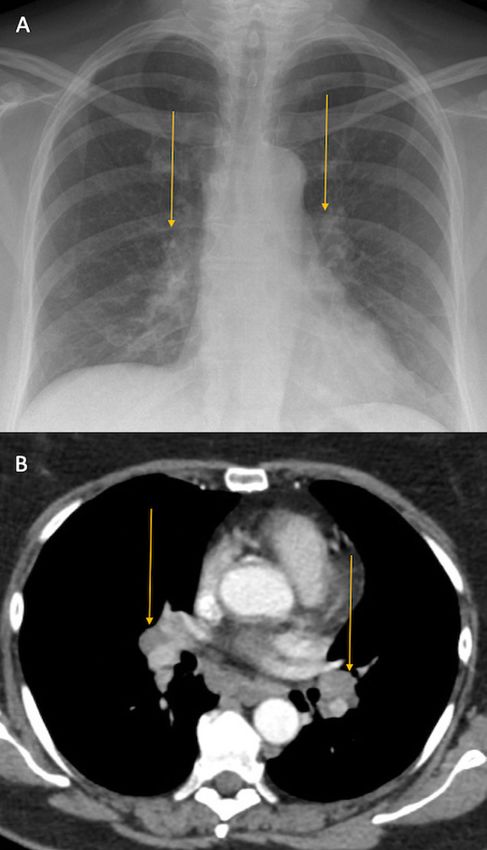
Case report
BMJ Case Rep: first published as 10.1136/bcr-2021-242312 on 26 August 2021. Downloaded from http://casereports.bmj.com/ on October 18, 2021 by guest. Protected by copyright.
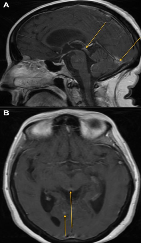
Figure 2 Images (A) and (B) show bilateral hilar lymphadenopathy on
chest X-ray and computerised tomography (CT) of the chest, respectively
(October 2020).
Figure 4 Images (A) and (B) show tectal plate and occipital
allowed us to arrive at the diagnosis of neurosarcoidosis. The lack of enhancement on sagittal and axial T1 postcontrast MRI of the brain,
a confirmatory test in diagnosing sarcoidosis means that it remains respectively (October 2020).
a diagnosis of exclusion. It is notoriously difficult to ensure that
tuberculosis has been excluded, with lymphoma being the other
Hence, we did not feel that subjecting this patient to an invasive
major diagnosis to rule out. The advent of CSF flow cytometry has
CNS biopsy was justified at this time.
been very helpful in the latter. Excluding lymphoma was partic-
ularly important here as there is a two-way statistical association
between an individual and a first-degree relative for breast cancer TREATMENT
and non-Hodgkin’s lymphoma.5 The Neurosarcoidosis Consortium The patient was treated with 3 days of 1 mg methylprednisolone
Consensus Group published diagnostic criteria in 2018 for possible, intravenously, followed by 60 mg of daily oral prednisolone.
probable and definite neurosarcoidosis.6 As per those criteria, the After 5 days of steroid treatment, her ACE-R score increased to
index case qualifies for a diagnosis of probable neurosarcoidosis. 75/100. Her mobility also improved such that it was possible
To establish a diagnosis of definite neurosarcoidosis, a CNS biopsy
would have been required. However, this is an invasive procedure
that carries risk. As we had rigorously excluded other diagnoses, we
felt confident that the diagnosis we had established was accurate.
Figure 5 The lymph node core biopsy demonstrates effacement of the
nodal architecture by closely packed non-caseating granulomas seen
mainly in the upper core. The lower core shows zones of associated
Figure 3 CT of the abdomen shows right inguinal lymphadenopathy hyalinising fibrosis (star symbol). Numerous Langhans-type giant cells
which was biopsied (October 2020). are seen (insert, October 2020).
2 Pandey A, et al. BMJ Case Rep 2021;14:e242312. doi:10.1136/bcr-2021-242312
Case report
BMJ Case Rep: first published as 10.1136/bcr-2021-242312 on 26 August 2021. Downloaded from http://casereports.bmj.com/ on October 18, 2021 by guest. Protected by copyright.
Table 1 Evaluation and exclusion of differential diagnoses
Differential diagnosis Features in favour How diagnosis was excluded
Lymphoma ►► CSF findings of lymphocytosis with raised protein ►► Absence of atypical cells on CSF flow cytometry
►► Widespread lymphadenopathy on CT of the chest, abdomen and pelvis ►► No evidence of lymphoma on tissue biopsy
►► Peripheral lymphopaenia since the onset of symptoms ranging between
0.5×10⁹/L and 1.0×10⁹/L (NR: 1.4–4.8×10⁹/L)
►► Maternal history of breast cancer
Chronic pathogenic ►► Subacute history with neurological symptoms ►► Negative CSF, serum, urine and sputum analysis for pathogens, including
infections ►► Lymph node biopsy showing granulomatous lymphadenitis acid-fast bacillus stains, prolonged culture and mycobacterium tuberculosis
►► CSF showing raised lymphocytes, low glucose and raised protein PCR on CSF
suggestive of tuberculosis or fungal infection ►► Negative staining for microorganisms on lymph node biopsy
Carcinomatous meningitis ►► Subacute history with neurological symptoms and aseptic meningitis ►► Absence of malignancy identified on CT of the chest, abdomen and pelvis/
MRI of the brain and spine
►► No malignant cells in CSF
Autoimmune vasculitis ►► Lymph node biopsy showing granulomatous lymphadenitis ►► Negative auto-antibody screen
Phaeochromocytoma ►► Headache and visual disturbances ►► Normal 24-hour urinary metanephrines
►► Labile blood pressures ranging between 191/114 mm Hg and 220/142 mm ►► Absence of adrenal mass on CT of the chest, abdomen and pelvis
Hg
CSF, cerebrospinal fluid; NR, normal range.
to discharge her home. At the time of discharge, the patient’s ventriculo-peritoneal (VP) shunts, meaning that the decision
cognitive problems had significantly improved although she to offer early neurosurgical intervention should be taken care-
continued to experience mild gait instability. She was discharged fully. Nonetheless, on discharge, this patient was placed under
on prednisolone 60 mg daily, and this was reduced to 40 mg daily the care of a specialist multidisciplinary team that did include
over the next 4 weeks. Four weeks after discharge, she was also a neurosurgical opinion. The plan was to consider neurosur-
started on azathioprine 25 mg daily with a plan to increase to gical intervention if medical management failed to control her
150 mg daily. hydrocephalus- related symptoms. Although hydrocephalus is
In the context of sarcoid-related hydrocephalus, we could reported to be present in only 6% of cases of neurosarcoidosis,4
not find any published data as to whether medical or surgical it is becoming increasingly recognised as a feature of this condi-
management or a combination of the two, would be the most tion. From our literature review, we found 21 cases of hydro-
appropriate treatment. In her case, the rapid response to cephalus identified as a presenting feature of neurosarcoidosis.
steroids meant there was no indication for acute neurosurgical Of these 21 patients, 17 underwent both medical and surgical
intervention. Moreover, active inflammation can also block management for their hydrocephalus, 2 underwent surgery alone
Table 2 Summary of treatment of published cases of neurosarcoidosis with hydrocephalus (adapted from Saban et al27)
Author Treatment Outcome
Pandey et al (2021, index case) Corticosteroids + azathioprine with provisional plan for surgical intervention Partial recovery
Saban et al27 (2020) Corticosteroids + methotrexate, followed by VP shunt months later Partial recovery
McKeever et al11 (2019) Case 1: Corticosteroids + azathioprine Case 1: Complete recovery
Case 2: Initial endoscopic third ventriculostomy followed by multiple neurosurgical procedures Case 2: Partial recovery after the initial procedure but
7–10 years later, including a VP shunt insertion, two shunt revisions and endoscopic fenestration significant neurological disabilities after 10 years
of the third and fourth ventricles
Sugiyama et al28 (2016) Corticosteroids + VP shunt Partial recovery
Chandna et al29 (2015) Corticosteroids + VP shunt Death
Hitti et al30 (2015) Corticosteroids + VP shunt + mycophenolate mofetil Unknown
Matsuda et al31 (2015) Corticosteroids + ventriculostomy followed by VP shunt Complete recovery
Sano et al32 (2015) Corticosteroids + VP shunt + methotrexate + infliximab Partial recovery
Yoshitomi et al33 (2015) Corticosteroids + endoscopic fenestration foramen of Magendie, followed by VP shunt Complete recovery
Tabuchi et al34 (2013) Corticosteroids + VP shunt Partial recovery
Zoja et al7 (2012) Not applicable as diagnosis established at autopsy Death
Kim et al35 Corticosteroids + VP shunt Complete recovery
(2012)
van Rooijen et al8 (2011) VP shunt + corticosteroids Partial recovery
Berhouma et al36 (2009) Corticosteroids + right temporal tip lobectomy Complete recovery
Brouwer et al37 (2009) Ventriculoscopy-assisted fenestration of lateral ventricle cyst Complete recovery
Westhout et al38 (2008) Corticosteroids + VP shunt Complete recovery
Benzagmout et al20 (2007) Corticosteroids + external ventricular drain Partial recovery
Muayqil et al39 (2006) Corticosteroids + VP shunt Partial recovery
Muniesa et al40 (2006) Corticosteroids + VP shunt Complete recovery
Onoda et al41 (2004) Corticosteroids + VP shunt Death from nosocomial pneumonia
Chiang et al42 (2002) Corticosteroids + VP shunt Unknown
VP, ventriculo-peritoneal.
Pandey A, et al. BMJ Case Rep 2021;14:e242312. doi:10.1136/bcr-2021-242312 3Case report
BMJ Case Rep: first published as 10.1136/bcr-2021-242312 on 26 August 2021. Downloaded from http://casereports.bmj.com/ on October 18, 2021 by guest. Protected by copyright.
Finally, some of the more common manifestations of neuro-
Table 3 Sequential ACE-R assessment scores.
sarcoidosis include cranial neuropathy, peripheral neuropathy,
ACE-R domain October 2020 November 2020 January 2021 mononeuropathy, myopathy, psychiatric disorders and cerebellar
Attention (/18) 4 14 13 ataxia.4 The pathophysiological mechanisms leading to these
Memory (/26) 11 18 18 manifestations are not fully understood, although upregulation
Fluency (/14) 1 5 2 of inflammatory cytokines such as tissue necrosis factor α, oxida-
Language (/26) 20 24 26 tive damage and alterations in neurotransmitter metabolism
Visuospatial (/16) 9 14 16 are thought to contribute to cognitive deficits.18 19 Inflamma-
Total (/100) 45 75 76 tion of the arachnoid villi may lead to reduced CSF absorption,
causing a communicating hydrocephalus and its associated clin-
ACE-R, Addenbrooke’s Cognitive Examination–Revised.
ical features.20 21 The patient’s elevated blood pressure (ranging
between 191/114 mm Hg and 220/142 mm Hg) was felt to be
and 1 underwent medical management alone. The remaining due to her poor compliance with antihypertensives secondary
patient died suddenly and did not receive any treatment as her
hydrocephalus was established at autopsy.7 These results are
summarised in table 2 along with the index case. Despite the lack
of data on the management of sarcoid-related hydrocephalus, Patient’s perspective
most authors employed the use of medical management prior to
surgical management, as in this case. One author even suggested My husband and I started to notice over a year ago that my
that earlier use of steroids may have precluded the need for movement was becoming languid and whilst in [censored
surgical placement of a VP shunt altogether.8 location] for my son’s graduation, I tripped on a pavement
In general, the management of sarcoidosis can vary depending outside our hotel and could not prevent my head from colliding
on the organ system involved. For example, the British Thoracic with the pavement, which caused an injury to the bridge of my
Society recommends that patients with pulmonary sarcoidosis nose. In the months after this, my movement became worse.
can be managed without treatment if they remain asymptomatic My husband and I are [censored sporting event] ticket holders;
due to high rates of spontaneous remission.9 However, neuro- it became very uncomfortable to walk from the car park to the
sarcoidosis rarely undergoes spontaneous remission and remains stadium. I visited the doctor on a few occasions as my joints,
a severe illness, often requiring long-term treatment. A step- particularly knees, shoulders and ankles, became extremely
wise approach to management includes using steroids as initial painful. I was almost constantly also suffering from a severe
management. The next step up includes methotrexate, myco- headache. Various tests could not find any problems to explain
phenolate mofetil, leflunomide and azathioprine, before finally these pains. During early May, I woke with blurred vision and
escalating to biological agents such as infliximab, adalimumab an extremely bad headache; we were told to go to [censored
and rituximab.10 location] where they would carry out a CT scan. My memory
was getting worse at this point and my employer was raising
OUTCOME AND FOLLOW-UP concerns. My general practitioner (GP) arranged for a mental
The patient was reviewed 7 weeks after being discharged. At
assessment, more blood tests and an MRI scan. We met with the
this time, her ACE-R score was 76/100. Her sequential ACE-R
neurologist, and they suggested that the problems were due to
assessments with breakdown of scores are shown in table 3.
migraines. My employer decided that I was not able to continue
to work and wrote to my GP at the end of August expressing
concern that there was something seriously wrong which needed
DISCUSSION
to be looked into.
It is important to reiterate that establishing a diagnosis of neuro-
In September, I became more confused and found that I was
sarcoidosis can be challenging and time-consuming. We identified
not able to do basic things like write greetings cards or emails;
one case where a diagnosis of neurosarcoidosis was established
my husband thought my driving was less controlled and I was
over 10 years after the identification of hydrocephalus. Unfor-
very light headed and in a lot of pain in my joints. I fell over in
tunately, that patient developed progressive disabilities that
our bedroom and was too feeble to be able to get to my feet.
did not respond to initial therapy. The authors concluded that
About a week after this, I went for a hair appointment and got
establishing a swifter diagnosis could have prevented irrevers-
confused and very light headed whilst in the shopping centre.
ible neuronal damage, thus highlighting the importance of a
My husband and my employer all realised that something was
timely diagnosis.11 Histologically, sarcoidosis is characterised
very wrong and in late September contacted my GP again. My
by the presence of non- caseating granulomas. A number of
GP said that [(s)he] wanted me to be admitted to hospital, so
immune cell types are found in these granulomas, including
my husband took me in. I spent almost 9 weeks in hospital; my
macrophages, epithelioid cells and multinucleated giant cells, as
memory became much worse and I was telling my husband that
well as lymphocytes that are the predominant cell type in the
I was thinking I was in a spa on some days and often mentioned
central part of a sarcoid granuloma.12 Accumulation of activated
that I had been talking to my late mum and dad. I contracted
T cells to the sites of inflammation causes a peripheral lymph-
COVID-19 in hospital. Although I did fall over a few times whilst
opaenia which is seen in the majority of patients with sarcoid-
in hospital, and on a couple of occasions was left in a very
osis including this case.13–15 The epithelioid cells secrete ACE,
uncomfortable position for a considerable time, I have to say
and this enzyme is widely used as a biomarker in the work-up
that the care I received was absolutely brilliant and I want to
for sarcoidosis. However, its use is limited, as quoted sensitivity
thank all involved for getting to the bottom of my illness and
and specificity for elevated ACE in diagnosing sarcoidosis are
diagnosing my condition. Everybody at [censored location] was
41.4% and 89.9%, respectively.16 Another series of 128 patients
so caring and professional despite these very difficult times. I am
with neurosarcoidosis found that CSF protein was raised in 76%
at home now; I am still having problems with my mobility, but
of samples, as in this case, with a median CSF protein level of
my memory and confusion are vastly improving.
0.8 g/L, though it may be significantly elevated.17
4 Pandey A, et al. BMJ Case Rep 2021;14:e242312. doi:10.1136/bcr-2021-242312Case report
BMJ Case Rep: first published as 10.1136/bcr-2021-242312 on 26 August 2021. Downloaded from http://casereports.bmj.com/ on October 18, 2021 by guest. Protected by copyright.
10 Voortman M, Drent M, Baughman RP. Management of neurosarcoidosis: a clinical
Learning points challenge. Curr Opin Neurol 2019;32:475–83.
11 McKeever A, Cox A, Garnett M, et al. Hydrocephalus as the first presenting symptom
►► This case demonstrates an important sequela of sarcoidosis. of neurosarcoidosis in two patients: a diagnosis more forthcoming in the context of
systemic disease. BMJ Case Rep 2019;12:e229903.
►► The lack of a confirmatory test and the multisystemic nature
12 Mitchell DN, Scadding JG, Heard BE. Sarcoidosis: histopathological definition and
of sarcoidosis can make its diagnosis very challenging. clinical diagnosis. J Clin Pathol 1977;30:395–408.
►► There is a lack of evidence as to whether medical or surgical 13 Hedfors E, Holm G, Pettersson D. Lymphocyte subpopulations in sarcoidosis. Clin Exp
management or a combination of the two, would be the most Immunol 1974;17:219–26.
appropriate treatment in sarcoid-related hydrocephalus. 14 Lower EE, Smith JT, Martelo OJ, et al. The anemia of sarcoidosis. Sarcoidosis
1988;5:51–5.
►► Prompt diagnosis may result in more favourable outcomes, 15 Selroos O, Koivunen E. Prognostic significance of lymphopenia in sarcoidosis. Acta
and a multidisciplinary approach towards diagnosis and Med Scand 1979;206:259–62.
management is recommended. 16 Ungprasert P, Carmona EM, Crowson CS, et al. Diagnostic utility of angiotensin-
converting enzyme in sarcoidosis: a population-based study. Lung 2016;194:91–5.
17 Kidd DP. Sarcoidosis of the central nervous system: clinical features, imaging, and CSF
results. J Neurol 2018;265:1906-1915.
to her cognitive impairment. Hypertension can also occur in
18 McAfoose J, Baune BT. Evidence for a cytokine model of cognitive function. Neurosci
sarcoidosis secondary to autonomic dysfunction caused by a Biobehav Rev 2009;33:355–66.
small fibre neuropathy22–25 or due to renal dysfunction caused 19 Hoitsma E, Faber CG, Drent M, et al. Neurosarcoidosis: a clinical dilemma. Lancet
by interstitial granulomatous nephritis or other glomerular Neurol 2004;3:397–407.
pathologies.26 There was no evidence of renal involvement in 20 Benzagmout M, Boujraf S, Góngora-Rivera F, et al. Neurosarcoidosis which manifested
as acute hydrocephalus: diagnosis and treatment. Intern Med 2007;46:1601–4.
this patient at the time this report was written. 21 Nakayama T, Ouchi Y, Yoshikawa E, et al. Striatal D2 receptor availability after
shunting in idiopathic normal pressure hydrocephalus. J Nucl Med 2007;48:1981–6.
Acknowledgements M. Ahtsham Zafar and Abhinav Jha (radiology registrars) 22 Bakkers M, Merkies ISJ, Lauria G, et al. Intraepidermal nerve fiber density and its
assisted in providing the radiological descriptions of the imaging shown in this application in sarcoidosis. Neurology 2009;73:1142–8.
manuscript. Zachary Moulder (medical student at University College London) assisted 23 Hoitsma E, Marziniak M, Faber CG, et al. Small fibre neuropathy in sarcoidosis. Lancet
with the literature review. 2002;359:2085–6.
24 Hoitsma E, Reulen JPH, de Baets M, et al. Small fiber neuropathy: a common and
Contributors Anmol Pandey (neurology senior house officer), Thomas Stoker important clinical disorder. J Neurol Sci 2004;227:119–30.
(neurology registrar) and Sybil Stacpoole (neurology consultant) had direct 25 Hoitsma E, Drent M, Verstraete E, et al. Abnormal warm and cold sensation
clinical contact with the patient during the index admission. Lukasz A Adamczyk thresholds suggestive of small-fibre neuropathy in sarcoidosis. Clin Neurophysiol
(histopathology consultant) provided the tissue biopsy report. Sybil Stacpoole was 2003;114:2326–33.
responsible for the overall care of the patient. Anmol Pandey wrote the manuscript 26 Hilderson I, Laecke SV, Wauters A. Treatment of renal sarcoidosis: is there a
and obtained written consent from the patient. All authors were involved in guideline?Overview of the different treatment options, Nephrology Dialysis
organising the relevant investigations. All authors approved the manuscript before Transplantation. Nephrol Dial Transplant 2014;29:1841–7.
submission. 27 Saban RJ, Berns MM, Al-Hakim MM, et al. Hydrocephalus as the presenting symptom
Funding The authors have not declared a specific grant for this research from any of sarcoidosis: a case report and review of literature. Clin Case Rep 2020;8:363–8.
funding agency in the public, commercial or not-for-profit sectors. 28 Sugiyama A, Kobayashi M, Agatsuma K, et al. Hydrocephalus mimicking idiopathic
normal pressure hydrocephalus as the first manifestation of neurosarcoidosis. Brain
Competing interests None declared. Nerve 2016;68:1477–82.
Patient consent for publication Obtained. 29 Chandna A, Todd C, Murphy D, et al. Sarcoidosis presenting with acute hydrocephalus
in a New Zealand European female. N Z Med J 2015;128:110–3.
Provenance and peer review Not commissioned; externally peer reviewed. 30 Hitti F, Kennedy B, Odia Y, et al. Isolated neurosarcoidosis presenting with recurrent
Open access This is an open access article distributed in accordance with the hydrocephalus. Neuroimmunol Neuroinflamm 2015;2:287–90.
Creative Commons Attribution Non Commercial (CC BY-NC 4.0) license, which 31 Matsuda R, Nishimura F, Motoyama Y, et al. [A case of intraventricular isolated
permits others to distribute, remix, adapt, build upon this work non-commercially, neurosarcoidosis diagnosed by neuroendoscopic biopsy]. No Shinkei Geka
and license their derivative works on different terms, provided the original work 2015;43:247–52.
is properly cited and the use is non-commercial. See: http://creativecommons.org/ 32 Sano H, Deguchi I, Fukuoka T, et al. Intractable neurosarcoidosis effectively treated
licenses/by-nc/4 .0/. with infliximab. Intern Med 2016;55:811–4.
33 Yoshitomi M, Uchikado H, Hattori G, et al. Endoscopic biopsy for the diagnosis of
neurosarcoidosis at the fourth ventricle outlet with hydrocephalus. Surg Neurol Int
REFERENCES 2015;6:S633–6.
1 Parkes SA, Baker SB, Bourdillon RE, et al. Incidence of sarcoidosis in the Isle of man. 34 Tabuchi S, Uno T. Hydrocephalus with panventricular enlargement as the primary
Thorax 1985;40:284–7. manifestation of neurosarcoidosis: a case report. J Med Case Rep 2013;7:240.
2 Kidd DP. The epidemiology of systemic sarcoidosis in Eastern Hertfordshire, UK. Annals 35 Kim SH, Lee SW, Sung SK, et al. Treatment of hydrocephalus associated
of Public Health Reports 2018;2:22–5. with neurosarcoidosis by multiple shunt placement. J Korean Neurosurg Soc
3 Spencer TS, Campellone JV, Maldonado I, et al. Clinical and magnetic resonance 2012;52:270–2.
imaging manifestations of neurosarcoidosis. Semin Arthritis Rheum 2005;34:649–61. 36 Berhouma M, Abderrazek K, Krichen W, et al. Apropos of an unusual and menacing
4 Allen RKA, Sellars RE, Sandstrom PA. A prospective study of 32 patients with presentation of neurosarcoidosis: the space-occupying trapped temporal horn. Clin
neurosarcoidosis. Sarcoidosis Vasc Diffuse Lung Dis 2003;20:118–25. Neurol Neurosurg 2009;111:196–9.
5 Zheng G, Yu H, Hemminki A, et al. Familial associations of female breast cancer with 37 Brouwer MC, de Gans J, Willemse RB, et al. Sarcoidosis presenting with
other cancers. Int J Cancer 2017;141:2253–9. hydrocephalus. J Neurol Neurosurg Psychiatry 2009;80:550–1.
6 Stern BJ, Royal W, Gelfand JM, et al. Definition and consensus diagnostic criteria for 38 Westhout FD, Linskey ME. Obstructive hydrocephalus and progressive psychosis: rare
neurosarcoidosis: from the neurosarcoidosis consortium consensus group. JAMA presentations of neurosarcoidosis. Surg Neurol 2008;69:288–92.
Neurol 2018;75:1546–53. 39 Muayqil T, Hussain MS, Saqqur M. A patient with neurosarcoidosis. Can J Neurol Sci
7 Zoja R, Andreola S, Gentile G, et al. Sudden death from systemic sarcoidosis: a case of 2006;33:92–4.
legal medicine. Sarcoidosis Vasc Diffuse Lung Dis 2012;29:62–8. 40 Muniesa C, Marcoval J, Moreno A, et al. Hydrocephalic neurosarcoidosis diagnosed by
8 van Rooijen JM, Mijnhout GS, Aalders TTA, et al. Hydrocephalus, a rare manifestation cutaneous lesions. J Am Acad Dermatol 2006;55:S125–6.
of sarcoidosis. Clin Pract 2011;1:e66:136–8. 41 Onoda K, Tsuchimoto S, Katsumata A. A case of neurosarcoidosis presented with
9 Bradley B, Branley HM, Egan JJ, et al. Interstitial lung disease guideline: the British hydrocephalus and marked exacerbations. Japanese J Neurosurg 2004;13:669–73.
thoracic society in collaboration with the thoracic society of Australia and New 42 Chiang JK, Ortiz-Ferrer LC, Remlinger K, et al. Subcutaneous nodules in a patient with
Zealand and the Irish thoracic society. Thorax 2008;63 Suppl 5:v1–58. hydrocephalus. Arch Dermatol 2002;138:259–64.
Pandey A, et al. BMJ Case Rep 2021;14:e242312. doi:10.1136/bcr-2021-242312 5Case report
BMJ Case Rep: first published as 10.1136/bcr-2021-242312 on 26 August 2021. Downloaded from http://casereports.bmj.com/ on October 18, 2021 by guest. Protected by copyright.
Copyright 2021 BMJ Publishing Group. All rights reserved. For permission to reuse any of this content visit
https://www.bmj.com/company/products-services/rights-and-licensing/permissions/
BMJ Case Report Fellows may re-use this article for personal use and teaching without any further permission.
Become a Fellow of BMJ Case Reports today and you can:
►► Submit as many cases as you like
►► Enjoy fast sympathetic peer review and rapid publication of accepted articles
►► Access all the published articles
►► Re-use any of the published material for personal use and teaching without further permission
Customer Service
If you have any further queries about your subscription, please contact our customer services team on +44 (0) 207111 1105 or via email at support@bmj.com.
Visit casereports.bmj.com for more articles like this and to become a Fellow
6 Pandey A, et al. BMJ Case Rep 2021;14:e242312. doi:10.1136/bcr-2021-242312You can also read