Non-invasive Evaluation for Epilepsy Surgery - J-Stage
←
→
Page content transcription
If your browser does not render page correctly, please read the page content below
R EVIEW A RTICLE doi: 10.2176/nmc.ra.2016-0186
Neurol Med Chir (Tokyo) 56, 632–640, 2016 Online September 14, 2016
Non-invasive Evaluation for Epilepsy Surgery
Masaki Iwasaki,1,2 Kazutaka Jin,3 Nobukazu Nakasato,3 and Teiji Tominaga2
1
Department of Neurosurgery, National Center Hospital of Neurology and Psychiatry,
Kodaira, Tokyo, Japan, Department of 2Neurosurgery and 3Epileptology,
Tohoku University Graduate School of Medicine, Sendai, Miyagi, Japan
Abstract
Epilepsy surgery is aimed to remove the brain tissues that are indispensable for generating patient’s epi-
leptic seizures. There are two purposes in the pre-operative evaluation: localization of the epileptogenic
zone and localization of function. Surgery is planned to remove possible epileptogenic zone while pre-
serving functional area. Since no single diagnostic modality is superior to others in identifying and local-
izing the epileptogenic zone, multiple non-invasive evaluations are performed to estimate the location
of the epileptogenic zone after concordance between evaluations. Essential components of non-invasive
pre-surgical evaluation of epilepsy include detailed clinical history, long-term video-electroencephalog-
raphy monitoring, epilepsy-protocol magnetic resonance imaging (MRI), and neuropsychological testing.
However, a significant portion of drug-resistant epilepsy is associated with no or subtle MRI lesions or
with ambiguous electro-clinical signs. Additional evaluations including fluoro-deoxy glucose positron
emission tomography (FDG-PET), magnetoencephalography and ictal single photon emission computed
tomography can play critical roles in planning surgery. FDG-PET should be registered on three-dimensional
MRI for better detection of focal cortical dysplasia. All diagnostic tools are complementary to each other
in defining the epileptogenic zone, so that it is always important to reassess the data based on other results
to pick up or confirm subtle abnormalities.
Key words: epilepsy surgery, evaluation, electroencephalography, magnetic resonance imaging, semiology
Introduction epileptogenic zone and how epilepsy surgery is
considered on multiple evaluations. Then, several
Epilepsy surgery is indicated for patients with recent topics on non-invasive evaluations are
‘drug-resistant’ epilepsy. Current practical definition reviewed, although cyclopedic coverage is beyond
of drug-resistance is defined as failure of seizure the scope of this review.
control after adequate medical therapy with two or
more appropriate anti-epileptic drugs.1,2) The goal Epileptogenic Zone and Concept of
of epilepsy surgery is the improvement of patient’s Pre-surgical Evaluation in Epilepsy
quality of life by seizure control with or without
continuing anti-epileptic medications. Albeit not Complete removal of epileptogenic zone is aimed
the goal of treatment, it is always kept in mind in epilepsy surgery. The concept, epileptogenic
that surgical treatment is an only strategy that can zone, is defined as the area of cortex that is indis-
remove the cause of, or ‘cure,’ epilepsy. Epilepsy pensable for the generation of epileptic seizures.4)
surgery can be indicated earlier when drug-resistance This concept is built on the notion that no single
is highly expected such as in the mesial temporal diagnostic modality is currently available to identify
lobe epilepsy with hippocampal sclerosis.3) or when the accurate brain area generating patient’s seizures.
adverse effect of poor seizure control is expected For example, cavernous malformation certainly causes
on patient’s development in young children.2) patient’s epilepsy, but the brain region responsible for
Surgical treatment is planned after comprehen- generating seizures usually exists in the surrounding
sive evaluation of the patient’s epilepsy. In the brain, where enduring hyperexcitability was acquired
first part of this review, we introduce a concept of by degeneration and hemosiderin deposition induced
by the malformation. It is currently hard to know
Received June 17, 2016; Accepted July 29, 2016 pre-operatively to what extent, the surrounding brain
632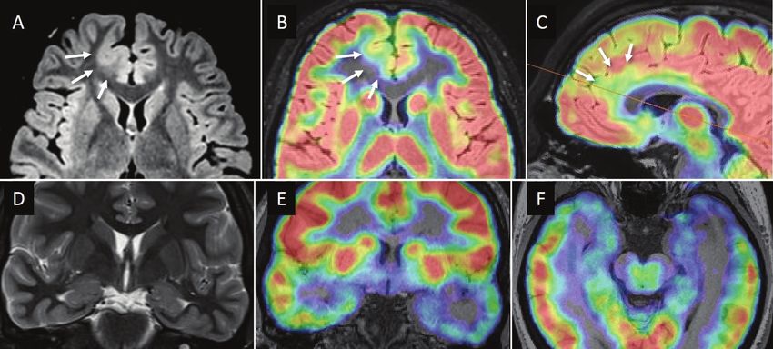
Non-invasive Evaluation for Epilepsy Surgery                                  633

should be removed to achieve seizure control.5,6) This            secondary generalized tonic-clonic seizure strongly
is also the case in other etiologies, including focal             indicates seizure originating in the left hemisphere.
cortical dysplasia, brain tumors, stroke, and traumatic           Similarly, the focal tonic-clonic seizure in the left
brain injury, although intrinsic epileptogenicity is              leg strongly indicates the epileptogenic zone nearby
proved in certain lesions with neuronal components,               the right primary motor cortex of the leg level. These
such as focal cortical dysplasia, ganglioglioma, and              are highly diagnostic even in the absence of clear
cortical tubers.7,8) Therefore, the epileptogenic zone            MRI lesion and clear EEG (electroenphalography)
is a theoretical concept.                                         substrates.10) All diagnostic results are complemen-
   Epileptogenic zone is estimated after results of               tary to each other, and are interpreted on their own
multiple evaluations. A variety of diagnostic tools               sensitivity and specificity.
define different cortical zones of epileptic abnor-
mality (Table 1). These cortical zones can overlap                  Non-invasive Pre-surgical Evaluation
with high concordance or can be discordant each                                 of Epilepsy
other, because each diagnostic method has their
own sensitivity and specificity for defining the                  There are two purposes in the pre-operative evalu-
location and extent of epileptogenic zone. When                   ation: localization of the epileptogenic zone and
the concordance is high, estimated location and                   localization of function. Surgery is planned to
the extent of epileptogenic zone becomes accurate                 remove possible epileptogenic zone while preserving
and confident. Currently, the presence of a struc-                functional area. Non-invasive evaluations of drug-
tural epileptogenic lesion in MRI is most reliable                resistant epilepsy are listed in Table 1. Detailed
and accurate information for epileptogenic zone.                  history taking, structural MRI, long-term video-EEG
Complete removal of MRI-visible epileptogenic                     monitoring, and neuropsychological testing are
lesion is associated with seizure freedom after                   considered as essential for pre-surgical evaluation
epilepsy surgery.9)                                               in clinical practice. Optional evaluations can be
   Information obtained by a single evaluation can                performed on necessity. Because the evaluation is
be fragmented and spatially not clear, but highly                 aimed to make surgical decision, no routine sets
specific for laterality or possible location of the               exist and evaluations can be tailored to the patient’s
epileptogenic zone. For example, the presence of                  needs. For example, anterior temporal lobectomy
head version to the right side immediately before                 can be indicated for a patient with mesial temporal
                                                                  lobe epilepsy only based on structural MRI, routine
                                                                  EEG, and a detailed clinical history, after confirming
Table 1 Non-invasive pre-surgical evaluations for
epilepsy
                                                                  the presence of unilateral hippocampal atrophy and
                                                                  typical history for mesial temporal lobe epilepsy
                                Cortical zones of epileptic       ‘syndrome,’11) On the other hand, optional evalua-
Evaluations
                                abnormalitya
                                                                  tions such as MEG and ictal SPECT can be critical
Detailed clinical history*      —                                 for surgical decision making in patients with ‘MRI-
Video-EEG monitoring*           Seizure onset zone,               negative’ focal epilepsy.
                                Symptomatogenic zone,
                                Irritative zone
                                                                       Long-term Video-EEG Monitoring
MRI*                            Epileptongenic lesion
                                                                       (LTVEEG) with Scalp Electrodes
Neuropsychological              Functional deficit zone
evaluation*                                                       The purpose of LTVEEG is 1) to rule out non-
FDG-PET                         Epileptogenic lesionb,            epileptic seizures and 2) to analyze ictal semiology /
                                Functional deficit zone           EEG for localization of epilepsy. Even if a patient
Magnetoencephalography          Irritative zone                   has established diagnosis of epilepsy, the presence
(MEG)                                                             of concomitant non-epileptic seizures should be care-
Iomazenil-SPECT                 Epileptogenic lesionb,            fully ruled out. A significant proportion of medical
                                Functional deficit zone           intractability is caused by misdiagnosis of non-
Ictal ECD-SPECT                 Seizure onset zone                epileptic seizures.2) Moreover, 10–30% of patients
Functional MRI /                Eloquent cortex                   with drug-resistant seizures have both epileptic
Functional MEG                                                    and non-epileptic seizures. Although epilepsy can
*Essential evaluations. aRosenow F, Lüders H. Presurgical
                                                                  be affirmed by clear history of witnessed seizures
evaluation of epilepsy. Brain 124:1683–700, 2001. bMRI-           and interictal epileptic EEG, further documentation
negative epileptogenic lesion could be detected as functionally   of the patient’s habitual seizures with LTVEEG is
impaired region.                                                  routinely recommended.

Neurol Med Chir (Tokyo) 56, October, 2016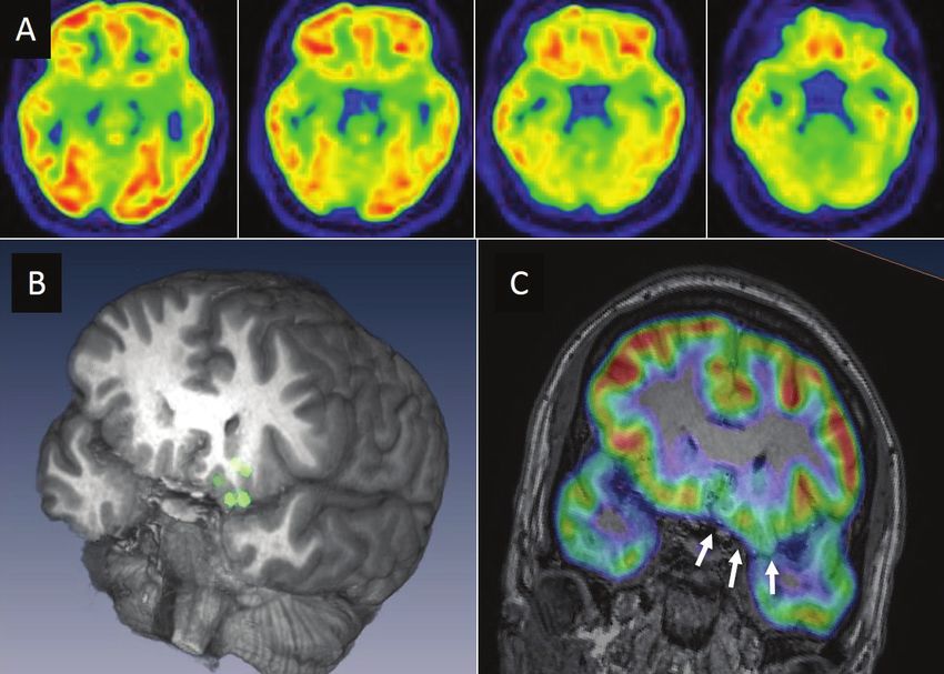
634                                                M. Iwasaki et al.

   The sensitivity and specificity of localizing/             Table 2 Essential six sequences of magnetic resonance
lateralizing features in seizure semiology have been          imaging for epilepsy patients (Wellmer J, et al. Epilepsia
reported.12,13) Localizing/lateralizing features are often    54(11): 1977–87, 2013)
only supportive for other evaluations, and lack of
                                                              Sequence           Slice thickness   Cut-plane orientation
those features does not mean non-localizable or
non-lateralizable epilepsy. However, seizure semi-            T1 / MPRAGE        1 mm isotropic    3-dimensional
ology can be highly diagnostic when the epileptic             T2 / STIR
Non-invasive Evaluation for Epilepsy Surgery 635
Recently, small middle-fossa encephalocele is recog- PET-positive TLE’.34,35) The surgical outcome of MRI-
nized as a cause of ‘non-lesional’ TLE.30–32) Thin-sliced negative PET-positive TLE is comparable with mesial
examination of the middle fossa would be recommended TLE with hippocampal sclerosis, although dichotic
for patients with MRI-negative temporal lobe epilepsy, response was pointed out in its post-operative seizure
not to overlook such under-recognized etiology. outcome, i.e. the patients were divided into those with
class I outcome and those with class III or IV outcome.
FDG-PET
Magnetoencephalography (MEG) and
Epileptic abnormality usually presents glucose ictal Single-photon Emission Computed
hypometabolism interictally. FDG-PET is indicated Tomography (SPECT)
especially when no structural lesions are identified
on MRI. The PET imaging should be registered on MEG and ictal SPECT are important localization
three-dimensional MRI (fusion image) for accurate tools for epileptogenic zone in patients with incon-
interpretation (Fig. 1A-C), because co-registration clusive findings in the above evaluations. Generators
of FDG-PET and MRI is known to improve detec- of interictal spikes are estimated with equivalent
tion of focal cortical dysplasia. 33) This method current dipole (ECD) modeling in MEG. Registered
especially enhances detection of MRI-negative images of ECDs on the patient’s MRI are called
type I cortical dysplasia, and complete removal magnetic source imaging (MSI). When interpreting
of PET-positive lesion is associated with excellent MSI, the following two limitations should be kept
seizure outcome. in mind. First, due to inherent limitation in MEG
FDG-PET is also useful in identification of surgically and ECD modeling, MSI inevitably has unknown
treatable MRI-negative temporal lobe epilepsy (TLE) amounts of errors in its spatial accuracy. Second,
(Fig. 1D–F). Anterior temporal glucose hypometabo- MSI is usually derived from interictal recording,
lism is recently recognized as a typical pattern in and interictal epileptic spikes can occur remote
non-lesional mesial TLE and denoted as ‘MRI-negative from the epileptogenic zone.36)
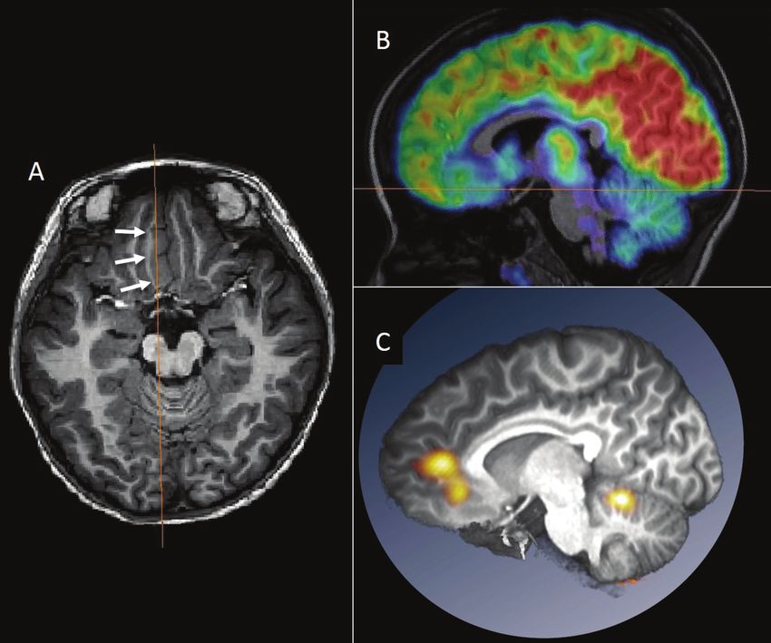
Fig. 1 Co-registration of FDG-PET and MRI. (A) Axial FLAIR image in a 19-year-old man with the right medial
frontal focal cortical dysplasia (FCD). Abnormally thickened cortex is associated with blurred gray-white matter
junction (arrows). (B, C) Co-registered FDG-PET images show focal glucose hypometabolism in the lesion (arrows).
Co-registration of FDG-PET and MRI improves detection and identification of FCD. The patient became seizure
free after lesionectomy. Histo-pathological diagnosis was FCD type 2b. (D) Coronal T2 weighted image shows
no remarkable abnormalities in a 27-year-old woman with the left temporal lobe epilepsy. (E, F) Co-registered
FDG-PET showed glucose hypometabolism in the left anterior temporal lobe. The patient underwent left anterior
temporal lobectomy, followed by seizure freedom. Histo-pathologically no specific abnormality was identified in
the hippocampus and temporal neocortex.
Neurol Med Chir (Tokyo) 56, October, 2016636 M. Iwasaki et al.
Nevertheless, MSI can provide crucial informa- co-registered to MRI (SISCOM) has higher predic-
tion in surgical decision making. Anterior temporal tive value for epileptogenic zone than side-by-side
distribution of ECDs is affirmative in the diagnosis comparison of ictal and interictal SPECT.40) Yield of
of mesial temporal lobe epilepsy.37) In extra-temporal ictal SPECT depends on the timing of tracer injec-
lobe epilepsy, complete removal of ‘clustered’ ECDs tion. Delayed injection may only detect secondary
is associated with better post-operative seizure hyperperfusion after seizure spread, producing diffuse
outcome.38,39) In contrast, diffusely distributed ECDs or non-localized findings. Early tracer injection is an
are suggestive of diffuse epileptogenic zone. The important factor for better localizability.41)
orientation of ECD provides an important clue for
determining the epileptogenic side of opposing cortices Combination of Multimodal Imaging
in the cerebral sulcus.36) At their peak, epileptic spikes
usually generate dipolar current oriented to the basal Multimodal evaluations should be reviewed and
side of the cortex. For example, in the central sulcus, interpreted together, because epilepsy-related
anteriorly oriented dipoles suggest activation of the abnormality is occasionally subtle, ambiguous, or
anterior, or frontal, bank of the sulcus. uncertain. Localizing information of one modality
Ictal SPECT visualizes the area of increased cerebral may enhance detection of or convince the presence
blood flow induced by an epileptic seizure. Although of subtle abnormalities in another modality. For
it is only feasible in patients with frequent seizures, example, reviewing the area of MEG dipoles may
Ictal SPECT is a powerful tool for localization of the detect small focal cortical dysplasia that was not
possible epileptogenic zone. Subtraction ictal SPECT previously found by routine MRI (Fig. 2).10) It is
Fig. 2 Magnetic source imaging enhanced detection of focal glucose hypometabolism caused by focal cortical
dysplasia. A 20-year-old man with the left orbito-frontal lobe epilepsy is presented. No remarkable abnormality
was noted in the conventional presentation of FDG-PET. (A) Magnetoencephalography recording revealed that
equivalent current dipoles (green circles in the 3-dimensional MR imaging) of his interictal spikes were localized
in the left orbito-frontal region. (B) Review of the dipole region led us to identify focal glucose hypometabolism in
the same region (arrows) in FDG-PET. (C) The patient underwent focal cortical resection of the orbito-frontal lobe
after the orbito-frontal seizure onset was confirmed by invasive evaluation with chronically implanted subdural
electrodes. Histo-pathological diagnosis of microdysgenesis was established.
Neurol Med Chir (Tokyo) 56, October, 2016Non-invasive Evaluation for Epilepsy Surgery 637
recommended that multimodal results are spatially For example, impairment of verbal memory perfor-
co-registered and reviewed in a common anatom- mance is caused by damage in the hippocampal
ical space, which is useful in planning surgery memory system, thus supportive of the diagnosis
(Fig. 3). Automated brain extraction, multimodal of mesial temporal lobe epilepsy, especially of the
image registration, and volume-rendered visualization language-dominant hemisphere.
are easily available by free software packages.42,43) The risk of post-operative cognitive impairment
depends on the ‘functional adequacy of the tissues
Neuropsychological Testing to be resected’ and ‘reserve capacity’.44) An important
principle is that the risk of post-operative cognitive
Neuropsychological evaluation of patient’s cognitive decline is minimum, when surgery is limited to tissues
capabilities is essential before and after epilepsy not involved in normal function. For example, removal
surgery. Neuropsychological deficits help not only of non-atrophic hippocampus in the left or language-
to localize or lateralize epileptogenic zone, but also dominant side carries high risk of post-operative decline
to estimate post-operative risks of cognitive decline. in verbal memory. In contrast, removal of atrophic
Fig. 3 Multimodal presentation in the anatomical space. A 6-year-old girl with the right medial frontal lobe
epilepsy is presented. Abnormally-thickened cortex was noted in the right medial frontal lobe (arrows in A).
FDG-PET in the sagittal section revealed the area of glucose hypometabolism corresponding to the lesion (B).
Subtraction ictal SPECT co-registered to MRI (SISCOM) images were registered on the patient’s MRI and presented
in the same section with the FDG-PET (C). Ictal hyperperfusion was found to occur in the dorsal part of the lesion.
Surgery was planned to remove both the lesion and the area of ictal hyperperfusion. Spatial co-registration of
multimodal images is useful in surgical planning.
Neurol Med Chir (Tokyo) 56, October, 2016638 M. Iwasaki et al.
hippocampus carries lower risk of post-operative cogni- 5) Sevy A, Gavaret M, Trebuchon A, Vaugier L,
tive decline.45) Better baseline cognitive performance Wendling F, Carron R, Regis J, Chauvel P, Gonigal
is indicative of both the ‘adequacy of tissues to be AM, Bartolomei F: Beyond the lesion: The epilepto-
resected’ and ‘reserve capacity.’ genic networks around cavernous angiomas. Epilepsy
Res 108: 701–708, 2014
6) Jehi LE, Palmini A, Aryal U, Coras R, Paglioli E:
Conclusion Cerebral cavernous malformations in the setting
of focal epilepsies: pathological findings, clinical
In pre-surgical evaluation of epilepsy, multiple characteristics, and surgical treatment principles.
diagnostic tools are used to estimate the location Acta Neuropathol 128: 55–65, 2014
of epileptogenic zone. Non-invasive evaluation of 7) Mohamed AR, Bailey CA, Freeman JL, Maixner W,
epilepsy includes detailed history taking, long- Jackson GD, Harvey AS: Intrinsic epileptogenicity
term video-EEG recording, epilepsy-protocol MRI, of cortical tubers revealed by intracranial EEG
neuropsychological testing, FDG-PET, MEG, and monitoring. Neurology 79: 2249–2257, 2012
SPECT. It is important to recognize that no single 8) Battaglia G, Colciaghi F, Finardi A, Nobili P: Intrinsic
diagnostic modality is superior to others in iden- epileptogenicity of dysplastic cortex: Converging
data from experimental models and human patients.
tifying and localizing the epileptogenic zone, and
Epilepsia 54: 33–36, 2013
that all evaluations are complementary to each other.
9) Berkovic SF, McIntosh AM, Kalnins RM, Jackson GD,
Epilepsy-related abnormality is occasionally subtle, Fabinyi GC, Brazenor GA, Bladin PF, Hopper JL:
ambiguous, and uncertain. Localizing information Preoperative MRI predicts outcome of temporal
of one modality may enhance detection of subtle lobectomy: an actuarial analysis. Neurology 45:
abnormality in another modality. 1358–1363, 1995
10) Itabashi H, Jin K, Iwasaki M, Okumura E, Kanno A,
Acknowledgment Kato K, Tominaga T, Kawashima R, Nakasato N:
Electro- and magneto-encephalographic spike source
This work was supported by Grant-in-Aid for localization of small focal cortical dysplasia in the
Scientific Research Nos. 16K10780 from the Japan dorsal peri-rolandic region. Clin Neurophysiol 125:
2358–2363, 2014
Society for the Promotion of Science.
11) Kasradze S, Alkhidze M, Lomidze G, Japaridze
G, Tsiskaridze A, Zangaladze A: Perspectives of
Conflicts of Interest Disclosure epilepsy surgery in resource-poor countries: a study
in Georgia. Acta Neurochir (Wien) 157: 1533–1540,
The authors report no conflicts of interest concerning 2015
with the materials or methods used in this study 12) Bleasel A, Kotagal P, Kankirawatana P, Rybicki
or the findings specified in this paper. The authors L: Lateralizing value and semiology of ictal limb
Masaki Iwasaki, Nobukazu Nakasato, Teiji Tominaga posturing and version in temporal lobe and extratem-
have registered online Self-reported COI Disclosure poral epilepsy. Epilepsia 38: 168–174, 1997
Statement Forms through the website for JNS members. 13) Kellinghaus C, Luders H: The symptomatogenic
zone - general principles. In: Luders H, ed. Textbook
References of Epilepsy Surgery. London: Informa Healthcare;
425–431, 2008
1) Kwan P, Arzimanoglou A, Berg AT, Brodie MJ, Allen 14) Bonini F, McGonigal A, Trébuchon A, Gavaret M,
Hauser W, Mathern G, Moshé SL, Perucca E, Wiebe Bartolomei F, Giusiano B, Chauvel P: Frontal lobe
S, French J: Definition of drug resistant epilepsy: seizures: from clinical semiology to localization.
consensus proposal by the ad hoc task force of Epilepsia 55: 264–277, 2014
the ILAE Commission on therapeutic strategies. 15) Leung H, Schindler K, Clusmann H, Bien CG,
Epilepsia 51: 1069–1077, 2010 Pöpel A, Schramm J, Kwan P, Wong LK, Elger
2) Kwan P, Schachter SC, Brodie MJ: Drug-resistant CE: Mesial frontal epilepsy and ictal body turning
epilepsy. N Engl J Med 365: 919–926, 2011 along the horizontal body axis. Arch Neurol
3) Engel J Jr, McDermott MP, Wiebe S, Langfitt JT, Stern 65: 71–77, 2008
JM, Dewar S, Sperling MR, Gardiner I, Erba G, Fried 16) von Lehe M, Wellmer J, Urbach H, Schramm J,
I, Jacobs M, Vinters HV, Mintzer S, Kieburtz K: Early Elger CE, Clusmann H: Insular lesionectomy for
Randomized Surgical Epilepsy Trial (ERSET) Study refractory epilepsy: management and outcome. Brain
Group. Early surgical therapy for drug-resistant 132: 1048–1056, 2009
temporal lobe epilepsy: a randomized trial. JAMA 17) Isnard J, Guénot M, Sindou M, Mauguière F:
307: 922–930, 2012 Clinical manifestations of insular lobe seizures: a
4) Rosenow F, Lüders H: Presurgical evaluation of stereo-electroencephalographic study. Epilepsia 45:
epilepsy. Brain 124: 1683–1700, 2001 1079–1090, 2004
Neurol Med Chir (Tokyo) 56, October, 2016Non-invasive Evaluation for Epilepsy Surgery 639
18) Alkawadri R, So NK, Van Ness PC, Alexopoulos AV: 31) Byrne RW, Smith AP, Roh D, Kanner A: Occult middle
Cingulate epilepsy: report of 3 electroclinical subtypes fossa encephaloceles in patients with temporal lobe
with surgical outcomes. JAMA Neurol 70: 1–8, 2013 epilepsy. World Neurosurg 73: 541–546, 2010
19) Von Oertzen J, Urbach H, Jungbluth S, Kurthen 32) Abou-Hamden A, Lau M, Fabinyi G, Berkovic SF,
M, Reuber M, Fernández G, Elger CE: Standard Jackson GD, Mitchell LA, Kalnins R, Fitt G, Archer
magnetic resonance imaging is inadequate for patients JS: Small temporal pole encephaloceles: a treatable
with refractory focal epilepsy. J Neurol Neurosurg cause of “lesion negative” temporal lobe epilepsy.
Psychiatry 73: 643–647, 2002 Epilepsia 51: 2199–2202, 2010
20) Wellmer J, Quesada CM, Rothe L, Elger CE, Bien CG, 33) Salamon N, Kung J, Shaw SJ, Koo J, Koh S, Wu
Urbach H: Proposal for a magnetic resonance imaging JY, Lerner JT, Sankar R, Shields WD, Engel J Jr,
protocol for the detection of epileptogenic lesions at Fried I, Miyata H, Yong WH, Vinters HV, Mathern
early outpatient stages. Epilepsia 54: 1977–1987, 2013 GW: FDG-PET/MRI coregistration improves detec-
21) Kim DW, Lee SK, Nam H, Chu K, Chung CK, Lee tion of cortical dysplasia in patients with epilepsy.
SY, Choe G, Kim HK: Epilepsy with dual pathology: Neurology 71: 1594–1601, 2008
surgical treatment of cortical dysplasia accompanied 34) Capraz IY, Kurt G, Akdemir Ö, Hirfanoglu T, Oner
by hippocampal sclerosis. Epilepsia 51: 1429–1435, Y, Sengezer T, Kapucu LO, Serdaroglu A, Bilir E:
2010 Surgical outcome in PET-positive, MRI-negative
22) Eriksson SH, Thom M, Bartlett PA, Symms MR, patients with temporal lobe epilepsy. Epilepsia 53:
McEvoy AW, Sisodiya SM, Duncan JS: Propeller 342–348, 2012
MRI visualizes detailed pathology of hippocampal 35) Kuba R, Tyrlíková I, Chrastina J, Slaná B, Pažourková
sclerosis. Epilepsia 49: 33–39, 2008 M, Hemza J, Brázdil M, Novák Z, Hermanová M,
23) Iwasaki M, Nakasato N, Suzuki H, Tominaga T: Rektor I: “MRI-negative PET-positive” temporal
Endfolium sclerosis in temporal lobe epilepsy diag- lobe epilepsy: invasive EEG findings, histopa-
nosed preoperatively by 3-tesla magnetic resonance thology, and postoperative outcomes. Epilepsy Behav
imaging. J Neurosurg 110: 1124–1126, 2009 22: 537–541, 2011
24) Jackson GD, Kuzniecky RI, Cascino GD: Hippocampal 36) Iwasaki M, Nakasato N: MEG in epilepsy and pre-
sclerosis without detectable hippocampal atrophy. surgical functional mapping. In: Supek S, Aine
Neurology 44: 42–46, 1994 CJ, eds. Magnetoencephalography from signals
25) Coan AC, Kubota B, Bergo FPG, Campos BM, to dynamic cortical networks. Berlin, Heidelberg:
Cendes F: 3T MRI Quantification of hippocampal Springer Berlin Heidelberg; 821–842, 2014
volume and signal in mesial temporal lobe epilepsy 37) Iwasaki M, Nakasato N, Shamoto H, Nagamatsu
improves detection of hippocampal sclerosis. AJNR K, Kanno A, Hatanaka K, Yoshimoto T: Surgical
Am J Neuroradiol 35: 77–83, 2014 implications of neuromagnetic spike localization
26) Morimoto E, Okada T, Kanagaki M, Yamamoto A, in temporal lobe epilepsy. Epilepsia 43: 415–424,
Fushimi Y, Matsumoto R, Takaya S, Ikeda A, Kunieda 2002
T, Kikuchi T, Paul D, Miyamoto S, Takahashi R, 38) Iida K, Otsubo H, Matsumoto Y, Ochi A, Oishi M,
Togashi K: Evaluation of focus laterality in temporal Holowka S, Pang E, Elliott I, Weiss SK, Chuang SH,
lobe epilepsy a quantitative study comparing double Snead OC 3rd, Rutka JT: Characterizing magnetic
inversion-recovery MR imaging at 3T with FDG-PET. spike sources by using magnetoencephalography-
Epilepsia 54: 2174–2183, 2013 guided neuronavigation in epilepsy surgery in
27) Wagner J, Weber B, Urbach H, Elger CE, Huppertz pediatric patients. J Neurosurg 102: 187–196, 2005
HJ: Morphometric MRI analysis improves detec- 39) Stefan H, Rampp S, Knowlton RC: Magnetoencepha-
tion of focal cortical dysplasia type II. Brain 134: lography adds to the surgical evaluation process.
2844–2854, 2011 Epilepsy Behav 20: 172–177, 2011
28) Wang ZI, Jones SE, Ristic AJ, Wong C, Kakisaka Y, 40) Matsuda H, Matsuda K, Nakamura F, Kameyama
Jin K, Schneider F, Gonzalez-Martinez JA, Mosher S, Masuda H, Otsuki T, Nakama H, Shamoto H,
JC, Nair D, Burgess RC, Najm IM, Alexopoulos AV: Nakazato N, Mizobuchi M, Nakagawara J, Morioka
Voxel-based morphometric MRI post-processing T, Kuwabara Y, Aiba H, Yano M, Kim YJ, Nakase H,
in MRI-negative focal cortical dysplasia followed Kuji I, Hirata Y, Mizumura S, Imabayashi E, Sato N:
by simultaneously recorded MEG and stereo-EEG. Contribution of subtraction ictal SPECT coregistered
Epilepsy Res 100: 188–193, 2012 to MRI to epilepsy surgery: a multicenter study.
29) Kim H, Bernasconi N, Bernhardt B, Colliot O, Ann Nucl Med 23: 283–291, 2009
Bernasconi A: Basal temporal sulcal morphology in 41) Lee JY, Joo EY, Park HS, Song P, Young Byun S,
healthy controls and patients with temporal lobe Seo DW, Hong SB: Repeated ictal SPECT in partial
epilepsy. Neurology 70: 2159–2165, 2008 epilepsy patients: SISCOM analysis. Epilepsia 52:
30) Saavalainen T, Jutila L, Mervaala E, Kälviäinen R, 2249–2256, 2011
Vanninen R, Immonen A: Temporal anteroinferior 42) Shattuck DW, Leahy RM: BrainSuite: an automated
encephalocele: An underrecognized etiology of temporal cortical surface identification tool. Med Image Anal
lobe epilepsy? Neurology 85: 1467–1474, 2015 6: 129–142, 2002
Neurol Med Chir (Tokyo) 56, October, 2016640 M. Iwasaki et al.
43) Reuter M, Schmansky NJ, Rosas HD, Fischl B: associated with hippocampal sclerosis. Neurol Med
Within-subject template estimation for unbiased Chir (Tokyo) 2016 (in press)
longitudinal image analysis. Neuroimage 61:
1402–1418, 2012
44) Helmstaedter C: Neuropsychological aspects
of epilepsy surgery. Epilepsy Behav 5: S45–S55, Address reprint requests to: Masaki Iwasaki, MD,
2004 Department of Neurosurgery, National Center Hospital
45) Khalil AF, Iwasaki M, Nishio Y, Jin K, Nakasato N, of Neurology and Psychiatry, 4-1-1 Ogawahigashi-
Tominaga T: Verbal dominant memory impairment machi, Kodaira, Tokyo 187-8551, Japan. Tel: +81-42-
and low risk for post-operative memory worsening 341-2711; Fax: +81-42-346-1793.
in both left and right temporal lobe epilepsy e-mail: iwa@ncnp.go.jp
Neurol Med Chir (Tokyo) 56, October, 2016You can also read