First multicenter experience using the Silk Vista flow diverter in 60 consecutive intracranial aneurysms: technical aspects
←
→
Page content transcription
If your browser does not render page correctly, please read the page content below
New devices and techniques
J NeuroIntervent Surg: first published as 10.1136/neurintsurg-2021-017421 on 8 April 2021. Downloaded from http://jnis.bmj.com/ on May 31, 2021 by guest. Protected by copyright.
Case series
First multicenter experience using the Silk Vista flow
diverter in 60 consecutive intracranial aneurysms:
technical aspects
Mario Martínez-Galdámez ,1 Yilmaz Onal,2 José E Cohen,3 Vladimir Kalousek ,4
Rodrigo Rivera ,5 Juan Gabriel Sordo,5 Daniel Echeverria,5 Vitor M Pereira,6
Jordi Blasco,7 Dikran Mardighian,8 Murat Velioglu,2 Brian van Adel,9 Bill Hao Wang,9
J Moshe Gomori,10 Andrei Filioglo ,11 Branimir Čulo,4 Jeremy Lynch,6
Ali Burak Binboga ,12 Mehmet Onay ,12 Jorge Galvan Fernandez,1
Miguel Schüller Arteaga ,1 Jose David Guio,7 Pervinder Bhogal ,13
Levan Makalanda,13 Ken Wong,13 Mohamed Aggour,13 Jean Christophe Gentric,14
Vladimir Gavrilovic,15 Pedro Navia ,16 Andrés Fernandez Prieto ,16
Eva González,17 Jesus Aldea,18 Jose Luis López,18 Antonio Lorenzo-Gorriz,19
Thomas Madelrieux,20,21 Aymeric Rouchaud ,20,21 Charbel Mounayer20,21
For numbered affiliations see ABSTRACT approach in many centers.1 Although the main indi-
end of article. Background The aim of this study was to assess the cation is wide-neck unruptured aneurysms arising
technical success and procedural safety of the new Silk from the paraclinoid or supraclinoid segments,
Correspondence to
Vista device (SV) by evaluating the intraprocedural and advancements in FD technology developed in
Dr Mario Martínez-Galdámez,
Interventional Neuroradiology/ periprocedural complication rate after its use in several recent years have enabled a broadening of the indi-
Endovascular Neurosurgery, institutions worldwide. cations, with treatment in smaller caliber vessels
Hospital Clínico Universitario Methods The study involved a retrospective review and in distal locations now feasible.2–5
de Valladolid, Valladolid, Spain; of multicenter data regarding a consecutive series of One of the main changes to the procedural set-up
mariomgaldamez@hotmail.com
patients with intracranial aneurysms, treated with the SV has been the use of triaxial systems which occurred
Received 11 February 2021 between September 2020 and January 2021. Clinical, as a consequence of the fact that nearly all available
Revised 18 March 2021 intra/periprocedural and angiographic data, including FDs require the use of 0.027 inch (0.69) microcath-
Accepted 22 March 2021 approach, materials used, aneurysm size and location, eters for the deployment of a device designed for a 4
device/s, technical details and initial angiographic or 5 mm vessel.6–8 The complexity of the procedure
aneurysm occlusion, were analyzed. is secondary to vessel tortuosity, where distal navi-
Results 60 aneurysms were treated with SV in 57 gation of the microcatheter can be cumbersome,
procedures. 66 devices were used, 3 removed and 63 requiring robust coaxial constructions,9 10 or the
implanted. The devices opened instantaneously in 60 opening/apposition of the braid may be affected.
out of 66 (91%) cases and complete wall apposition Increasing the number of devices and the proce-
was achieved in 58 out of 63 (92%) devices implanted. dural time have been related to intra/periprocedural
In 4 out of 66 (6%) devices a partial opening of complications.11 12
the distal end occurred, and in 5 (8%) devices Although low profile stents and low-profile FDs
incomplete apposition was reported. There were 3 have shown great benefits for the treatment of
(5%) intraprocedural thromboembolic events managed aneurysms located distal to the circle of Willis,12–14
successfully with no permanent neurological morbidity, the use of a lower profile FD for aneurysms arising
and 4 (7%) postprocedural events. There was no more proximally has not been assessed.
mortality in this study. The initial occlusion rates in the The Silk Vista device (SV) (Balt, Montmorency,
60 aneurysms were as follows: O’Kelly–Marotta (OKM) A France) was launched in 2020, and is the only FD
© Author(s) (or their in 34 (57%) cases, OKM B in 15 (25%) cases, OKM C in
employer(s)) 2021. Re-use with all sizes compatible with a 0.021 inch inner
permitted under CC BY-NC. No 6 (10%) cases, and OKM D in 5 (8%) cases. diameter (ID) microcatheter for vessels ranging
commercial re-use. See rights Conclusions Our study demonstrated that the use between 3.5 and 5 mm. The device has been
and permissions. Published of the new flow diverter Silk Vista for the treatment of redesigned from its predecessors Silk and Silk+,
by BMJ. intracranial aneurysms is feasible and technically safe. improving radiopacity and radial force.
To cite: Martínez- We present a multicenter retrospective series of
Galdámez M, Onal Y, 60 consecutive aneurysms treated with the new SV
Cohen JE, et al. FD. The aim of our study was to assess the technical
J NeuroIntervent Surg Epub
ahead of print: [please INTRODUCTION success and the procedural safety of this new device
include Day Month Year]. The use of flow diverters (FDs) for the treatment by evaluating the intraprocedural and periproce-
doi:10.1136/ of intracranial aneurysms has increased worldwide dural complication rate after its use in several insti-
neurintsurg-2021-017421 during the last decade, becoming the first- line tutions worldwide.
Martínez-Galdámez M, et al. J NeuroIntervent Surg 2021;0:1–8. doi:10.1136/neurintsurg-2021-017421 1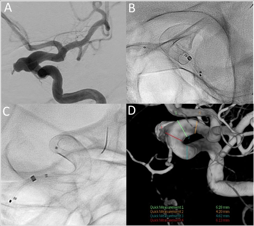
New devices and techniques
J NeuroIntervent Surg: first published as 10.1136/neurintsurg-2021-017421 on 8 April 2021. Downloaded from http://jnis.bmj.com/ on May 31, 2021 by guest. Protected by copyright.
Table 1 Characteristics of aneurysms Table 2 Clinical presentation and antiplatelet protocols
Results (n:60) Results (n:57)
Aneurysm characteristics total (%) Symptoms/antiplatelet regimen information Total (%)
Type Clinical presentation
Saccular 46 (77%) SAH 3 (5%)
Saccular partially thrombosed 2 (3%) Headache 11 (19%)
Dissecting 5 (8%) Blurred vision/diplopia/visual disturbances 7 (12, 3%)
Blister 3 (5%) Nerve paralysis/neuralgia 2 (4%)
Fusiform 4 (7%) Pontine stroke, tetraparesis, dizziness, dysarthria, diplopia 2 (4%)
Status Seizures 1 (2%)
Acutely ruptured (SAH) 3 (5%) Asymptomatic/incidental 31 (54%)
Subacute, partially coiled, SAH 1 month ago 1 (2%) Antiplatelet regimen
Recanalized, previous SAH more than 6 months ago 6 (10%) Aspirin + clopidogrel* 33 (58%)
Unruptured 50 (83%) Aspirin + ticagrelor* 14 (25%)
Location Aspirin + ticlopidine* 2 (4%)
A1 3 (5%) Aspirin + prasugrel* 4 (7%)
MCA 2 (3%) Integrillin ± iv aspirin 2 (4%)
ICA-distal bifurcation 1 (2%) Tirofiban ± iv aspirin 2 (4%)
ICA-cavernous 7 (12%) Platelet function testing
ICA-paraclinoid 4 (7%) Not performed 28 (49%)
ICA-paraophthalmic 26 (43%) Hyporesponse to clopidogrel 4 (7%)
ICA-supraclinoid 10 (17%) Hyperresponse to clopidogrel 2 (4%)
ICA-petrous-cavernous 1 (2%) Hyporesponse to aspirin 1 (2%)
ICA-extracranial 1 (2%) Normoresponders to aspirin and clopidogrel 22 (39%)
Mid-basilar 3 (5%) *A minimum of 3 days before the intervention.
iv, intravenous; SAH, sub-arachnoid hemorrhage.
V4 2 (3%)
ICA, internal carotid artery; MCA, middle cerebral artery; SAH, sub-arachnoid
hemorrhage.
Procedures
Antiplatelet therapy was mandatory before the procedure and
METHODS was administered according to the local institutional protocols.
The study involved a retrospective review of multicenter An antiplatelet reactivity test was not mandatory. Antiplatelet
data regarding a consecutive series of patients with intracra- therapy was continued after discharge per standard of care
nial aneurysms, treated with the SV between September 2020 (table 2).
and January 2021 at 19 institutions worldwide. Institutional Available SV sizes range from 3.5 to 4.75 mm diameters with
ethics committees approved this study. There were no exclu- lengths between 15 and 30 mm. The device has been designed
sion criteria based on aneurysm location, type, size or clinical for vessels from 3.5 to 5 mm in size. All device sizes are deliv-
presentation. ered through a 0.021 inch catheter . The implant is made of
Clinical, intra/periprocedural and angiographic data, including 48 drawn filled tubing (DFT) wires (matching Nitinol with a
approach, materials used, aneurysm size and location, device/s, platinum core into a single wire) which allows full radiopacity
technical details and initial angiographic aneurysm occlusion, without additional platinum wires compared with its predeces-
were analyzed. sors Silk or Silk+. It is 90% resheathable which is comparable to
All intra- and periprocedural events were evaluated. Clinical its smaller version, the Silk Vista Baby; however, the SV device
outcome was evaluated before treatment and at discharge using does not have flared-ends and the radial force is five times more.
the modified Rankin scale (mRS). Minor events were considered The stents were deployed through the following microca-
if symptoms resolved within 7 days and major events if symp- theters: Headway-21 (MicroVention, Tustin, CA) in 45 cases,
toms were present after 7 days. All events were evaluated during Rebar-18 (Medtronic Neurovascular, Irvine, CA) in 10 cases and
the hospitalization, focusing on the intra- and periprocedural Phenom-21 (Medtronic Neurovascular, Irvine, CA) in five cases.
period. Intermediate catheters were used in 38 out of 57 (67%) proce-
Aneurysm characteristics are summarized in table 1. Aneu- dures as follows: Navien 5/6F (Medtronic Neurovascular, Irvine,
rysms were categorized as saccular, dissecting, fusiform or blister CA) in 13 cases, Sofia 6F/Sofia EX (MicroVention, Tustin, CA)
type in nature. The aneurysm size in the subgroup of saccular in 13 cases, Cat 5 (Stryker Neurovascular, Fremont, CA) in four
aneurysms (48 out of 60) were: 22 (46%) cases were small cases, Fargo Max (Balt, Montmorency, France) in three cases,
(New devices and techniques
J NeuroIntervent Surg: first published as 10.1136/neurintsurg-2021-017421 on 8 April 2021. Downloaded from http://jnis.bmj.com/ on May 31, 2021 by guest. Protected by copyright.
Initial occlusion rates were graded intraoperatively on the
last angiographic run according to the O’Kelly–Marotta (OKM)
grading scale for assessment of cerebral aneurysms treated by
flow diversion (considering aneurysm filling as: A, total; B, sub-
total; C, entry remnant; D, no filling).
RESULTS
Sixty aneurysms were treated with SV in 57 procedures; 66
devices were used, three removed, and 63 implanted. The reasons
for removal were: incorrect sizing (one case) and distal-end non-
opening (two cases). On average 1.1 devices per aneurysm were
implanted in this series.
In nine cases (15%) adjunctive coiling was performed using
a jailing technique, seven (12%) cases were already coiled (in a
previous procedure), and in 43 (73%) cases no coils were used
(figure 1).
Among the 57 procedures, recapture and repositioning were
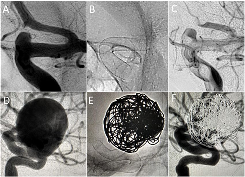
Figure 1 Paraophthalmic aneurysms treated with the Silk Vista device. performed in 18 cases. No microcatheter friction was noted.
(A) DSA, oblique view. A small unruptured paraophthalmic aneurysm. The stented parent vessels had an average diameter of 4.2 mm
(B) Fluoroscopy images which demonstrate the radiopacity of the (range 2.7–5.7 mm) proximally and 3.4 mm (range 2.2–4.93 mm)
4.5×20 mm braid despite the Nitinol design, and no flared-ends. (C) distally. The arterial proximal-distal discrepancy average was
Post-implant DSA showing aneurysm filling with contrast stagnation 0.8 mm (range 0–2.2 mm) (figure 2).
(OKM A). (D) DSA, lateral view. A giant unruptured paraophthalmic
aneurysm. The discrepancy of the proximal and distal arterial diameter
was significant (1.4 mm). (E, F) A 4×30 mm device was deployed in Braid opening
combination with coils achieving a complete aneurysm occlusion (OKM The devices opened instantaneously in 60 out 66 (91%) cases.
D). DSA, digital subtraction angiography; OKM, O’Kelly–Marotta. In four out of 66 (6%) devices a partial opening of the distal
end of the SV occurred. In three of the four cases of distal-end
partial opening, a significant discrepancy in the arterial diameter
(>1.3 mm) was noted. Additionally, in two cases of distal-end
The transfemoral approach was used in 48/57 (84%) of the
non-opening, the location of the aneurysms was on the A1
cases, transradial in eight (14%) cases and transulnar in one (2%)
segment resulting in braid oversizing. While two cases were
case.
managed intraoperatively, in the other two the operators decided
to remove the device.
In one case (2%) the mid- portion of the stent remained
partially constrained, in a 26 mm paraophthalmic aneurysm,
which required a prior coil deployment to prevent microcatheter
FD invagination. There was no restriction of flow and balloon
angioplasty was used to successfully fully open the device.
The proximal end of the device opened fully in all cases, but
in one case a slow opening was reported.
Wall apposition
A complete and full wall apposition was achieved in 58 out of 63
(92%) devices implanted.
In five (8%) devices, incomplete apposition was reported as
follows: an incorrect distal- end wall apposition in two (3%)
cases, one case related to a vessel angulation and another due to
inappropriate sizing, an in-complete mid-stent apposition in one
(2%) case (fully opened with a microwire internal massage), and
an incomplete proximal-end apposition in two (3%) cases—in
one case due to the vessel angulation and in the second case, a
cervical internal carotid artery (ICA) dissecting aneurysm with a
5.3 mm vessel; a 4.75 mm device was selected and after implan-
tation migrated into the aneurysm sac. Attempts were made to
Figure 2 Sample case of an unruptured aneurysm treated with a reposition the device; however, these proved unsuccessful and
single SV flow diverter and coils. (A) Dysplastic left ICA segment (6 mm) the patient was managed conservatively with no further compli-
and paraophthalmic aneurysm. Note distal ICA measures 4.2 mm and cations or repeat hemorrhage.
proximal landing zone measures 4.5 mm. The dysplastic ophthalmic In both cases of malapposition, secondary vessel angulation
segment of the left ICA at the level of the aneurysm neck measures and stent positioning balloon angioplasty failed to resolve the
6.1 mm. (B, C) A 4.75 mm x 25 mm SV device forshortened to 18.5 mm malapposition. One case was initially managed conservatively but
and fully opened to 6 mm through the dysplastic segment with excellent thrombosed 1 hour post-procedure despite adequate antiplatelet
wall apposition. (D) Three dimensional reconstruction of the stented medication and resulted in acute neurological deterioration. The
segment. ICA, internal carotid artery; SV, Silk Vista. patient underwent a repeat procedure with implantation of a
Martínez-G
aldámez M, et al. J NeuroIntervent Surg 2021;0:1–8. doi:10.1136/neurintsurg-2021-017421 3New devices and techniques
J NeuroIntervent Surg: first published as 10.1136/neurintsurg-2021-017421 on 8 April 2021. Downloaded from http://jnis.bmj.com/ on May 31, 2021 by guest. Protected by copyright.
Table 3 Intra- and periprocedural complications
Stent
Intraprocedural Clinical size Vessel mismatch
complication Management Result consequences (mm) (mm)* Aneurysm characteristics
Intrastent distal clotting Abciximab ia Solved None 4.5×20 1.3 Unruptured
Paraophthalmic/saccular
Distal-end device fishmouthing due to Nimodipine ia + Solved None 4×25 0.6 Unruptured
spasm abciximab ia A1/saccular
Distal-end device fishmouthing + PTA + tirofiban Solved None 3.75×20 1.6 Unruptured
clotting A1/saccular
Post/periprocedural complications
Stent thrombosis Tirofiban bolus + Solved Asymptomatic 3.5×25 0.4 Unruptured
(proximal-end malapposition) Telescoped Leo+ basal ganglia M1/dissecting
1 hour post-procedure stent stroke on 24 hours
CT
EVD tract hemorrhage Eptifibatide shut Stable on DAPT No neurological 4×20 (×2) 0.2 Ruptured
off. Continue DAPT deficits Supraclinoid/blister
Neurological deterioration 72 hours Conservative. Hemipontine Worsening on mRS 4×15 0.4 Unruptured, symptomatic mid-
after FD Continue DAPT stroke on DWI (0 to 2) Basilar/dissecting
Superficial groin hematoma Conservative. Solved None 4.75×25 1.4 Unruptured
Compression Paraophthalmic/saccular
Significant when >1 mm.
*Difference between parent artery proximal and distal to aneurysm.
DAPT, dual antiplatelet therapy; DWI, diffusion weighted imaging; EVD, external ventricular drain; FD, flow diverter; ia, intra-arterial; mRS, modified Rankin Scale; PTA,
percutaneous transarterial angioplasty.
Leo+ stent (Balt, Montmorency, France) proximally to fully
open the implanted SV, improving the clinical condition with no
neurological sequelae (table 3, figure 3). In the second case, an
Atlas stent (Stryker Neurovascular, Fremont, CA) was telescoped
distally with no clinical sequelae.
The initial occlusion rate in the 60 aneurysms were as follows:
OKM A in 34 (57%) cases, OKM B in 15 (25%) cases, OKM C
in six (10%) cases, and OKM D in five (8%) cases.
Intraprocedural events
There were three (5 %) intraprocedural thromboembolic events,
all of them related to stent malapposition as described above,
with no permanent neurological morbidity (table 3).
There were no aneurysm ruptures or dissections and no
mortality as a result of the procedure.
Periprocedural events
Four (7%) events were documented post-procedure as follows:
an immediate post-procedural stent thrombosis (table 3, figure 3)
resolved with a telescoped Leo+ stent (no clinical consequences
but an ischemic stroke in the left basal ganglia on 24 hours CT
was reported); one external ventricular drain tract hemorrhage
Figure 3 Left unruptured M1 dissecting/saccular aneurysm. (A) A post-procedure in a case of sub-arachnoid hemorrhage (SAH)
3.5×25 mm device was deployed from M1 to the supraclinoid ICA. (stable after eptifibatide shut off, resolved at time of discharge
The proximal landing zone was located into an arterial angulation (*), from hospital with no new focal neurological deficits); one
and the device did not appose fully to the ICA wall. After unsuccessful hemi-pontine stroke in a case of symptomatic dissecting mid-
attempts of repositioning with balloon angioplasty, it was managed basilar aneurysm; and one groin superficial hematoma with no
conservatively. (B) One hour after the procedure, the patient developed associated pseudoaneurysm.
right hemiparesis and a DSA showed stent thrombosis (arrow) There was no mortality in this study.
secondary to the proximal stent malapposition. (C) A bolus of tirofiban The median length of hospital stay was 3.4 days (range 1–23
was administered and a Leo+ stent was proximally telescoped allowing days). The mRS score at discharge from the hospital did not
a fully wall apposition. (D) Both stents remained opened, fully patent change from the admission mRS score except in three patients:
with no flow compromise. DSA, digital subtraction angiography; ICA, two patients with SAH were discharged with mRS 2 after
internal carotid artery. hospitalization; and one patient presenting with a symptomatic
4 Martínez-Galdámez M, et al. J NeuroIntervent Surg 2021;0:1–8. doi:10.1136/neurintsurg-2021-017421New devices and techniques
J NeuroIntervent Surg: first published as 10.1136/neurintsurg-2021-017421 on 8 April 2021. Downloaded from http://jnis.bmj.com/ on May 31, 2021 by guest. Protected by copyright.
dissecting midbasilar aneurysm was discharged with mRS 2, with In our series, 63 devices were deployed in 57 patients,
a favorable evolution at 1 month (mRS 1). with three thromboembolic complications and three devices
removed. No dissections and no procedure- related mortality
DISCUSSION were reported. All the thromboembolic complications were
Over the past decade, flow diversion has increasingly been related to stent malapposition and this was at least partly due
replacing conventional techniques as the first-line endovascular to the underlying vascular anatomy and the landing sites as well
treatment for many types of intracranial aneurysm.1 as the discrepancy between the diameters of the different vessel
Since the CE mark of the Silk FD device in 2008, three gener- segments where the device would be deployed.
ations of the device have been launched in the last 10 years. Although the safety and efficacy of the first two generations of
Some technical modifications have improved the visibility and the device have been demonstrated in large clinical series, a prin-
the radial force of the implant.15 16 cipal limitation of these early generation devices, as compared
The first generation Silk was constructed from 48 braided with cobalt- chromium FDs, was the radiopacity and radial
wires—44 Nitinol wires and four platinum wires and four plat- force.5
inum coils. The original Silk device was characterized by its flex- Although all FD devices follow the same hemodynamic
ibility and ability to adapt to the arterial anatomy; however, this concept, the companies embarked on the development and
limited the pushability and trackability in tortuous vessels, with manufacturing of a variety of braids with different proper-
poor opening on deployment due to lower radial force. ties during the last years. We could divide the FDs into two
The second generation device (CE mark in 2012), the so-called representative groups: cobalt-chromium and Nitinol devices
Silk+, was made of 48 wires with higher radial force, eight (table 4). While cobalt-chromium implants have the advan-
platinum wires and four platinum coils, designed to improve tage of a higher radial force and greater distal anchor stability
visibility and flared- ends to optimize wall apposition. It was during deployment, the Nitinol FDs are more trackable and
compatible with 0.021 inch and 0.025 inch microcatheters and require lower profile microcatheters (figure 4). Although the
could be resheathed up to 90%. stent design may have a theoretical impact, depending on
Lubicz et al15 reported a single center experience with the use anatomical tortuosity of the parent artery or aneurysm loca-
of both the Silk and Silk+ in 58 patients with 70 aneurysms: 32 tion, no significant clinical differences between FDs have been
patients were treated with the Silk and 26 with the Silk+. Clin- reported so far.
ical periprocedural complications occurred in 15%, the overall Our intra- and periprocedural results should be compared
permanent neurologic morbidity rate was 5.5%, and there was with the more recent literature evidence on FDs.
no procedure-related mortality. Embolization was successful in In 2020, Rice et al17 published the results of the large prospec-
54 patients (93%), and failure occurred in four patients (7%). tive study using the Pipeline Flex embolization device with Shield
In the DIVERSION16 prospective cohort study, 122 Silk technology for the treatment of 204 aneurysms. The average of
devices were deployed in 118 patients. The authors reported the devices was 1.1, the same as for our series. Four out of 252
three thromboembolic events, six incomplete deployments, one devices were removed and adjunctive coils were added in 18.6%.
dissection and two complications at the puncture site. Complete apposition was achieved in 93.1% of cases. Complete
Table 4 Overview of the characteristic of the current flow diverters
Microcatheter Recommended parent
compatibility Device diameter vessel
Device Number of wires Design/materials (inches) range (mm) (mm)
Silk Vista Baby 48 Single-layer. DFT (Nitinol + platinum) 0.017 2.25–3.25 1.5–3.5
Silk Vista 48 Single-layer. DFT (Nitinol + platinum) 0.021 3.5–4.75 3.5–5
Silk+ 48 Single-layer. Nitinol and platinum 0.021 and 0.025 2–5.5 1.5–5.75
Pipeline Shield 48 Single-layer. Cobalt-chromium-nickel alloy and platinum- 0.027 2.5–5 2–5
tungsten
P64 MW HPC 64 Single-layer. DFT (Nitinol + platinum) 0.021 3–5 2.5–5
P48 MW HPC 48 Single-layer. DFT (Nitinol + platinum) 0.021 2–3 1.75–3
Surpass Evolve 48 Single-layer. Cobalt-chromium and platinum-tungsten 0.027 2.5 2.0–2.5
Surpass Evolve 64 Single-layer. Cobalt-chromium and platinum-tungsten 0.027 3,25–5 2.5–5
Derivo 48 (24 wires looped) Single-layer. DFT (Nitinol + platinum) and platinum- 0.027 3.5–6 2.5–6
iridium
Derivo mini 48 (24 wires looped) Single-layer. DFT (Nitinol + platinum) and platinum- 0.021 2.5–3.5 1.5–3.5
iridium
FRED 48 (inner) + 16 (outer) Dual-layer. Nitinol and tantalum 0.027 3.5–5.5 2–5
FRED Jr 36 (inner) + 16 (outer) Dual-layer. Nitinol and tantalum 0.021 2.5–3 2–3
Tubridge 48 Single-layer. Nitinol and platinum-iridium 0.029 2.5–3 2–3
Tubridge 64 Single-layer. Nitinol and platinum-iridium 0.029 3.5–6.5 3–6.5
Silk Vista, Silk Vista Baby and Silk+ (Balt extrusion, Montmorency, France), Pipeline Shield (Medtronic Neurovascular, Irvine, CA), P48 MW HPC and P64 MW HPC (Phenox,
Bochum, Germany), Surpass Evolve (Stryker Neurovascular, Fremont, CA), Derivo and Derivo Mini (Acandis, Acandis GmbH, Pforzheim, Germany), FRED and FRED Jr
(Microvention, Tustin, CA), Tubridge (MicroPort NeuroTech, Shanghai, China).
DFT, drawn filled tubing.;
Martínez-G
aldámez M, et al. J NeuroIntervent Surg 2021;0:1–8. doi:10.1136/neurintsurg-2021-017421 5New devices and techniques
J NeuroIntervent Surg: first published as 10.1136/neurintsurg-2021-017421 on 8 April 2021. Downloaded from http://jnis.bmj.com/ on May 31, 2021 by guest. Protected by copyright.
the distal location, procedures without intermediate catheters/
triaxial systems were performed in 20 out of 41 procedures.
The SV can be considered to be a larger version of the SVB
and is designed for vessels with a diameter between 3.5 and
5 mm and compatible with a 0.021 inch microcatheter. The
design utilizes the same DFT technology as seen in the SVB and
is constructed from 48 DFT wires, but it does not have flared-
Figure 4 Paraophthalmic aneurysm, lateral view. (A) A Headway-21 ends. The radial force of the SV is considerably higher than that
microcatheter was intentionally navigated into the aneurysm lumen for of the SVB with approximately five times greater radial force
distal advance into the supraclinoid ICA due to the anatomical location than the SVB. The flared-ends concept was introduced in 2007
and the wide neck. An SV device was tracked without friction and with the Leo+ stents and continued with Silk, Silk+ and SVB,
opened by push-pull technique (B–E). Once the distal third of the SV with the intention to prevent a potential device migration. In
device was opened and anchored, the microcatheter was gently pulled the case of SV the design is purely tubular and may help with
back and centered into midline for deployment. ICA, internal carotid achieving a more precise wall apposition.
artery; SV, Silk Vista. FD malapposition is associated with an increased risk of
stroke-related complications.10 11 In our study we identified wall
malapposition in 8% of cases with affected segments at the prox-
occlusion of the aneurysm was achieved in 1% of the cases. imal, mid and distal ends of the device. The improved visibility
Periprocedural stroke occurred in 6.4% during the first 30 days. enabled by the DFT technology allowed this to be detected and
In our series wall apposition was achieved in 92%, complete would have gone unnoticed with the previous generation Silk or
aneurysm occlusion in 8% and thromboembolic events in 5%. Silk+ devices.
Also in 2020, Maus et al18 published a case series using the Most of the FDs used in proximal locations remain compat-
ible only with 0.027 inch ID microcatheters6–8 (table 4). While
new Surpass Evolve. Like the Pipeline Shield, devices were
in distal locations there is a clear benefit to the use of lower
deployed using a triaxial system with a 0.027 inch microcath-
profile stents and delivery catheters, the potential benefit of a
eter. Authors reported 46 aneurysms treated with the device, but
lower profile 0.021 inch compatible device for vessels of 4 or
the average was 1.2, slightly higher than in our series. Adjunc-
5 mm remains unclear. Beyond the evident benefits in terms of
tive coiling was performed in 37% of the cases; however, the
navigation through tortuous anatomies, it is our opinion that an
authors did not report the immediate OKM occlusion rate.
0.021 inch compatible FD could have several potential benefits
Intraprocedural events occurred in 2% (one patient suffering a
over the more conventional 0.027 inch systems. One hypothet-
stent thrombosis managed endovascularly without sequelae) and
ical benefit could be lowering the friction between the catheters,
mortality occurred in 2% (in a patient who presented with an
a source of polymer embolism. The excessive manipulation of
SAH). They reported two unsuccessful deployments, related to
tight-fitting devices in coaxial, triaxial and quadriaxial catheter-
tortuous vessel anatomy.
ization techniques within tortuous vessels, use of larger diameter
In our series, all cases were consecutive and there were no
devices, multiple or difficult catheterization through previously
unsuccessful deployments.
placed devices, repeat interventions, and the presence of calcific
In 2019, Pierot et al19 presented the results of the prospective atherosclerotic debris may ‘scrape’ the polymer coating layer or
SAFE study, using FRED and FRED Jr devices, for the treat- at times even the base coat layer off the device.21–23
ment of 103 aneurysms. Despite being made of Nitinol, FRED Other potential advantages could be the extra space within
requires a 0.027 inch microcatheter for delivery, while FRED the internal lumen of the guiding catheter that would allow for
Jr, designed for distal vessels ranging from 2.5 to 3 mm, is improved contrast injections and hence improved angiography,
designed for deployment through a 0.021 inch microcatheter. better detection of intra-aneurysmal stasis, earlier detection of
Treatment was successfully performed in 95% of cases. Throm- thrombus and distal complications, as well as more effective
boembolic complications occurred in 6.8%, three during the flushing which in turn may reduce the risk of thromboembolic
procedure or immediately after (within 6 hours) and four after complications. Similarly, in the case of telescoping, the naviga-
the procedure (1.4 and 7 days with a further event at 14 months tion of lower profile ID microcatheters through a previously
post-procedure). Intraoperative rupture occurred in two of 103 implanted device could reduce the risk of dislodging the initial
patients (1.9%). device.
The SV is the only FD made with 48 wires which is 0.021 Microcatheter size is an important factor to consider when
inch compatible and available for vessels up to 5 mm. Recently attempting to maneuver beyond the distal lip of an aneurysm.
another Nitinol FD with the same 0.021 inch profile has been Different techniques have been proposed for advancing a 0.027
launched, the P64 MW HPC; however, clinical experience for inch distal to an aneurysm for FD deployment.9 10 24 The inability
this device is limited at the moment.20 to navigate beyond challenging segments can result in the need
In 2018 the third generation device, the SVB, received CE to abandon the preferred treatment approach or change the
approval for the treatment of aneurysms located in small or distal components of the coaxial system. There has been a paradigm
vessels (1.5–3.5 mm). The device used DFT technology and shift in the design and approach to catheter support systems for
allowed full radio-opacity of the device. The SVB is constructed cases of FD from a classic biaxial set-up to a more robust triaxial
from 48 DFT wires and is the first FD that can be delivered system.9 10 Despite the safe profile, concern for catheter-induced
through a 0.017 inch microcatheter.3 4 arterial injury and proximal tortuosity limits the performance
Recently, the periprocedural outcomes of SVB in a series of 41 applicability of distal intermediate catheters . In our series, 34%
patients with 43 small aneurysms (mean 9.5 mm) at and beyond of the procedures were performed without the use of interme-
to the circle of Willis were assessed.5 The intraoperative complete diate catheters, highlighting the trackability of the SV device
occlusion rate was 18.6%. There were five cases of intrapro- (figure 4). Similarly, the requirement for extra delivery and
cedural complications with no clinical consequence. Despite access catheters also raises the cost of these procedures.
6 Martínez-Galdámez M, et al. J NeuroIntervent Surg 2021;0:1–8. doi:10.1136/neurintsurg-2021-017421New devices and techniques
J NeuroIntervent Surg: first published as 10.1136/neurintsurg-2021-017421 on 8 April 2021. Downloaded from http://jnis.bmj.com/ on May 31, 2021 by guest. Protected by copyright.
In braided stent FDs, the radial force is maximal in the center León, Spain
19
under nominal diameter, that is, the FD is not under constraint. Interventional Neuroradiology, Hospital General Universitario de Castellon,
Valencia, Castellon, Spain
The radial force drops rapidly toward both ends of the FD25 if 20
Interventional Neuroradiology, Centre Hospitalier Universitaire de Limoges,
the FD is under constraint when placed in an artery with a vessel Limoges, Limousin, France
diameter smaller than the nominal diameter. In our series, in 21
University Limoges, CNRS, XLIM, UMR 7252, Limoges, France
three cases of sub-optimal opening of the distal-end of the device
there was a significant arterial discrepancy and two of the cases Twitter Mario Martínez-Galdámez @Doctorgaldamez, Vladimir Kalousek @
occurred in a distal segment (A1) after braid oversizing. When VladoKZg, Rodrigo Rivera @neurofox, Miguel Schüller Arteaga @drschuller, Jose
a significant arterial discrepancy is present, the oversizing effect David Guio @jdavidguio and Pedro Navia @pnavia
of a single device may be minimized by placing two telescoped Contributors MMG: study concept, literature review, acquisition of data, draft
devices of different sizes. and review of the manuscript; PB: critical review. All authors have contributed to the
Physician experience with a device has been related to the risk authorship, and final review of the manuscript.
of complications, highlighting the need for a learning curve.26 Funding The authors have not declared a specific grant for this research from any
Despite the SV being a relatively new device, in this study the funding agency in the public, commercial or not-for-profit sectors.
average number of devices implanted was 1.1 and intrapro- Competing interests MMG is proctor and consultant for Balt, Medtronic and
cedural events occurred in 5%, suggesting that an extensive Stryker. PG is consultant for Phenox, Balt and Cerenovus. JGF is consultant for
Medtronic and Balt. MSA is consultant for Medtronic and Balt. PN is consultant and
learning curve is not required and the lessons of the past and proctor for Balt, Stryker and Penumbra. The rest of the co-authors have not declared
experience with FDs in general is sufficient for experienced any conflict of interesting regarding this manuscript.
operators. Patient consent for publication Not required.
Our study has limitations including the outcomes of a retro-
Ethics approval Institutional Review Board. CEIm Área de Salud Valladolid Este.
spective multicenter experience that is not randomized, which PI 21–2169.
leads to selection bias. This is a small sample size and long-
Provenance and peer review Not commissioned; externally peer reviewed.
term clinical outcomes are necessary for evaluating long-term
safety and efficacy. Angiographic findings, such as evaluation Open access This is an open access article distributed in accordance with the
Creative Commons Attribution Non Commercial (CC BY-NC 4.0) license, which
of in-stent stenosis27 or delayed parent artery occlusions,28 permits others to distribute, remix, adapt, build upon this work non-commercially,
need to be compared with previous generations. The sample and license their derivative works on different terms, provided the original work is
did not include a significant number of giant aneurysms, where properly cited, appropriate credit is given, any changes made indicated, and the use
complex maneuvers are required, including telescoping devices is non-commercial. See: http://creativecommons.org/licenses/by-nc/4.0/.
in tortuous anatomies. We did not perform a post-procedural ORCID iDs
diffusion weighted imaging, which could be helpful to evaluate Mario Martínez-Galdámez http://orcid.org/0000-0002-8024-4712
the potential emboli due to catheter frictions. Vladimir Kalousek http://orcid.org/0000-0002-1439-0930
Our results appear promising, but larger series with longer- Rodrigo Rivera http://orcid.org/0000-0001-8991-0972
Andrei Filioglo http://orcid.org/0000-0002-4200-3141
term follow-ups are needed to corroborate the effectiveness of Ali Burak Binboga http://orcid.org/0000-0001-5220-1232
this treatment method and its superiority to other devices or Mehmet Onay http://orcid.org/0000-0001-6220-3207
techniques. Miguel Schüller Arteaga http://orcid.org/0000-0003-3351-668X
Pervinder Bhogal http://orcid.org/0000-0002-5514-5237
Pedro Navia http://orcid.org/0000-0002-6516-6090
CONCLUSION Andrés Fernandez Prieto http://orcid.org/0000-0001-9207-6093
Our study demonstrated that the use of the new FD Silk Vista Aymeric Rouchaud http://orcid.org/0000-0003-0902-3375
for the treatment of intracranial aneurysms is feasible and tech-
nically safe. REFERENCES
1 Lv X, Yang H, Liu P, et al. Flow-diverter devices in the treatment of intracranial
Author affiliations aneurysms: a meta-analysis and systematic review. Neuroradiol J 2016;29:66–71.
1
Interventional Neuroradiology/Endovascular Neurosurgery, Hospital Clínico 2 Cagnazzo F, Perrini P, Dargazanli C, et al. Treatment of unruptured distal anterior
Universitario de Valladolid, Valladolid, Spain circulation aneurysms with flow-diverter stents: a meta-analysis. AJNR Am J
2
Radiology, Fatih Sultan Mehmet Training and Research Hospital, Istanbul, Turkey Neuroradiol 2019;40:687–93.
3
Neurosurgery & Radiology, Hadassah-Hebrew Univ Med Ctr, Jerusalem, Israel 3 Bhogal P, Wong K, Uff C, et al. The Silk Vista Baby: initial experience and report of two
4
Department of Radiology, Clinical Hospital Center "Sestre Milosrdnice", Zagreb, cases. Interv Neuroradiol 2019;25:530–8.
Croatia 4 Schob S, Hoffmann K-T, Richter C, et al. Flow diversion beyond the circle of Willis:
5
Neuroradiology, Instituto de Neurocirugia, Dr. Asenjo, Santiago, Chile endovascular aneurysm treatment in peripheral cerebral arteries employing a novel
6
Interventional Neuroradiology, Radiology Department, Toronto Western Hospital, low-profile flow diverting stent. J Neurointerv Surg 2019;11:1227–34.
Toronto, Ontario, Canada 5 Martínez-Galdámez M, Biondi A, Kalousek V, et al. Periprocedural safety and technical
7
Neurointerventional Department C.D.I, Hospital Clinic de Barcelona, Barcelona, outcomes of the new Silk Vista Baby flow diverter for the treatment of intracranial
Spain aneurysms: results from a multicenter experience. J Neurointerv Surg 2019;11:723–7.
8
Neuroradiology, Radiological imaging department, Spedali Civili of Brescia, Brescia, 6 Chiu AHY, Phillips TJ. Future directions of flow diverter therapy. Neurosurgery
Italy 2020;86:S106–16.
9
Department of Surgery/Medicine, McMaster University, Hamilton, Ontario, Canada 7 Dandapat S, Mendez-Ruiz A, Martínez-Galdámez M, et al. Review of current
10
Radiology, Hadassah-Hebrew Univ Med Ctr, Jerusalem, Israel intracranial aneurysm flow diversion technology and clinical use. J Neurointerv Surg
11
Neurology, Hadassah-Hebrew Univ Med Ctr, Jerusalem, Israel 2021;13:54–62.
12
Radiology, Dr Ersin Arslan Training and Research Hospital, Sahinbey, Gaziantep, 8 Maragkos GA, Dmytriw AA, Salem MM, et al. Overview of different flow diverters and
Turkey flow dynamics. Neurosurgery 2020;86:S21–34.
13
Department of Interventional Neuroradiology, Royal London Hospital, London, UK 9 Colby GP, Lin L-M, Xu R, et al. Utilization of a novel, multi-durometer intracranial
14
CHRU de Brest, Brest, Bretagne, France distal access catheter: nuances and experience in 110 consecutive cases of aneurysm
15
Interventional Radiology, Azienda Sanitaria Universitaria Friuli Centrale, UDINE, Ud, flow diversion. Interv Neurol 2017;6:90–104.
Italy 10 Lin L-M, Colby GP, Iyer RR, et al. Pentaxial access platform for ultra-distal intracranial
16
Radiology- Interventional Neuroradiology, Hospital Universitario La Paz, Madrid, delivery of a large-bore hyperflexible DIC (distal intracranial catheter): a technical
Spain note. Interdisciplinary Neurosurgery 2016;6:29–34 https://www.researchgate.net/
17
Interventional Neuroradiology. Radiology, Hospital de Cruces, Barakaldo, País publication/304107815_Pentaxial_a ccess_platform_for_ultra-d istal_intracranial_
Vasco, Spain delivery_of_a_large-bore_hyperflexible_DIC_distal_intracranial_c atheter_A_
18
Interventional Neuroradiology, Hospital Universitario de Burgos, Burgos, Castilla y technical_note
Martínez-G
aldámez M, et al. J NeuroIntervent Surg 2021;0:1–8. doi:10.1136/neurintsurg-2021-017421 7New devices and techniques
J NeuroIntervent Surg: first published as 10.1136/neurintsurg-2021-017421 on 8 April 2021. Downloaded from http://jnis.bmj.com/ on May 31, 2021 by guest. Protected by copyright.
11 Jabbour P, Chalouhi N, Tjoumakaris S. The Pipeline embolization device: learning 19 Pierot L, Spelle L, Berge J, et al. SAFE study (Safety and efficacy Analysis of FRED
curve and predictors of complications and aneurysm obliteration. Neurosurgery Embolic device in aneurysm treatment): 1-year clinical and anatomical results. J
2013;73:113–20. discussion 120. Neurointerv Surg 2019;11:184–9.
12 Tan LA, Keigher KM, Munich SA, et al. Thromboembolic complications with Pipeline 20 Petrov A, Rentsenkhuu G, Nota B, et al. Initial experience with the novel p64MW HPC
embolization device placement: impact of procedure time, number of stents and pre- flow diverter from a cohort study in unruptured anterior circulation aneurysms under
procedure P2Y12 reaction unit (PRU) value. J Neurointerv Surg 2015;7:217–21. dual antiplatelet medication. Interv Neuroradiol 2021;27:42-50.
13 Sirakov A, Bhogal P, Möhlenbruch M, et al. Endovascular treatment of patients 21 Chopra AM, Mehta M, Bismuth J, et al. Polymer coating embolism from intravascular
with intracranial aneurysms: feasibility and successful employment of a medical devices - a clinical literature review. Cardiovasc Pathol 2017;30:45–54.
new low profile visible intraluminal support (LVIS) EVO stent. Neuroradiol J 22 Geisbush TR, Marks MP, Heit JJ. Cerebral foreign body reaction due to hydrophilic
2020;33:377–85. polymer embolization following aneurysm treatment by Pipeline flow diversion device.
14 Martínez-Galdámez M, Orlov K, Kadziolka K, et al. Safety and efficacy of intracranial Interv Neuroradiol 2019;25:447–53.
aneurysm embolization using the "combined remodeling technique": low-profile 23 Tokunaga K, Hatano T, Nakahara I, et al. Factors associated with postprocedural
stents delivered through double lumen balloons: a multicenter experience. diffusion-weighted imaging-positive lesions in endovascular treatment for unruptured
Neuroradiology 2019;61:1067–72. cerebral aneurysms. World Neurosurg 2019;130:e457–62.
15 Lubicz B, Van der Elst O, Collignon L, et al. Silk flow-diverter stent for the treatment 24 Kellner CP, Chartrain AG, Schwegel C, et al. The bumper technique for advancing a
of intracranial aneurysms: a series of 58 patients with emphasis on long-term results. large profile microcatheter. J Neurointerv Surg 2017;9:e38.
AJNR Am J Neuroradiol 2015;36:542–6. 25 Kühn AL, Wakhloo AK, Gounis MJ, et al. Use of self-expanding stents for better
16 Gariel F, Marnat G, Barreau X, et al. Safety and efficacy of the Silk flow diverter: intracranial flow diverter wall apposition. Interv Neuroradiol 2017;23:129–36.
insight from the DIVERSION prospective cohort study. J Neuroradiol 2020;29:S0150- 26 Jabbour P, Chalouhi N, Tjoumakaris S, et al. The Pipeline embolization device: learning
9861:30203–0. curve and predictors of complications and aneurysm obliteration. Neurosurgery
17 Rice H, Martínez Galdámez M, Holtmannspötter M, et al. Periprocedural to 1-year 2013;73:113–20.
safety and efficacy outcomes with the pipeline embolization device with Shield 27 Essbaiheen F, AlQahtani H, Almansoori TM, et al. Transient in-stent stenosis
technology for intracranial aneurysms: a prospective, post-market, multi-center study. at mid-term angiographic follow-up in patients treated with Silk flow diverter
J Neurointerv Surg 2020;12:1107–12. stents: incidence, clinical significance and long-term follow-up. J Neurointerv Surg
18 Maus V, Weber W, Berlis A, et al. Initial experience with Surpass Evolve flow diverter in 2019;11:166–70.
the treatment of intracranial aneurysms. Clin Neuroradiol 2020. doi:10.1007/s00062- 28 Macdonald IR, Shankar JJS. Delayed parent artery occlusions following use of Silk flow
020-00972-5 diverters for treatment of intracranial aneurysms. J Neurointerv Surg 2019;11:690–3.
8 Martínez-Galdámez M, et al. J NeuroIntervent Surg 2021;0:1–8. doi:10.1136/neurintsurg-2021-017421You can also read