Characterization of laser speckle flowgraphy pulse waveform parameters for the evaluation of the optic nerve head and retinal circulation
←
→
Page content transcription
If your browser does not render page correctly, please read the page content below
www.nature.com/scientificreports
OPEN Characterization of laser speckle
flowgraphy pulse waveform
parameters for the evaluation
of the optic nerve head and retinal
circulation
Nobuko Enomoto1*, Ayako Anraku1, Goji Tomita1, Aiko Iwase2, Takashi Sato1,
Nobuyuki Shoji3, Tomoaki Shiba4, Toru Nakazawa5, Kazuhisa Sugiyama6, Koji Nitta7 &
Makoto Araie8
To characterize laser speckle flowgraphy (LSFG) pulse waveform parameters for ocular circulation
evaluation, a multicenter, prospective, cross-sectional study was conducted in 111 eyes of 86
healthy Japanese individuals. Optic nerve head (ONH) tissue-area, vessel-area mean blur rate (MT
and MV, respectively), and MT and MV pulse waveform parameters were obtained using LSFG and
ONH structural parameters using planimetry. Multivariate linear mixed-effects modeled regression
analysis identified factors contributing to MT- or MV-waveforms using age, gender, smoking history,
body mass index, systolic and diastolic blood pressure, heart rate, intraocular pressure, axial length,
disc, rim, and β-peripapillary atrophy areas, MT or MV, central retinal artery, and vein equivalents
(CRAE and CRVE) as explanatory variables. MT- and MV-waveforms significantly correlated with
one or more systemic factors, consistent with previous studies. Following confounding factor
adjustment, MT-Skew significantly negatively correlated with β-PPA area (P = 0.026); MT- and MV-flow
acceleration index positively correlated with CRAE, MT, and MV (P = 0.041–< 0.001), compatible with
these parameters’ observed correlations to systemic factors. Significantly negative correlations of
the blowout score and acceleration time index to CRAE partly conflicted with their correlations to
systemic factors, and other waveform parameters showed little correlation to ocular factors. Thus,
Skew and flow acceleration index assisted the in vivo ocular circulation characterization.
Disturbed circulation in the optic nerve head (ONH) is related to the development and/or progression of
glaucoma1–3, and many investigators have studied blood flow impairment in the ONH in glaucoma using vari-
ous methods, such as scanning laser Doppler flowmetry, color Doppler imaging, or laser speckle flowgraphy
(LSFG)4. LSFG utilizes the laser speckle phenomenon for the measurement of ocular blood flow in a noninvasive
manner5 and provides the mean blur rate (MBR), which is a quantitative index of blood flow velocity in the
target tissues. For assessing the ONH circulation, the ONH tissue-area MBR (MT) corresponding to the ONH
micro-circulation, the ONH vessel-area MBR (MV) corresponding to blood velocity through large visible ves-
sels in the ONH area, and all-area MBR (MA), which is mean MBR in all area of the ONH, can be derived using
LSFG6–9. Animal experiments using the m icrosphere10 or hydrogen gas clearance m
ethod11,12 have shown that
MT is a quantitative index of the ONH tissue blood flow, and a comparison of the measurement results from
LSFG to those from laser Doppler velocimetry has shown that MV indicates blood flow through the major retinal
vessels13–15. Furthermore, LSFG enables the recording of changes in the pulse waveforms of the measured MBR,
1
Department of Ophthalmology, Toho University Ohashi Medical Center, 2‑22‑36, Ohashi Meguro‑ku,
Tokyo 153‑8515, Japan. 2Tajimi Iwase Eye Clinic, Tajimi, Gifu, Japan. 3Department of Ophthalmology, Kitasato
University School of Medicine, Sagamihara, Kanagawa, Japan. 4Department of Ophthalmology, School of
Medicine, Toho University, Tokyo, Japan. 5Department of Ophthalmology, Tohoku University Graduate School
of Medicine, Sendai, Japan. 6Department of Ophthalmology, Kanazawa University Graduate School of Medical
Science, Kanazawa, Japan. 7Fukui-ken Saiseikai Hospital, Fukui, Japan. 8Kanto Central Hospital of the Mutual Aid
Association of Public School Teachers, Tokyo, Japan. *email: nobuko.enomoto@med.toho-u.ac.jp
Scientific Reports | (2021) 11:6847 | https://doi.org/10.1038/s41598-021-86280-5 1
Vol.:(0123456789)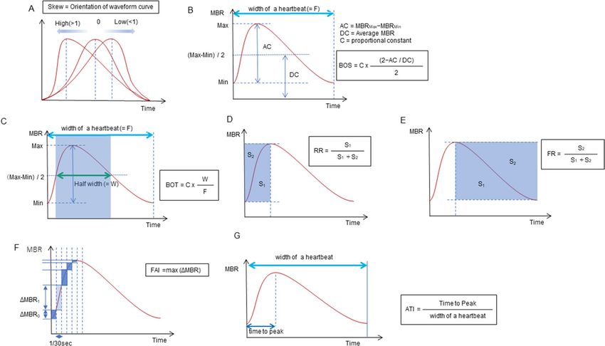
www.nature.com/scientificreports/
Parameters 111 eyes, 86 participants Range
Age, years 47.1 ± 12.2 30–75
Gender: Men/Woman, no. (%) 57/54 (51.4 / 48.6)
Smoking history: Yes/No, no. (%) 17/94 (15.3 / 87.4)
BMI, kg /m 22.1 ± 2.8 17.8–29.1
Systolic blood pressure, mmHg 116.7 ± 14.1 83–148
Diastolic blood pressure, mmHg 72.7 ± 9.4 51–89
Pulse pressure, mmHg 44.0 ± 10.2 24–78
Heart rate, bpm 71.6 ± 8.6 54–96
Spherical equivalent refractive error, diopters − 1.32 ± 1.97 − 5.8–2.0
Axial length, mm 24.1 ± 1.0 22.2–26.4
Central corneal thickness, µm 529.5 ± 37.2 443–617
Intraocular pressure, mmHg 14.5 ± 2.1 8.0–20.0
Disc area, mm 2.35 ± 0.39 1.50–3.37
Rim area, mm 1.56 ± 0.42 0.75–2.98
β-PPA area, mm 0.40 ± 0.46 0.00–2.19
CRAE, µm 141.5 ± 17.6 106–191
CRVE, µm 222.8 ± 24.8 176–276
Table 1. Characteristics of participants. The values are represented as mean ± standard deviation. AU arbitrary
unit, CRAE central retinal artery equivalent, CRVE central retinal vein equivalent, MBR mean blur rate, MT
MBR in the optic nerve head tissue area, MV MBR in the optic nerve head vessel area, PPA peripapillary
atrophy.
such as MT and MV, which are synchronized with the cardiac cycle6,7,16–21. Because laser speckle phenomenon is
an interference event observed when lasers are scattered by a diffusing surface22, the LSFG-measured MBR should
also be influenced by reflection, absorption, and penetration depth of the laser in the target tissue5,13, and needs
to be adjusted not only for systemic factors but also for local ocular factors such as disc, cup, β-PPA area or axial
length23 for inter-individual comparison. Conversely, LSFG pulse waveforms themselves are relatively free from
such effects; therefore, they could be more suited for inter-individual comparison of the ONH or retinal circula-
tory status than the MT or MV. Several LSFG pulse waveform parameters reportedly showed significant differ-
ences between normal and glaucoma e yes24–26, between various disease stages of g laucoma24, and a significant
change after t rabeculectomy27,28, and water d rinking29. Further, a significant correlation of leukocyte glutathione
levels, an indicator of mitochondrial dysfunction, was found with one LSFG pulse waveform parameter for MT
in glaucoma patients30. These previous studies have suggested the potential of LSFG pulse waveform parameters
in investigating the circulatory status changes in the ONH in relation to glaucomatous damages. In an attempt to
characterize the pathophysiological implications of the LSFG pulse waveform parameters, previous studies have
correlated them to the circulatory parameters of the systemic circulation. For example, blow-out time (BOT)
was reportedly related to the stiffness of large a rteries18–21, left ventricular f unction19, and systemic vascular
resistance31; blow-out score (BOS) to the stiffness of large arteries20 and left ventricular f unction32; Skew to the
stiffness of large arteries21; and acceleration time index (ATI) to left ventricular f unction32.
While several studies have looked at the differences of LSFG pulse waveform parameters between the glau-
coma and normal e yes24–26 or between s exes6,7,17, it is still largely unclear which LSFG pulse waveform param-
eters reflect the pathophysiology of ocular fundus circulation more effectively. We assumed that there should be
particularly useful waveform parameters for ocular circulation studies. The objective of this study is to investi-
gate the ophthalmologic implications of seven MT- and MV-LSFG pulse waveform parameters through careful
examination of healthy Japanese according to the predetermined uniform measurement protocol and investigate
how the known quantitative indices of ocular fundus circulation, that is, MT, MV, and central retinal artery and
vein equivalents (CRAE and CRVE) were correlated with seven pulse waveform parameters after adjustment for
systemic parameters6,16,17 and the ONH structural parameters.
Results
A total of 111 eyes from 86 healthy participants (57 men and 54 women) with an average age of 47.1 ± 12.2 years
were included in the study. Tables 1 and 2 show the baseline characteristics and LSFG parameters of the study
participants, respectively. In a preliminary study including 40 normal eyes of a separate group, seven MT-
and MV-waveform parameters were twice obtained as described above at an interval of 7 days (by T.S.) and
intra-class correlation coefficients between them were calculated. The intra-class correlation coefficients were
between 0.967 (95% confidence interval: 0.938–0.982) and 0.999 (95% confidence interval: 0.998–1.000) for
MT-waveform parameters and between 0.963 (95% confidence interval: 0.931–0.980) and 0.995 (95% confidence
interval: 0.990–0.997) for MV-waveform parameters. Among the MT- and MV-waveform parameters, very high
inter-correlations were observed between MT- or MV-BOS and MT- or MV-resistivity index (Spearman’s cor-
relation coefficient: − 0.997– − 0.991) and between MT- or MV-BOS and MT- or MV-Fluctuation (Spearman’s
Scientific Reports | (2021) 11:6847 | https://doi.org/10.1038/s41598-021-86280-5 2
Vol:.(1234567890)www.nature.com/scientificreports/
MT MV
LSFG parameter (AU) (range) (range)
12.6 ± 2.3 48.8 ± 7.4
MBR
(7.4–20.4) (33.9–71.6)
12.0 ± 1.7 10.7 ± 1.7
Skew
(6.3–15.8) (5.1–14.4)
76.9 ± 4.8 80.0 ± 4.4
BOS
(62.4–85.1) (65.6–89.1)
50.6 ± 4.2 55.0 ± 4.3
BOT
(39.4–59.4) (45.2–67.7)
12.7 ± 1.0 13.1 ± 1.2
RR
(9.9–15.3) (9.8–16.1)
13.1 ± 0.9 12.4 ± 0.8
FR
(11.2–15.6) (10.7–14.5)
1.5 ± 0.4 5.5 ± 1.6
FAI
(0.8–3.2) (1.9–10.0)
29.5 ± 3.3 29.4 ± 4.7
ATI
(21.0–40.5) (15.6–40.8)
Table 2. LSFG parameters of 111 eyes. The values are represented as mean ± standard deviation. ATI
acceleration time index, AU arbitrary unit, BOS blowout score, BOT blowout time, FAI flow acceleration index,
FR falling rate, MBR mean blur rate, MT MBR in tissue area, MV MBR in vessel area, RR rising rate.
Parameter
(AU) BOS BOT RR FR FAI ATI RI Fluctuation (%)
MT − 0.700* − 0.776* 0.153 0.735* 0.429* − 0.329* 0.706* 0.708*
Skew
MV − 0.569* − 0.745* 0.042 0.630* 0.391* − 0.399* 0.588* 0.617*
MT – 0.696* − 0.164 − 0.718* − 0.550* 0.058 − 0.997* − 0.981*
BOS
MV – 0.489* − 0.136 − 0.479* − 0.557* 0.172 − 0.991* − 0.967*
MT – – 0.089 − 0.883* − 0.185 − 0.042 − 0.697* − 0.658*
BOT
MV – – 0.178 − 0.805* − 0.056 0.028 − 0.503* − 0.487*
MT – – – 0.031 0.244* 0.112 0.177 0.205†
RR
MV – – – 0.009 0.163 0.305* 0.149 0.197†
†
MT – – – – 0.221 0.147 0.711* 0.653*
FR
MV – – – – 0.047 0.120 0.472* 0.418*
MT – – – – – − 0.303* 0.560* 0.597*
FAI
MV – – – – – − 0.488* 0.554* 0.595*
MT – – – – – – − 0.069 − 0.085
ATI
MV – – – – – – − 0.179 − 0.175
MT – – – – – – – 0.986*
RI
MV – – – – – – – 0.971*
Table 3. Spearman’s rank correlation coefficient between MT- and MV-waveform parameters. ATI
acceleration time index, AU arbitrary unit, BOS blowout score, BOT blowout time, FAI flow acceleration index,
FR falling rate, RI resistivity index, RR rising rate. *P < 0.01, †P < 0.05.
correlation coefficient: − 0.981– − 0.967), indicating that these two parameters were represented by MT- or MV-
BOS (Table 3). Table 4 shows the results of the multivariate linear mixed-effect modeled regression analysis
assessing the contribution of each factor to the pulse waveforms for MT and MV.
MT-Skew significantly negatively correlated with diastolic blood pressure (DBP), heart rate (HR), and β-PPA
area, and positively with age, while MV-Skew significantly negatively correlated with DBP and HR, and positively
with age when DBP was adopted as an explanatory variable.
Both MT- and MV-BOS were significantly negatively correlated with age and CRAE and positively corre-
lated with HR, when the systolic blood pressure (SBP) was adopted as an explanatory variable, and significantly
negatively correlated with age and CRAE and positively correlated with DBP and HR when DBP was adopted
as an explanatory variable.
MT-BOT significantly negatively correlated with age and positively with HR and MV-BOT negatively cor-
related with age.
MT-rising rate (RR) was significantly negatively correlated with HR when SBP or DBP was adopted as an
explanatory variable, and MV-RR was significantly negatively correlated with gender (greater in women), smok-
ing history (greater with no smoking history), and HR, when DBP was adopted as an explanatory variable.
Scientific Reports | (2021) 11:6847 | https://doi.org/10.1038/s41598-021-86280-5 3
Vol.:(0123456789)www.nature.com/scientificreports/
SBP includeda DBP includedb SBP and DBP not includedc
Coefficient (P-value) Coefficient (P-value) Coefficient (P-value)
Parameters Explanatory variable MT-Skew MV-Skew MT-Skew MV-Skew MT-Skew MV-Skew
0.062 ± 0.012
Age, years NS NS 0.038 ± 0.013 (0.005)
(< 0.001)
− 0.040 ± 0.017 − 0.047 ± 0.018
DBP, mmHg
(0.022) (0.011)
Skew
− 0.061 ± 0.019 − 0.044 ± 0.020
HR, bpm NS NS
(0.001) (0.031)
− 0.617 ± 0.273
β-PPA area, mm2 NS NS NS
(0.026)
SBP included a
DBP included b
SBP and DBP not includedc
Coefficient (P-value) Coefficient (P-value) Coefficient (P-value)
Parameters Explanatory variable MT-BOS MV-BOS MT-BOS MV-BOS MT-BOS MV-BOS
− 0.149 ± 0.036 − 0.079 ± 0.037 − 0.173 ± 0.035 − 0.099 ± 0.036
Age, years
(< 0.001) (0.037) (< 0.001) (0.007)
DBP, mmHg 0.119 ± 0.049 (0.017) 0.150 ± 0.050 (0.003)
BOS 0.186 ± 0.051
HR, bpm 0.161 ± 0.052 (0.003) 0.139 ± 0.051 (0.008) 0.111 ± 0.052 (0.038)
(< 0.001)
− 0.042 ± 0.017 − 0.047 ± 0.018 − 0.038 ± 0.017 − 0.040 ± 0.018
CRAE, µm
(0.015) (0.012) (0.026) (0.029)
SBP includeda DBP includedb SBP and DBP not includedc
Coefficient (P-value) Coefficient (P-value) Coefficient (P-value)
Parameters Explanatory variable MT-BOT MV-BOT MT-BOT MV-BOT MT-BOT MV-BOT
− 0.193 ± 0.033 − 0.144 ± 0.032
Age, years
BOT (< 0.001) (< 0.001)
HR, bpm 0.122 ± 0.043 (0.006) NS
SBP includeda DBP includedb SBP and DBP not includedc
Coefficient (P-value) Coefficient (P-value) Coefficient (P-value)
Parameters Explanatory variable MT-RR MV-RR MT-RR MV-RR MT-RR MV-RR
Gender (Men: 1/ − 0.552 ± 0.231
NS NS NS
Woman: 0) (0.019)
Smoking history (Yes: − 0.905 ± 0.014
RR NS NS NS
1/No: 0) (0.006)
− 0.040 ± 0.011 − 0.041 ± 0.011 − 0.048 ± 0.014
HR, bpm NS
(< 0.001) (< 0.001) (0.001)
SBP includeda DBP includedb SBP and DBP not includedc
Coefficient (P-value) Coefficient (P-value) Coefficient (P-value)
Parameters Explanatory variable MT-FR MV-FR MT-FR MV-FR MT-FR MV-FR
0.035 ± 0.006
Age, years 0.026 ± 0.006 (< 0.001)
(< 0.001)
FR
− 0.026 ± 0.009
HR, bpm NS
(0.005)
SBP includeda DBP includedb SBP and DBP not includedc
Coefficient (P-value) Coefficient (P-value) Coefficient (P-value)
Parameters Explanatory variable MT-FAI MV-FAI MT-FAI MV-FAI MT-FAI MV-FAI
− 0.029 ± 0.013
Age, years NS NS NS
(0.023)
− 0.007 ± 0.002 − 0.032 ± 0.010
SBP, mmHg
(0.001) (0.002)
− 0.014 ± 0.004 − 0.067 ± 0.016
DBP, mmHg
(< 0.001) (< 0.001)
− 0.036 ± 0.017
HR, bpm NS NS NS
FAI (0.041)
0.111 ± 0.012 0.111 ± 0.012
MT, AU
(< 0.001) (< 0.001)
0.067 ± 0.017 0.074 ± 0.016
MV, AU
(< 0.001) (< 0.001)
− 0.168 ± 0.083 − 0.175 ± 0.081
Disc area, mm2
(0.046) (0.034)
CRAE, µm 0.004 ± 0.001 (0.007) 0.015 ± 0.007 (0.041) 0.004 ± 0.001 (0.015) NS
Scientific Reports | (2021) 11:6847 | https://doi.org/10.1038/s41598-021-86280-5 4
Vol:.(1234567890)www.nature.com/scientificreports/
SBP includeda DBP includedb SBP and DBP not includedc
Coefficient (P-value) Coefficient (P-value) Coefficient (P-value)
Parameters Explanatory variable MT-ATI MV-ATI MT-ATI MV-ATI MT-ATI MV-ATI
Age, years NS NS 0.051 ± 0.025 (0.046) NS
Gender (Men: 1/ − 2.244 ± 0.612 − 3.738 ± 0.902
NS NS
Woman: 0) (< 0.001) (< 0.001)
ATI
DBP, mmHg 0.099 ± 0.034 (0.005) 0.131 ± 0.047 (0.006)
− 0.031 ± 0.014
CRAE, µm NS NS NS
(0.027)
Table 4. Results of the multivariate linear mixed effect model analysis evaluating the contributing factors to
pulse waveforms of MT and MV. The values indicate estimated partial correlation coefficients ± standard error
and those in parentheses indicate P values. ATI acceleration time index, AU arbitrary unit, BOS blowout score,
BOT blowout time, CRAE central retinal artery equivalent, DBP diastolic blood pressure, FAI flow acceleration
index, FR falling rate, HR heart rate, MT mean blur rate (MBR) in tissue area, MV MBR in vessel area, PPA
peripapillary atrophy, RR rising rate, SBP systolic blood pressure, NS not significant. a Only SBP was adopted as
an explanatory variable since univariate analysis yielded a P value less than 0.2 for both SBP and DBP. b Only
DBP was adopted as an explanatory variable since univariate analysis yielded a P value less than 0.2 for both
SBP and DBP. c SBP and DBP were not adopted as explanatory variables since univariate analysis yielded P
values > 0.2 for both SBP and DBP.
MT- falling rate (FR) significantly negatively correlated with HR and positively with age, and MV- FR was
positively correlated with age.
MT-flow acceleration index (FAI) was significantly negatively correlated with SBP and disc area and positively
correlated with MT and CRAE, when SBP was adopted as an explanatory variable, and was significantly negatively
correlated with DBP and disc area and positively with MT and CRAE, when DBP was adopted as an explanatory
variable. MV-FAI was significantly negatively correlated with age, SBP, and HR and positively correlated with
MV and CRAE, when SBP was adopted as an explanatory variable, and significantly negatively correlated with
DBP and positively with MV when DBP was adopted as an explanatory variable.
MT-ATI significantly negatively correlated with gender (greater in women) and CRAE and positively with age
and DBP, while MV-ATI was significantly negatively correlated with gender (greater in women) and positively
correlated with DBP when DBP was adopted as an explanatory variable.
Discussion
In the current study, we found some of the LSFG parameters to be significantly correlated with the quantitative
indices of ocular circulation, that is, MT, MV, CRAE, or the ONH structural parameters, such as disc and β-PPA
area, after adjustment for systemic parameters such as age or blood pressure.
Skew. Skew quantifies the asymmetry of the waveform distribution. If the distribution of the waveform is
leftward, Skew is higher (Fig. 1A). The value of Skew also increases as the slope of the waveform after the peak
becomes steeper, indicating a quicker drop-off in the blood flow after the peak. Both MT- and MV-Skew showed
a significant positive correlation with age and a negative correlation with DBP and HR (Table 4). Since the
buffering capacity of the large arteries (i.e., the Windkessel effect) diminishes with increasing age, because of
arteriosclerosis and reduction of elastic fiber, peripheral ejection of arterial blood occurs mostly during systole
and is decreased during diastole33,34, resulting in higher Skew values, being compatible with the previous results
obtained for age and H R16,21. An inverse correlation of the MT- or MV-Skew to age and DBP warrants discus-
sion. In general, there was a rise in SBP and DBP with age. However, after the age of 50–60 years, DBP declined,
yielding a rise in the pulse pressure (SBP–DBP)35. It was considered that the initial increase in SBP and DBP was
due to an increase in peripheral vascular resistance, and a decrease in DBP observed after the age of 50 years
was due to the increase in aortic stiffness35. Therefore, it seems reasonable that the opposite direction of cor-
relation of MT- or MV-Skew to age and DBP was seen under certain conditions. Higher MV-Skew was showed
to be associated with lower HR in the current study, being compatible with the water-drinking test (WDT)
result24, because MV-Skew reportedly increased 40 min after the W DT24, which probably reflected a decrease
36
in HR associated with the WDT . Regarding the correlation to ocular factors, we found that the MT-Skew was
significantly negatively correlated with the β-PPA area (Table 4). In previous reports, MT-Skew was significantly
lower in patients with normal-tension glaucoma (NTG)24 and primary open-angle glaucoma (POAG)26 than in
those with normal eyes. It is well known that a greater β-PPA area is a risk for progression of glaucoma37,38, being
associated with the extent of glaucomatous visual field d amage39–41. Therefore, lower MT-Skew associated with
a greater β-PPA area seems compatible with an increase in glaucomatous damage or risk for glaucoma, and also
with the previous results of lower MT-Skew reported in NTG24 and POAG26. It is possible that after adjustment
for age, DBP, and HR, MT-Skew could reflect circulatory changes in the ONH tissue associated with the β-PPA
area. MT is an indicator of blood flow in the ONH peripheral circulation supplied by the short posterior ciliary
artery10–12 and MV is an indicator of blood flow in the major retinal vessels supplied by central retinal artery13–15.
Thus, the difference in the correlation of β-PPA area between MT and MV-Skew is thought to be, at least in part,
associated with the difference in the supplying arterial system.
Scientific Reports | (2021) 11:6847 | https://doi.org/10.1038/s41598-021-86280-5 5
Vol.:(0123456789)www.nature.com/scientificreports/
Figure 1. The pulse waveform analysis of the seven parameters. Skew represents the asymmetry of the waveform.
A skew value of 0 describes a perfectly symmetrical waveform shape. If the peak comes faster and the distribution
is leftward, the skew increases, and if the peak is slower and the distribution is rightward, the skew decreases (A).
The blowout score (BOS) is considered an index of the blood flow that is maintained in one heartbeat (width of a
heartbeat) and is calculated using the difference of the maximum and the minimum MBR as well as the average
MBR. A high BOS indicates a high constancy of blood flow during the cardiac cycle (B). The blowout time
(BOT) represents the ratio of the half-width in one heartbeat (width of a heartbeat). High BOT is an indicator of
well-maintained perfusion during the cardiac cycle (C). The rising rate (RR) is the proportion of the area of S1 to
S1 + S2 (D). The falling rate (FR) is the proportion of the area of S2 to S1 + S2 (E). The RR and FR characterize the
steepness of the ascending and descending portion of the waveform curve, respectively. Higher values indicate a
more sudden increase or decrease of MBR. The flow acceleration index (FAI) represents the maximum change
in the increasing MBR in 1/30 s (F). The acceleration time index (ATI) is the ratio of the duration of the time to
reach a peak (width to reach peak) in one heartbeat (width of a heartbeat) (G).
Blowout score. The BOS indicates the variation in the MBR during the systolic and diastolic periods, and
a higher BOS indicates a higher constancy of blood flow during the cardiac cycle (Fig. 1B). In the current study,
MT- and MV-BOS showed a significant negative correlation with age and a positive correlation with DBP and
HR in healthy participants (Table 4). These results suggest that the decrease in MT- or MV-BOS systemically
reflects an age-related increase in the stiffness of large arteries, consistent with the previous r esults6,17, and are
compatible with a very strong negative correlation of MT- or MV-BOS to the resistivity index (Spearman’s cor-
relation coefficient: − 0.997– − 0.991). The opposite direction of correlation of MT- or MV-BOS to age and DBP
may be explained as previously described for MT- or MV-Skew. Thus, MT- and MV-BOS are considered to be
useful waveform parameters to obtain information on the systemic circulatory status.
In addition, we newly found that MT-BOS was also significantly negatively correlated with CRAE (Table 4).
Since many studies have shown that a narrower CRAE is associated with aging and cardiovascular d iseases42 and
also with glaucoma and its development43–46, the current finding implied that a higher MT-BOS may be sugges-
tive of compromised retinal circulation. Gardiner et al.26 reported that a lower MT-BOS in glaucoma suspect/
fellow eyes without a functional loss than in normal eyes was associated with a higher MT or higher blood flow
in these eyes than in normal eyes. Conversely, Takeshima et al.27 reported that MT-BOS increased significantly
after trabeculectomy, which could suggest an increased ONH circulation resulting from the ONH vascular bed
reaction (in which the autoregulation mechanism was compromised47,48) to increased perfusion pressure due to
IOP reduction. A strong negative correlation of MT-BOS to the resistivity index also supports the hypothesis of
Takeshima et al. Thus, it seems currently difficult to use a lower MT-BOS value as an indicator of compromised
ONH circulation.
Blowout time. The BOT represents the length of time that the wave maintained more than half of the
mean of the maximum and minimum MBR during a heartbeat (Fig. 1C). In the current study, MT- and MV-
Scientific Reports | (2021) 11:6847 | https://doi.org/10.1038/s41598-021-86280-5 6
Vol:.(1234567890)www.nature.com/scientificreports/
BOT showed a significant negative correlation with age, and MT-BOT showed a positive correlation with HR
(Table 4). Shiba et al. suggested that the decrease in MT-BOT reflected the age-related increase in the stiffness
in large arteries and/or systemic vascular r esistance21, being compatible with its negative correlation with age in
the current and previous r esults6,16,17,21. Since no significant correlation to ocular parameters was currently found
for MT- and MV-BOT, these waveform parameters may be more sensitively reflect systematic vascular changes
associated with aging rather than ocular circulatory status.
Rising rate and falling rate. The RR is defined as the ratio of the waveform area before the peak (S1) to
the entire area (S1 + S2) before the peak (Fig. 1D). The FR is defined as the ratio of the waveform area after the
peak (S2) to the entire area after the peak (S1 + S2) (Fig. 1E). Higher values indicate a more sudden increase or
decrease in the MBR. Currently, we found that both MT- and MV-RR were significantly negatively correlated
with HR, and MV-RR to gender (higher in women) and smoking history (Table 4). The HR is strongly associ-
ated with LSFG pulse waveforms because the frame number (the total number of frames was 118 frames/4 s in
the present study) reflects the duration of a heartbeat. Accordingly, the higher the HR, the shorter the duration
of one heartbeat and the lower the number of frames per heartbeat. Thus, changes in the MT- or MV-RR will
be correlated with changes in HR. Previous studies have reported that women have higher MT-6 and MV-RR17
compared to men7–9, which was consistent with the current result.
In the current study, both MT- and MV-FR showed a significant positive correlation with age, and MT-FR was
negatively correlated with HR (Table 4), consistent with the previous results obtained for correlations between
both MT-6,16,17 and MV-FR17 and age. An increase in MT- or MV-FR, indicating a more sudden drop-off in the
blood flow after the peak, may reflect age-related stiffness in the large arteries, as in the cases of Skew and the
BOS. The reported increase in MT-FR 10 min after WDT in healthy participants may be explained by a significant
negative correlation of MT-FR with HR, since it decreased after WDT36,49. The current study could not find a
significant correlation between MT-, MV-RR, and FR and any of the ocular parameters, which suggested limited
usefulness of these parameters in studying ocular circulatory status.
Flow acceleration index. The FAI describes the maximum change among all frames in a rising curve
(Fig. 1F). The correlation of MT- or MV-FAI with systemic parameters has not been reported yet. We found that
both MT- and MV-FAI showed significantly negative correlations with SBP and DBP and positive correlations
with MT and MV, respectively, and with CRAE. Further, MT-FAI showed a significantly negative correlation
with disc area, and MV-FAI was negatively correlated with age and HR (Table 4). As discussed above, a negative
correlation of MT- or MV-FAI with SBP and DBP is considered to represent the effects of an age-related increase
in the stiffness of large arteries on the waveform parameters. Many previous studies have reported a lower MT in
NTG eyes25,50,51, and MT-FAI was also reported to be significantly lower in NTG eyes than in normal eyes25. Both
MT and MT-FAI were reported to be significantly higher in glaucoma suspect/fellow eyes without a functional
loss than in normal eyes26. These results are compatible with the significant positive correlation between MT-
FAI and MT found in the current study. A previously reported negative correlation between CRAE and SBP or
DBP42 also seems consistent with the significant negative correlation of MT- or MV-FAI with SBP and DBP, and
the positive correlation of MT- or MV FAI with CRAE observed in the current study. Taken together, MT- and
MV-FAI could be ophthalmologically useful LSFG pulse waveform parameters to further characterize the ONH
tissue circulation complementing MT, a quantitative index of the ONH tissue blood flow10–12, and MV, the blood
flow through major retinal vessels13–15, respectively. That is, higher values of MT- and MV-FAI indicate advanta-
geous conditions in the ONH tissue circulation and retinal blood flow, respectively. A significant negative cor-
relation between MT-FAI and disc area may be difficult to explain. Histological studies have demonstrated that
optic nerve fiber count significantly increased with the enlargement of the optic disc size, but the nerve fiber
density per disc area decreased when the disc area increased52,53. If local circulation is associated with the density
of nerve fibers, it may be possible that the ONH tissue circulation, that is, the MBR measurement results from
a unit area (one pixel) of the ONH or MT, and consequently its waveform parameter, MT-FAI, may be affected
by the disc size. Whatever the causes for the correlation between MT-FAI and disc area, this result suggests
that MT-FAI needs correction for disc area for inter-individual comparison. A significant negative correlation
between MV-FAI and HR may be explained by the partial dependency of the FAI on the number of frames per
heartbeat, as in the case of MT- or MV-RR.
Acceleration time index. The ATI is derived from the duration of time taken before reaching the peak,
and a higher MT-ATI represents a delay in the peak of the waveform (Fig. 1G). MT-ATI showed a significantly
positive correlation with age and a negative correlation with CRAE, and both MT- and MV-ATI showed a sig-
nificantly positive correlation with DBP and a negative correlation with gender (higher in women) (Table 4). A
positive correlation of MT-ATI with age and DBP would be compatible with an age-related increase in the stiff-
ness in large arteries, suggesting that a higher MT-ATI is associated with an unfavorable status of the systemic
ass32 and a higher
circulation. Conversely, MV-ATI reportedly showed a negative correlation to left ventricular m
MT- or MV-ATI in women was reported to be associated with lower left ventricular m ass11. Since increased
left ventricular mass, indicating increased left ventricular hypertrophy, was associated with increased risk of
cardiovascular disease morbidity and mortality54,55, a higher MT-6,17 or MV-ATI17 in women currently and pre-
viously found suggests that higher MT- or MV-ATI favored left ventricular function, which is not compatible
with a positive correlation of MT- and MV-ATI to age currently and previously found6,11. MT and MV have
been reported to be higher in women than in men7–9. Thus, a higher MT- or MV-ATI in women may suggest
that a higher MT- or MV-ATI favors the ONH tissue circulation and blood flow through major retinal vessels,
respectively. On the other hand, a higher MT-ATI was reported in NTG e yes24, and MT was generally lower in
Scientific Reports | (2021) 11:6847 | https://doi.org/10.1038/s41598-021-86280-5 7
Vol.:(0123456789)www.nature.com/scientificreports/
NTG eyes25,50,51. A higher MT-ATI was currently found to be significantly associated with lower CRAE. Taken
together, these results suggest that a higher MT-ATI may be associated with a compromised ONH or retinal
circulation. Thus, as far as the current and previous results are concerned, it seems difficult to determine how
MT- or MV-ATI reflects systemic or ophthalmic circulatory status, and further studies are needed to character-
ize MT- or MV-ATI as an indicator of systemic or ophthalmic circulatory status.
Our study had several limitations. First, we used disc parameters that were evaluated by planimetric methods.
The current photographically determined β-PPA area included the γ-zone PPA and disc area could be better
evaluated using spectral-domain optical coherence tomography (OCT)56,57. However, until now, the effects of
β-PPA area on glaucoma have been investigated using photographs in many studies. Moreover, it is not common
to measure the β-PPA area using OCT in routine clinical practice, but rather to evaluate the β-PPA area using
photographs or ophthalmoscopy. Therefore, we believe that the current findings obtained for photographically
determined β-PPA have clinical and practical significance. Second, the ellipsoidal bands needed to be fitted to
determine the ONH margins in our participants. Thus, participants for whom the contours deviated from the
ellipsoid, such as those with highly myopic discs, were not included in this study. Therefore, these results may
not be relevant especially for individuals with high myopia, which is relatively common in Japan. Finally, the
average age of the participants in the current study was relatively young. Therefore, the influence of age or blood
pressure might not have been sensitively evaluated in the current study.
In summary, caution is needed to adopt some of the LSFG pulse waveform parameters, such as the BOS and
ATI, in studying ocular circulation, since the results reported so far, including those from the current study,
have yielded conflicting correlations between these waveform parameters and the ocular circulatory status. The
BOS, BOT, RR, and FR may be used to obtain information on the systemic circulatory status, as a correlation to
a quantitative index of ocular circulation such as MT, MV, or CRAE could not be detected as far as the current
study was concerned. Conversely, MT-Skew was found to significantly correlate with β-PPA area, which was
closely related to glaucoma d amage24,26,37,38 and the FAI were found to significantly correlate with the quantita-
tive indices of ocular circulation after adjustment for other confounding factors, which was compatible with the
correlation of this parameters to the systemic circulatory status. Therefore, Skew and FAI were considered to
have the potential to yield additional information which has ophthalmological implication.
Methods
Participants. This was a prospective cross-sectional study conducted at multiple facilities. The participating
research facilities in Japan were the Fukui-ken Saiseikai Hospital (Fukui), Kanazawa University Hospital (Kanaz-
awa), Kitazato University Hospital (Kanagawa), Tajimi Iwase Eye Clinic (Gifu), Toho University Ohashi Medical
Center (Tokyo), Toho University Omori Medical Center (Tokyo), and Tohoku University Hospital (Sendai). This
study was approved by the ethics committee of Toho University Medical Center Ohashi Hospital (No.15–86),
a representative facility, and was also approved by the institutional review boards of each facility. All study
conduct adhered to the tenets of the Declaration of Helsinki. Written informed consent was obtained from all
participants.
Self-reportedly healthy participants, between 30 and 80 years of age, underwent a comprehensive screening
examination, including a slit-lamp examination, indirect dilated fundoscopy, and measurement of IOP using a
Goldmann applanation tonometer. The exclusion criteria were as follows: best-corrected visual acuity ≤ 20/40;
spherical refractive errors > ± 6.0 diopters (D); refractive cylindrical errors > 2.0 D; axial length > 26.5 mm;
IOP > 21 mmHg; narrow peripheral anterior chamber with a Van Herick grade of ≤ 2; significant opacities of
the optical media (e.g., corneal scars, clinically significant cataract according to the lens opacities classification
system (LOCS) III c riteria58); an abnormal visual field test result according to the Anderson-Patella c riteria59
or an unreliable visual field test result (false positives or false negatives > 20%, or fixation loss > 30%); a history
of intraocular eye diseases and intraocular surgery; a history of diabetes mellitus or cardiovascular disease;
SBP > 150 mmHg and/or DBP > 90 mmHg; and intake of oral medications that may affect ocular circulation
(calcium antagonists, α-1 or β blockers, or sildenafil).
Measurement protocol. The LSFG measurement protocol was as follows. (1) Participants were inter-
viewed to record medical history, including oral medication, and smoking history to ensure that they did not
meet the exclusion criteria. (2) On measurement days, smoking was prohibited, and participants were instructed
to abstain from caffeine-containing beverages. (3) Height and weight were measured. (4) Ocular examinations
including measurements of refraction, best-corrected visual acuity, corneal curvature, axial length (AL), IOP,
standard automated perimetry, OCT, and color fundus photography were conducted. (5) The pupils were dilated
by topical instillation of 0.4% tropicamide 30 min before the LSFG examination. Measurements were obtained
in the afternoon, and examination within 2 h after a meal was avoided. (6) BP measurement was performed after
a 10 min resting period. After a further 10 min resting period in a dark room, three consecutive LSFG meas-
urements were performed. During the measurement period, participants were encouraged to keep their breath
steady. Artificial tear drops were instilled if the tear film was unstable because of dryness of the eye.
Measurements of pulse waveform parameters in laser speckle flowgraphy. ONH blood flow
was evaluated using LSFG (LSFG-NAVI; Softcare Ltd., Kyushu, Japan), and the parameters were calculated by
LSFG Analyzer software (ver. 3.2.3.0, Softcare Co.). The principle and methods of LSFG have been described in
previous studies5. Briefly, the instrument comprises a fundus camera equipped with a diode laser (wavelength,
830 nm) as the light source and a digital charge-coupled device camera (resolution, 750 × 360 pixels). LSFG auto-
matically detects errors due to blinking and fixation. And we have further deleted data for which measurement
results were not available due to heart rate analysis errors. The ONH margin was manually drawn with an ellip-
Scientific Reports | (2021) 11:6847 | https://doi.org/10.1038/s41598-021-86280-5 8
Vol:.(1234567890)www.nature.com/scientificreports/
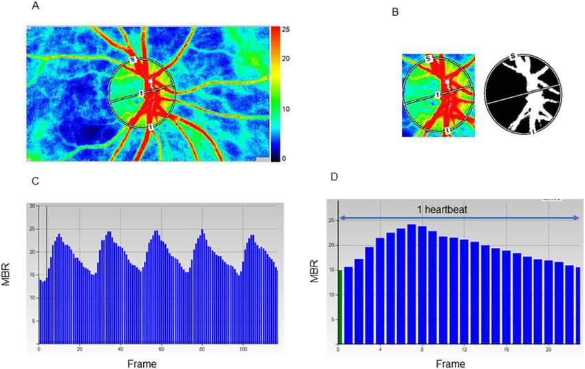
Figure 2. Analysis of pulse waveforms for the optic nerve head (ONH) using laser speckle flowgraphy (LSFG).
Representative color-coded composite map (A). The mean blur rate (MBR) was automatically calculated with
the help of the circle rubber band at the ONH. A binary format image for segmentation between the tissue area
(black area) and the vessel area (white area) on the ONH (B). Pulse waves showing changes in the MBR, which
is tuned to the cardiac cycle for 4 s. The total number of frames is 118 in a scan (C). Normalization of the change
in the MBR in one heartbeat. This shape of MBR is a pulse waveform (D).
soidal band (Fig. 2A), and the position of the ONH was saved on the system. The accompanying LSFG software
automatically divided the ONH area into the large visible vessels and capillary (tissue) area using a binarization
(cross-section analysis) (Fig. 2B) and provided the values for the ONH tissue-area MBR (MT), ONH vessel-area
MBR (MV), and all-area MBR (MA). The primary output parameter of LSFG, the MBR, represents the relative
blood flow velocity and is expressed in arbitrary units (AUs)5. After collecting the LSFG data from each facility,
the ONH margins for all participants were determined with the ellipsoidal bands by a single experienced opera-
tor (T.S.) while referring to the fundus photograph. The time changes in MT or MV with the cardiac cycle were
also recorded automatically for 4 s with a total number of frames of 118 (Fig. 2C) and images corresponding
to identical phases within the duration of one heartbeat were synthesized to one image sequence (Fig. 2D). By
delineating an MT or MV waveform by plotting the MTs or MVs derived from each frame (Fig. 2D), several
shape parameters of the MT or MV pulse waveform, which are synchronized with the cardiac c ycle7,16, were out-
put by LSFG. We focused on seven MT and MV pulse waveform parameters in the current study: Skew (Fig. 1A),
BOS (Fig. 1B), BOT (Fig. 1C), RR (Fig. 1D), FR (Fig. 1E), FAI (Fig. 1F), and ATI (Fig. 1G) in MT and MV. We
adopted an average of three outputs of the MT and MV waveform parameters in the analysis. The resistivity
index and fluctuation were considered to be represented by the BOS because of their high inter-correlations with
the BOS (Spearman’s correlation coefficient: − 0.997– − 0.967).
Measurement of clinical parameters. Standard automated perimetry was performed with the Hum-
phrey visual field analyzer 24–2 Swedish interactive threshold algorithm-standard strategy test (Carl Zeiss Med-
itec, Dublin, USA). The AL was measured with an optical biometer (IOLMaster; Carl Zeiss Meditec) or OA-2000
(Tomey, Nagoya, Japan). The data from OA-2000 were transformed to values yielded by the IOLMaster accord-
ing to previous studies60,61. Color fundus photographs were acquired with TRC-50DX (Topcon, Tokyo, Japan)
or TRC-NW7SF (Topcon). OCT measurements were performed with one of the following instruments based
on the availability in each facility: RS-3000 (NIDEK, Tokyo, Japan), 3D OCT-2000 (Topcon), DRI OCT Triton
(Topcon), and RTVue-XR Avanti (Optovue Inc., Fremont, CA, USA). Only the cup/disc area ratio values were
used for the analysis of the OCT measurements.
Scientific Reports | (2021) 11:6847 | https://doi.org/10.1038/s41598-021-86280-5 9
Vol.:(0123456789)www.nature.com/scientificreports/
Disc and β‑peripapillary atrophy (PPA) area measurements. The details of the planimetric method
used in this study have been reported previously62–64. An experienced ophthalmologist (A.I.) examined all color
fundus photographs. After correcting for magnification based on a modification of Littman’s method provided
by the manufacturer (Topcon), planimetric parameters, disc, and β-PPA areas were calculated using image anal-
ysis software (JGSTKDiscAnalysisSoft; Topcon). In the current study, the OCT measurements were performed
using one of the following instruments: RS-3000 (NIDEK), 3D OCT-2000 (Topcon), DRI OCT Triton (Topcon),
or RTVue-XR Avanti (Optovue Inc.). Different OCT instruments might employ different algorithms to deter-
mine the clinical optic disc margin and to correct magnification of the fundus image; thus, we used the disc
area of each eye determined using a fundus photograph and used only the cup/disc area ratio yielded with each
OCT instrument to calculate the cup and rim areas for each participant’s eye using the following formula: cup
area = disc area provided by the fundus photograph × cup-to-disc area ratio provided by each OCT instrument;
rim area = disc area − cup area of the same eye.
CRAE and CRVE measurements. CRAE and CRVE were determined using the photographs obtained
with the Topcon fundus cameras, according to a previously reported method65.
Statistical analyses. All data are shown as the mean ± standard deviation. The normality of the data was
examined using the Kolmogolov-Smirnov test. We evaluated the contributing factors for the seven LSFG pulse
waveforms: Skew, BOS, BOT, RR, FR, FAI, and ATI in MT and MV using univariate and multivariate linear
mixed-effect modeled regression analyses to adjust for the confounding effects of other factors and the correla-
tion between two eyes of a subject. Independent variables were age, gender, smoking history, body mass index,
SBP, DBP, HR, IOP, AL, disc area, rim area, β-PPA area, MT for MT-waveforms, MV for MV-waveforms, CRAE,
and CRVE. The factors that showed P values less than 0.2 in the univariate analyses were included as independ-
ent variables in the multivariate linear mixed-effect modeled regression analysis. If SBP and DBP simultane-
ously yielded P values < 0.2 in the univariate analyses, they were included separately in the multivariate analyses.
Multivariate analyses included 9 or less independent variables. It was confirmed that no independent variables
included in the multivariate analysis showed correlation coefficients > 0.7. Since 10 samples or more are required
for one dependent variable in multivariate a nalysis66, the sample size of 111 eyes is considered to be within the
appropriate range. All analyses were performed using the statistical software SPSS version 24.0 for Windows
(IBM Corp., Armonk, NY). Statistical significance was considered at P < 0.05.
Data availability
The datasets generated during and/or analyzed during the current study are available from the corresponding
author on reasonable request.
Received: 20 December 2020; Accepted: 12 March 2021
References
1. Leske, M. C. et al. Risk factors for incident open-angle glaucoma: the Barbados eye studies. Ophthalmology 115, 85–93 (2008).
2. Yanagi, M. et al. Vascular risk factors in glaucoma: a review. Clin. Exp. Ophthalmol. 39, 252–258 (2011).
3. Caprioli, J. & Coleman, A. L. Blood flow in glaucoma discussion. Blood pressure, perfusion pressure, and glaucoma. Am. J. Oph-
thalmol. 149, 704–712 (2010).
4. Prada, D. et al. Autoregulation and neurovascular coupling in the optic nerve head. Surv. Ophthalmol. 61, 164–186 (2016).
5. Sugiyama, T., Araie, M., Riva, C. E., Schmetterer, L. & Orgul, S. Use of laser speckle flowgraphy in ocular blood flow research. Acta
Ophthalmol. 88, 723–729 (2010).
6. Luft, N. et al. Ocular blood flow measurements in healthy white subjects using laser speckle flowgraphy. PLoS ONE 11, e0168190
(2016).
7. Yanagida, K. et al. Sex-related differences in ocular blood flow of healthy subjects using laser speckle flowgraphy. Invest. Ophthalmol.
Vis. Sci. 56, 4880–4890 (2015).
8. Iwase, T. et al. Investigation of causes of sex-related differences in ocular blood flow in healthy eyes determined by laser speckle
flowgraphy. Sci. Rep. 7, 13878 (2017).
9. Aizawa, N. et al. Age- and sex-dependency of laser speckle flowgraphy measurements of optic nerve vessel microcirculation. PLoS
ONE 11, e0148812 (2016).
10. Wang, L., Cull, G. A., Piper, C., Burgoyne, C. F. & Fortune, B. Anterior and posterior optic nerve head blood flow in nonhuman
primate experimental glaucoma model measured by laser speckle imaging technique and microsphere method. Invest. Ophthalmol.
Vis. Sci. 53, 8303–8309 (2012).
11. Takahashi, H. et al. Comparison of CCD-equipped laser speckle flowgraphy with hydrogen gas clearance method in the measure-
ment of optic nerve head microcirculation in rabbits. Exp. Eye Res. 108, 10–15 (2013).
12. Aizawa, N. et al. Laser speckle and hydrogen gas clearance measurements of optic nerve circulation in albino and pigmented
rabbits with or without optic disc atrophy. Invest. Ophthalmol. Vis. Sci. 55, 7991–7996 (2014).
13. Nagahara, M., Tamaki, Y., Tomidokoro, A. & Araie, M. In vivo measurement of blood velocity in human major retinal vessels using
the laser speckle method. Invest. Ophthalmol. Vis. Sci. 52, 87–92 (2011).
14. Shiga, Y. et al. Relative flow volume, a novel blood flow index in the human retina derived from laser speckle flowgraphy. Invest.
Ophthalmol. Vis. Sci. 55, 3899–3904 (2014).
15. Luft, N. et al. Measurements of retinal perfusion using laser speckle flowgraphy and Doppler optical coherence tomography. Invest.
Ophthalmol. Vis. Sci. 57, 5417–5425 (2016).
16. Tsuda, S. et al. Pulse-waveform analysis of normal population using laser speckle flowgraphy. Curr. Eye Res. 39, 1207–1215 (2011).
17. Kobayashi, T. et al. Influence of age and gender on the pulse waveform in optic nerve head circulation in healthy men and women.
Sci. Rep. 9, 17895 (2019).
18. Shiba, T., Takahashi, M., Hori, Y. & Maeno, T. Pulse-wave analysis of optic nerve head circulation is significantly correlated with
brachial-ankle pulse-wave velocity, carotid intima-media thickness, and age. Graefes. Arch. Clin. Exp. Ophthalmol. 250, 1275–1281
(2012).
Scientific Reports | (2021) 11:6847 | https://doi.org/10.1038/s41598-021-86280-5 10
Vol:.(1234567890)www.nature.com/scientificreports/
19. Shiba, T., Takahashi, M., Hori, Y., Maeno, T. & Shirai, K. Optic nerve head circulation determined by pulse wave analysis is signifi-
cantly correlated with cardio ankle vascular index, left ventricular diastolic function, and age. J. Atheroscler. Thromb. 19, 999–1005
(2012).
20. Rina, M., Shiba, T., Takahashi, M., Hori, Y. & Maeno, T. Pulse waveform analysis of optic nerve head circulation for predicting
carotid atherosclerotic changes. Graefes Arch. Clin. Exp. Ophthalmol. 253, 2285–2291 (2015).
21. Shiba, T., Takahashi, M., Matsumoto, T., Shirai, K. & Hori, Y. Arterial stiffness shown by the cardio-ankle vascular index is an
important contributor to optic nerve head microcirculation. Graefes Arch. Clin. Exp. Ophthalmol. 255, 99–105 (2017).
22. Françon, M. in Laser Speckle and Application in Optics. (ed Arsenault, H. H.) 1–72 (Academic Press, 1979).
23. Anraku, A. et al. Ocular and systemic factors affecting laser speckle flowgraphy measurements in the optic nerve head. Trans. Vis.
Sci. Tech. 10, 13 (2021).
24. Shiga, Y. et al. Waveform analysis of ocular blood flow and the early detection of normal tension glaucoma. Invest. Ophthalmol.
Vis. Sci. 54, 7699–7706 (2013).
25. Mursch-Edlmayr, A. S. et al. Laser speckle flowgraphy derived characteristics of optic nerve head perfusion in normal tension
glaucoma and healthy individuals: a pilot study. Sci. Rep. 8, 5343 (2018).
26. Gardiner, S. K., Cull, G., Fortune, B. & Wang, L. Increased optic nerve head capillary blood flow in early primary open-angle
glaucoma. Invest. Ophthalmol. Vis. Sci. 60, 3110–3118 (2019).
27. Takeshima, S. et al. Effects of trabeculectomy on waveform changes of laser speckle flowgraphy in open angle glaucoma. Invest.
Ophthalmol. Vis. Sci. 60, 677–684 (2019).
28. Masai, S., Ishida, K., Anraku, A., Takumi, T. & Tomita, G. Pulse waveform analysis of the ocular blood flow using laser speckle
flowgraphy before and after glaucoma treatment. J. Ophthalmol. 2019, 1980493 (2019).
29. Bhatti, M. S., Tang, T. B. & Laude, A. Effects of water drinking test on ocular blood flow waveform parameters: a laser speckle
flowgraphy study. PLoS ONE 12, e0181512 (2017).
30. Yabana, T. et al. The relationship between glutathione levels in leukocytes and ocular clinical parameters in glaucoma. PLoS ONE
14, e0227078 (2019).
31. Shiba, T., Takahashi, M., Hashimoto, R., Matsumoto, T. & Hori, Y. Pulse waveform analysis in the optic nerve head circulation
reflects systemic vascular resistance obtained via a Swan-Ganz catheter. Graefes Arch. Clin. Exp. Ophthalmol. 254, 1195–1200
(2016).
32. Shiba, T., Takahashi, M., Shiba, C., Matsumoto, T. & Hori, Y. The relationships between the pulsatile flow form of ocular microcir-
culation by laser speckle flowgraphy and the left ventricular end-diastolic pressure and mass. Int. J. Cardiovasc. Imag. 34, 1715–1723
(2018).
33. Frank, O. The basic shape of arterial pulse. First treatise: mathematical analysis. J. Mol. Cell. Cardiol. 22, 255–257 (1990).
34. London, G. M. & Guerin, A. P. Influence of arterial pulse and reflected waves on blood pressure and cardiac function. Am. Heart
J. 138, 220–224 (1999).
35. Franklin, S. S. et al. Hemodynamic patterns of age-related changes in blood pressure. The Framingham heart study. Circulation
96, 308–315 (1997).
36. Gameiro, G., Monsalve, P., Golubev, I., Ventura, L. & Porciatti, V. Neuro-vascular changes associated with the water drinking test.
J. Glaucoma 27, 429–432 (2018).
37. Araie, M., Sekine, M., Suzuki, Y. & Koseki, N. Factors contributing to the progression of visual field damage in eyes with normal-
tension glaucoma. Ophthalmology 101, 1440–1444 (1994).
38. Teng, C. C. et al. β-Zone parapapillary atrophy and the velocity of glaucoma progression. Ophthalmology 117, 909–915 (2010).
39. Buus, D. R. & Anderson, D. R. Peripapillary crescents and halos in normal-tension glaucoma and ocular hypertension. Ophthal-
mology 96, 16–19 (1989).
40. Park, K. H., Tomita, G., Liou, S. Y. & Kitazawa, Y. Correlation between peripapillary atrophy and optic nerve damage in normal-
tension glaucoma. Ophthalmology 103, 1899–1906 (1996).
41. Tezel, G., Kass, M. A., Kolker, A. E. & Wax, M. B. Comparative optic disc analysis in normal pressure glaucoma, primary open-
angle glaucoma, and ocular hypertension. Ophthalmology 103, 2105–2113 (1996).
42. Sun, C., Wang, J. J., Mackey, D. A. & Wong, T. Y. Retinal vascular caliber: systemic, environmental, and genetic associations. Surv.
Ophthalmol. 54, 74–95 (2009).
43. Jonas, J. B. & Naumann, G. O. Parapapillary retinal vessel diameter in normal and glaucoma eyes. II. Correlations. Invest. Oph-
thalmol. Vis. Sci. 30, 1604–1611 (1989).
44. Mitchell, P. et al. Retinal vessel diameter and open-angle glaucoma: the Blue Mountains eye study. Ophthalmology 112, 245–250
(2005).
45. Amerasinghe, N. et al. Evidence of retinal vascular narrowing in glaucomatous eyes in an Asian population. Invest. Ophthalmol.
Vis. Sci. 49, 5397–5402 (2008).
46. Kawasaki, R. et al. Retinal vessel caliber is associated with the 10-year incidence of glaucoma: the Blue Mountains eye study.
Ophthalmology 120, 84–90 (2013).
47. Flammer, J. et al. The impact of ocular blood flow in glaucoma. Prog. Retin. Eye Res. 21, 359–393 (2002).
48. Hafez, A. S., Bizzarro, R. L. & Lesk, M. R. Evaluation of optic nerve head and peripapillary retinal blood flow in glaucoma patients,
ocular hypertensives, and normal subjects. Am. J. Ophthalmol. 136, 1022–1031 (2003).
49. Jordan, J. et al. The pressor response to water drinking in humans: a sympathetic reflex?. Circulation 101, 504–509 (2000).
50. Aizawa, N., Kunikata, H. & Nakazawa, T. Diagnostic power of laser speckle flowgraphy-measured optic disc microcirculation for
open-angle glaucoma: analysis of 314 eyes. Clin. Exp. Ophthalmol. 47, 680–683 (2019).
51. Shiga, Y. et al. Preperimetric glaucoma prospective study (PPGPS): predicting visual field progression with basal optic nerve head
blood flow in normotensive PPG eyes. Trans. Vis. Sci. Technol. 7, 11 (2018).
52. Jonas, J. B., Schmidt, A. M., Müller-Bergh, J. A., Schlötzer-Schrehardt, U. M. & Naumann, G. O. Human optic nerve fiber count
and optic disc size. Invest. Ophthalmol. Vis. Sci. 33, 2012–2018 (1992).
53. Jonas, J. B., Schmidt, A. M., Müller-Bergh, J. A. & Naumann, G. O. Optic nerve fiber count and diameter of the retrobulbar optic
nerve in normal and glaucomatous eyes. Graefes Arch. Clin. Exp. Ophthalmol. 233, 421–424 (1995).
54. Levy, D., Garrison, R. J., Savage, D. D., Kannel, W. B. & Castelli, W. P. Prognostic implications of echocardiographically determined
left ventricular mass in the Framingham heart study. N. Engl. J. Med. 322, 1561–1566 (1990).
55. Haider, A. W., Larson, M. G., Benjamin, E. J. & Levy, D. Increased left ventricular mass and hypertrophy are associated with
increased risk for sudden death. J. Am. Coll. Cardiol. 32, 1454–1459 (1998).
56. Jonas, J. B. et al. Parapapillary gamma zone and axial elongation-associated optic disc rotation: the Beijing eye study. Invest. Oph-
thalmol. Vis. Sci. 57, 396–402 (2016).
57. Chauhan, B. C. & Burgoyne, C. F. From clinical examination of the optic disc to clinical assessment of the optic nerve head: a
paradigm change. Am. J. Ophthalmol. 156, 218-227.e2 (2013).
58. Chylack, L. T. Jr. et al. The lens opacities classification system III. The longitudinal study of cataract study group. Arch. Ophthalmol.
111, 831–836 (1993).
59. Anderson, D. R. & Patella, V. M. Automated Static Perimetry (Mosby, 1999).
60. Goebels, S. et al. Comparison of 3 biometry devices in cataract patients. J. Cataract. Refract. Surg. 41, 2387–2393 (2015).
Scientific Reports | (2021) 11:6847 | https://doi.org/10.1038/s41598-021-86280-5 11
Vol.:(0123456789)www.nature.com/scientificreports/
61. Hua, Y., Qiu, W., Xiao, Q. & Wu, Q. Precision (repeatability and reproducibility) of ocular parameters obtained by the Tomey
OA-2000 biometer compared to the IOLMaster in healthy eyes. PLoS ONE 13, e0193023 (2018).
62. Saito, H., Tsutsumi, T., Iwase, A., Tomidokoro, A. & Araie, M. Correlation of disc morphology quantified on stereophotographs to
results by Heidelberg retina tomograph II, GDx variable corneal compensation, and visual field tests. Ophthalmology 117, 282–289
(2010).
63. Iwase, A. et al. Optic disc, rim and peripapillary chorioretinal atrophy in normal Japanese eyes: the Kumejima study. Jpn. J. Oph-
thalmol. 61, 223–229 (2017).
64. Mataki, N., Tomidokoro, A., Araie, M. & Iwase, A. Morphology of the optic disc in the Tajimi study population. Jpn. J. Ophthalmol.
61, 441–447 (2017).
65. Iwase, A. et al. A new method of magnification correction for accurately measuring retinal vessel calibers from fundus photographs.
Invest. Ophthalmol. Vis. Sci. 58, 1858–1864 (2017).
66. Peduzzi, P. et al. A simulation study of the number of events per variable in logistic regression analysis. J. Clin. Epidemiol. 49,
1373–1379 (1996).
Acknowledgements
This research received no specific Grant from any funding agency in the public, commercial or not-for-profit
sectors.
Author contributions
The design and conduct of the study (N.E. and M.A.); collection of data (N.E., A.A., A.I, T.S., N.S., T.S., T.N.,
K.S., and K.N.), the management, analysis, and interpretation of the data (N.E., A.A, G.T., A.I., and M.A.); the
preparation of manuscript (N.E. and M.A.); the review and final approval of the manuscript, all authors.
Competing interests
M.A. has consulted for Topcon and received compensation. A.I. has a patent licensed to Topcon Medical System
without any royalties. Topcon was not involved in study design, data acquisition or data analysis. N.E., A.A.,
G.T., T.S., N.S., T.S., T.N., K.S., K.N. declare no competing interests.
Additional information
Correspondence and requests for materials should be addressed to N.E.
Reprints and permissions information is available at www.nature.com/reprints.
Publisher’s note Springer Nature remains neutral with regard to jurisdictional claims in published maps and
institutional affiliations.
Open Access This article is licensed under a Creative Commons Attribution 4.0 International
License, which permits use, sharing, adaptation, distribution and reproduction in any medium or
format, as long as you give appropriate credit to the original author(s) and the source, provide a link to the
Creative Commons licence, and indicate if changes were made. The images or other third party material in this
article are included in the article’s Creative Commons licence, unless indicated otherwise in a credit line to the
material. If material is not included in the article’s Creative Commons licence and your intended use is not
permitted by statutory regulation or exceeds the permitted use, you will need to obtain permission directly from
the copyright holder. To view a copy of this licence, visit http://creativecommons.org/licenses/by/4.0/.
© The Author(s) 2021
Scientific Reports | (2021) 11:6847 | https://doi.org/10.1038/s41598-021-86280-5 12
Vol:.(1234567890)You can also read