Unilateral Intervention in the Sinuses of Rabbits Induces Bilateral Inflammatory and Microbial Changes
←
→
Page content transcription
If your browser does not render page correctly, please read the page content below
ORIGINAL RESEARCH
                                                                                                                                        published: 14 September 2021
                                                                                                                                       doi: 10.3389/fcimb.2021.585625

                                             Unilateral Intervention in the
                                             Sinuses of Rabbits Induces Bilateral
                                             Inflammatory and Microbial Changes
                                             Christian A. Lux 1,2, James J. Johnston 2, Sharon Waldvogel-Thurlow 2, Camila Dassi 3,
                                             Richard G. Douglas 2*, Do-Yeon Cho 4, Michael W. Taylor 1 and Kristi Biswas 2
                                             1 School of Biological Sciences, University of Auckland, Auckland, New Zealand, 2 Department of Surgery, School of

                                             Medicine, University of Auckland, Auckland, New Zealand, 3 Department of Otorhinolaryngology, Pontifı´cia Universidade
                                             Católica do Paraná (PUCPR), Curitiba, Brazil, 4 Department of Otolaryngology-Head and Neck Surgery, University of Alabama
                                             at Birmingham and Veteran Affairs Medical Center, Birmingham, AL, United States

                                             Background: Chronic rhinosinusitis (CRS) is a globally prevalent inflammatory condition
                                             of the paranasal sinuses which severely impairs patients’ quality of life. An animal model of
                                             unilateral sinusitis by transient sinus occlusion has been described previously in rabbits.
                           Edited by:
                        Emily K. Cope,
                                             The aim of this study was to characterise the sinusitis rabbit model by investigating
           Northern Arizona University,      temporal and bilateral changes in the bacterial community and mucosal inflammation.
                         United States
                                             Methods: Development of sinusitis was achieved by endoscopically placing Merocel®, a
                      Reviewed by:
                  Robert Craig Kern,         sterile nasal packing material, in the left middle meatus of six New Zealand white rabbits for
Northwestern Medicine, United States         four weeks. After a total period of 14 weeks, rabbits were assessed for sinusitis by
                   Michael Kohanski,
          University of Pennsylvania,
                                             endoscopic examination, magnetic resonance imaging (MRI) and histology. Swabs from
                        United States        the left and right middle meatus were obtained for bacterial community analysis at three
                  *Correspondence:           time points (week 0, week 4, week 14) during the study.
                  Richard G. Douglas
      richard.douglas@auckland.ac.nz         Results: Endoscopic evaluation showed unilateral inflammation in all animals examined
                                             after the 4-week blocking period and at week 14. Notably, inflammatory changes were
                  Specialty section:         also seen in the contralateral sinus of all animals at week 4. MRI images demonstrated
        This article was submitted to
   Microbiome in Health and Disease,
                                             unilateral sinus opacification at week 4 in two rabbits, and partial unilateral sinus
              a section of the journal       opacification at week 14 in one rabbit only. Histological analyses revealed substantial
                   Frontiers in Cellular
                                             spatial heterogeneity of mucosal inflammation with inconsistent findings across all
           and Infection Microbiology
                                             animals. No significant differences in mucosal inflammatory markers (such as goblet cell
             Received: 21 July 2020
         Accepted: 23 August 2021            hyperplasia, epithelial denudation and oedema) could be identified between nostrils at
      Published: 14 September 2021           week 14. The bacterial community in the rabbit sinuses was heavily dominated by
                                 Citation:   Helicobacter at week 0 (baseline). At the end of the blocking period (week 4), bacterial
                    Lux CA, Johnston JJ,
        Waldvogel-Thurlow S, Dassi C,
                                             alpha and beta diversity were significantly increased in both nostrils. The bacterial
    Douglas RG, Cho D-Y, Taylor MW           community composition at week 14 had primarily returned to baseline, reflecting the
        and Biswas K (2021) Unilateral
                                             endoscopic and radiological results.
          Intervention in the Sinuses of
                Rabbits Induces Bilateral    Conclusion: This study reaffirmed the ability for development of sinusitis without
 Inflammatory and Microbial Changes.
Front. Cell. Infect. Microbiol. 11:585625.
                                             inoculation of any pathogens in a rabbit model. We were able to demonstrate bilateral
       doi: 10.3389/fcimb.2021.585625        sinonasal mucosal inflammation, by inducing unilateral sinus blockage, which resulted in

Frontiers in Cellular and Infection Microbiology | www.frontiersin.org              1                                   September 2021 | Volume 11 | Article 585625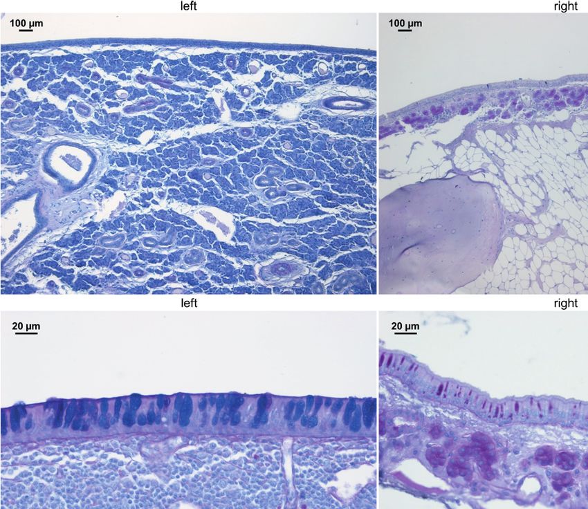
Lux et al.                                                                                                        Bilateral Changes in a Unilateral Sinusitis Model

                                            significant changes to the sinonasal bacterial community. These findings may explain
                                            some of the clinical observations seen in CRS and warrant further research to reveal
                                            potential implications for its therapeutic management.
                                            Keywords: sinusitis, chronic rhinosinusitis, inflammation, microbiota, dysbiosis, rabbit model, animal model

INTRODUCTION                                                                        with obstruction of the sinus outflow tract (Lee and Lane, 2011).
                                                                                    Therefore, sinus blockage, especially if applied reversibly, is
The pathogenesis of chronic rhinosinusitis (CRS) has been under                     advantageous when trying to mimic the physiological
intense research scrutiny, with several hypotheses put forward.                     development of CRS. Temporary blockage has been shown to
Many studies suggest that the sinus microbial community plays a                     re-create the distinctive immune response and characteristic
role in the pathobiology of CRS (Abreu et al., 2012; Boase et al.,                  microbial community changes that are associated with the
2013; Biswas et al., 2015; Paramasivan et al., 2020), with bacterial                disease (Liang et al., 2008; Migliavacca et al., 2014). A rabbit
dysbiosis (a deleterious shift in the microbiota) being reported                    model of unilateral CRS with excellent potential for microbiome
(Hoggard et al., 2017; Chalermwatanachai et al., 2018). In                          research has been described previously (Cho et al., 2018).
addition, host immunity is considered a contributor to chronic                          In general, CRS is considered as a bilateral disease, although a
mucosal inflammation (Van Crombruggen et al., 2011; Fokkens                          subset of patients presents with single-sided symptoms (Fokkens
et al., 2020). Increased numbers of T and B lymphocytes and                         et al., 2020). Kennedy and his colleagues previously demonstrated
macrophages in the sinus mucosal tissue were identified as                           that a rabbit infected with Pseudomonas aeruginosa in the sinuses
characteristic of CRS (Biswas et al., 2017; Yin et al., 2018) and                   can show signs of inflammation at sites other than the site of
interleukins, such as IL-5 and IL-13, play a role in the initial                    primary infection (Perloff et al., 2000). However, to our limited
inflammation and its progression in this disease, especially with                    knowledge, none of the animal models explored the development
nasal polyposis (Kato, 2015; Jiao et al., 2016; Bachert et al., 2018;               of bilateral sinonasal mucosal inflammation by inducing unilateral
Chowdhury et al., 2019). The complex interplay between the                          sinus blockage. In this study, we have attempted to induce CRS in
immune response, sinonasal microbial community, medical                             rabbits by unilateral transient sinus obstruction for four weeks and
treatment and sinus epithelial integrity complicates the                            investigated whether this could result in bilateral changes to the
investigation of underlying mechanisms in the development of                        sinus bacterial communities and mucosal inflammation.
CRS. The high inter-individual variation of disease severity and
observed symptoms, coupled with the difficulty in controlling
antecedent medical interventions and lifestyle components such                      METHODS
as smoking and diet, make it challenging to gain insights from
clinical studies.                                                                   Animal Model
    Animal models can help overcome many of these issues and                        This study was approved by the University of Auckland Animal
several species have been used successfully to study the                            Ethics Committee (AEC# 001910). All animals used in this study
pathophysiology of CRS (London and Lane, 2016; Shin, 2016;                          were farm-sourced male and female New Zealand white rabbits
Lux et al., 2019). Animal models are particularly helpful when                      (4-6 kg). A pilot study was conducted to establish study protocols
studying the efficacy of novel treatments (Chiu et al., 2007;                        and set up surgical procedures using three animals (data not
Tamashiro et al., 2009; Jia et al., 2017; Yoruk et al., 2017). In a                 shown). For the current study, six rabbits were used. Animals
recent CRS-focused review of animal models for the study of                         were kept at the animal facility at the University of Auckland.
chronic mucosal inflammation, rabbits were found to be a                             Rabbits were housed in individual cages with dry food and water
particularly suitable model in regards to animal handling and                       provided ad libitum. Animal wellbeing was monitored
features of sinus anatomy and physiologic characteristics (Lux                      throughout the study using an animal welfare scoring sheet
et al., 2019). Methods for inducing inflammation of the sinus                        based on the rabbit grimace scale. Before initiation, rabbits
mucosa include microbial inoculation (Abreu et al., 2012; Jia                       were acclimatised at the animal facility for at least one week.
et al., 2014), administration of inflammatory agents (Gocea et al.,                     Rabbits were anaesthetised using a combined injectable and
2013; Khalmuratova et al., 2016) and mechanical obstruction of                      inhalational approach. A mix of medetomidine (0.25 mg/kg, SVS
the sinus ostia (Ha et al., 2007; Migliavacca et al., 2014; Cho et al.,             Veterinary Supplies Ltd, Hamilton, New Zealand), ketamine
2018), or a combination of these (Boase et al., 2011). An                           (5 mg/kg, SVS) and buprenorphine (0.03 mg/kg, Onelink,
important pathway that underlies the pathogenesis and                               Auckland, New Zealand) was given subcutaneously 30-45 min
morbidity of CRS appears to be the impaired mucociliary                             prior to placing the animals on oxygen for 1-2 min via facemask.
clearance (Lusk, 1998). This results in the accumulation of                         At that point, the airway was accessed via a supraglottic airway
hyper-viscous mucus, which restricts oxygen, promotes                               device (V-Gel, Docsinnovent Ltd, London, United Kingdom). All
inflammation and supports microbial colonization (Cho et al.,                        rabbits were maintained (as needed) on 1-2.5% isoflurane at an
2020). Ultimately, regardless of the etiology, a pathophysiological                 oxygen flow rate of 0.8-1 L/min. Medetomidine was reversed
characteristic of CRS is inflammation of the sinonasal mucosa                        with intramuscular atipamezole at 0.5-1 mg/kg. Rabbits were

Frontiers in Cellular and Infection Microbiology | www.frontiersin.org          2                                September 2021 | Volume 11 | Article 585625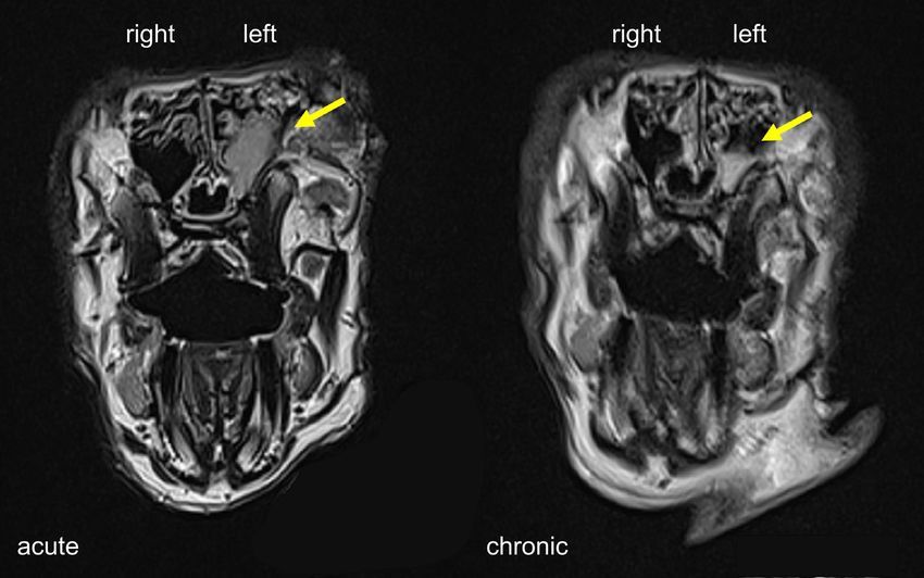
Lux et al.                                                                                             Bilateral Changes in a Unilateral Sinusitis Model

maintained on oxygen via facemask until aware of their                       inflammation and 4 severe inflammation with ulceration or
surroundings and able to hold their head up. All rabbits were                polyps and luminal opacification (Marks, 1997).
monitored continuously until they were able to hop adequately
and showed an interest in food and water. Rabbits did not receive
any other medication before or during the study.                             Magnetic Resonance Imaging
                                                                             MRI scanning was performed using a 3T Skyra MRI system
                                                                             (Siemens Healthineers, Erlangen, Germany), with a 15 cm
Induction of Unilateral Sinus Inflammation                                    diameter 16-channel transmit-receive radiofrequency coil. Both
Sinusitis was induced using a transnasal endoscopic technique as             coronal and transverse images, each with 30 contiguous 1.5 mm
previously described by Cho et al. (2018). In brief, sterile nasal           thick slices, were acquired using a T2-weighted turbo spin echo
packing material (Merocel ® , Medtronic, Auckland, New                       sequence so that all sinus cavities were covered and could be
Zealand) was placed in the left middle meatus (unilateral)                   examined for sinus opacification. The images had a 120 x 120
under endoscopic guidance to achieve complete obstruction of                 mm field of view, acquired with a 320 x 256 matrix,
this sinus ostium. All procedures were carried out by an                     reconstructed to give an in-plane resolution of 0.4 x 0.4 mm.
experienced otorhinolaryngology surgeon with the assistance of
a veterinary anaesthetist and an animal welfare officer to ensure
the best possible care for the rabbits during surgery. Our pilot             Histological Analysis
study indicated that two weeks of sinus obstruction was                      Animals were euthanised by the administration of an overdose of
insufficient to achieve significant and persistent sinus                       pentobarbital (SVS). After death was confirmed, the rabbits’
inflammation (data not shown). Accordingly, the blockage                      snouts were removed from the heads by making two
period was extended to four weeks. After the removal of                      intersecting cuts using an electric saw. Tissue samples were
Merocel®, all animals were observed for a further ten weeks,                 placed in Carnoy’s fixative (60% ethanol, 30% chloroform, and
i.e., up to 14 weeks from the time point of obstruction. A timeline          10% glacial acetic acid) immediately after harvesting and
of the study is illustrated in Figure 1.                                     incubated for 24 h. Snouts were decalcified subsequently in
                                                                             RDO Rapid Decalcifier (Apex Engineering, Illinois, USA).
Outcome Measures                                                             Decalcified snouts were then separated into sections as
Animals were examined for features of CRS throughout the                     previously described (Pereira et al., 2011) and embedded in
study, and samples were collected at baseline (insertion of                  paraffin. Sections were stained with haematoxylin and eosin
Merocel®), during the acute phase (Merocel® removal) and the                 (H&E) to assess the structural integrity of the epithelium and
chronic phase (culling of the animal). Examination and sample                submucosa, as well as to document immune cell infiltration. In
collection at each time point included endoscopic scoring and                addition, Periodic Acid – Schiff (PAS) stain was used to detect
sinus swab collection from both nostrils for microbiota analysis.            mucin-producing cells and structures (goblet cells and
Additional MRI scans at the acute and chronic time points were               submucosal glands). The evaluation of tissue samples was
performed. Finally, the rabbit snout was collected for histological          conducted by two independent examiners and includes
analysis at the end of the study.                                            qualitative observation of inflammatory signs as well a semi-
                                                                             quantitative analysis. To quantify inflammatory markers and
Endoscopic Score Grading for Sinus Inflammation                               mucosal integrity in all animals, a set of parameters, including
A 1.9 mm endoscope (HOPKINS® Straight Forward Telescope                      epithelial hypertrophy and thickness of the base membrane and
0°, Karl Storz, Tuttlingen, Germany) was used to examine the                 submucosa (Table S1), were scored on multiple representative
nasal cavities as previously described (Cho et al., 2017). A scale of        sinus histology sections for each rabbit. The parameters were
0 to 4 was used to grade inflammation with 0 representing                     scored with zero, one, two or three to reflect whether changes
normal, 1 mild inflammation, 2 moderate inflammation, 3 severe                 between sides were absent, mild, moderate or severe.

 FIGURE 1 | Timeline of model development.

Frontiers in Cellular and Infection Microbiology | www.frontiersin.org   3                             September 2021 | Volume 11 | Article 585625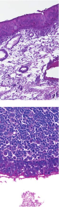
Lux et al.                                                                                              Bilateral Changes in a Unilateral Sinusitis Model

Bacterial Community Analysis                                                 subsequent analysis, and data were rarefied to an even
Sample Acquisition and DNA Extraction                                        sequencing depth of 2000 reads per sample. Analyses of
Pairs of swabs were collected under endoscopic guidance from                 bacterial community diversity as well as pairwise comparisons
the middle meatus of each nostril at three time points (as                   of single ZOTU abundances were conducted on samples of the
indicated above). Swabs were placed in a nucleic acid                        left sinus only.
preservative solution (RNAlater, Thermo Scientific, New                           Alpha diversity (diversity within a sample) was calculated for
Zealand) and put on ice immediately. In accordance with the                  Shannon and Simpson indices within the USEARCH pipeline.
manufacturer’s guidelines, samples were incubated with                       The Shannon and Simpson diversity metrics measure richness as
RNAlater for 24 h at 4°C before being stored at -20°C until                  well as evenness (relative abundance of ZOTUs and their
further processing. DNA was extracted from the swabs using                   distribution in a sample). Beta diversity (diversity between
sterile Lysing Matrix E bead tubes (MP Biomedicals, Seven Hills,             samples) was calculated as Bray-Curtis (BC) dissimilarity in R
NSW, Australia) and the AllPrep DNA/RNA Mini Kit (Qiagen,                    (version 3.6.0) using the vegdist command from the vegan
Hilden, Germany), as described previously (Biswas et al., 2015).             package. The BC dissimilarity index quantifies the
A negative DNA extraction control containing 200 mL sterile                  compositional similarity of the bacterial communities based on
water was also carried out simultaneously.                                   both presence/absence and the relative abundance of ZOTUs
                                                                             within the community.
Bacterial Community Sequencing
To evaluate bacterial communities the V3-V4 region of the                    Statistical Analysis
bacterial 16S rRNA gene was amplified using 341F and 806R                     Statistical analyses were carried out in R (version 3.6.0).
primers (Herlemann et al., 2011) together with Nextera DNA                   Histological scores were evaluated using a paired t-test. Overall
library prep kit adapters. PCR reactions and DNA purification                 differences in alpha diversity were examined with an analysis of
were carried out as described elsewhere (Hoggard et al., 2017). In           variance (ANOVA) followed by Tukey’s honest significant
brief, approximately 100 ng of genomic template DNA was used                 difference test. Permutational multivariate analyses of variance
in duplicate PCRs, each consisting of 35 cycles. Negative PCR                (PERMANOVA) based on BC distance matrices were conducted
controls were included in all PCR reactions as well as eluate from           using the adonis command in the vegan package. A principal
the negative extraction control, which yielded no detectable                 coordinate analysis (PCoA) was performed using the cmdscale
products. Amplicons from duplicate PCRs were pooled to a                     command from the stats package for visualisation of the BC
final volume of 50 uL and purified using Agencourt AMPure                      dissimilarity matrix. The resulting PCoA plot images the beta
magnetic beads (Beckman Coulter Inc., USA). Purified PCR                      diversity data in two-dimensional space with samples that are
products were quantitatively assessed with Qubit dsDNA high-                 more dissimilar being spaced further apart from each other.
sensitivity kits (Life Technologies, New Zealand) and                        Pairwise comparisons of single ZOTU abundances were
standardised to ~ five ng per sample. Purified products were                   conducted using Kruskal-Wallis (KW) and post-hoc Dunn’s test
submitted to Auckland Genomics Ltd for library preparation and               with Benjamini-Hochberg adjustment for multiple comparisons.
sequencing using Illumina MiSeq (2 x 300 bp paired-end
chemistry). Raw sequences are publicly available on the SRA-
NCBI database (BioProject ID: PRJNA639396).                                  RESULTS
Bioinformatics                                                               Establishing Sinonasal Inflammation
Raw sequences from 32 samples originating from six rabbits were              Two of the six rabbits (Rabbit #1 and #3) used in this study died
merged and quality filtered using USEARCH (Edgar, 2010)                       from unknown cause during the post-sedation observation
version 11 with settings as previously described (Hoggard                    period after the surgical procedure. The first animal (#3) died
et al., 2017). For direct comparisons of bacterial community                 eight days after Merocel® placement and the second (#1) 24 h
composition between a previous study and the current one,                    after Merocel® removal. Sample collections were still performed
rabbit sinonasal swab samples from the previous study (Cho                   from the first animal. Acute sinus inflammation was established
et al., 2018) were bioinformatically processed alongside the raw             in all six rabbits. Endoscopic examination showed moderate to
sequence reads obtained during our study. To account for                     severe sinus inflammation on the left (blocked) side of all rabbits
differences in sequence length between the two studies, our                  at the acute stage (week four). Of note, endoscopy also revealed
merged sequences were trimmed to 256 bp, so as to overlap                    mild to moderate inflammation in the right (control) side at
precisely the sequenced V4 16S rRNA gene region from the                     week four. At the chronic stage (week 14), obstructed sinuses
earlier study. Zero-radius operational taxonomic units (ZOTUs)               remained mildly inflamed while all examined control sides
representing 100% sequence similarity for each ZOTU were                     presented normal (Table 1). MRI at week four showed
generated using the unoise3 algorithm within USEARCH                         unilaterally blocked sinuses in two rabbits, with one of them
(Edgar, 2016b). Each ZOTU was taxonomically assigned using                   still showing partial opacification at week 14 (Figure 2). The
the sintax classifier in USEARCH (Edgar, 2016a) with the RDP                  overall success rate for establishing radiographic evidence of
16S rRNA gene database (version 16) (Cole et al., 2014).                     sinus opacification in the rabbits as determined by MRI at week 4
Sequences mapping to eukaryotic genomes were removed from                    was 40% (2 out of 5 rabbits) and week 14, 25% (1 out of 4

Frontiers in Cellular and Infection Microbiology | www.frontiersin.org   4                              September 2021 | Volume 11 | Article 585625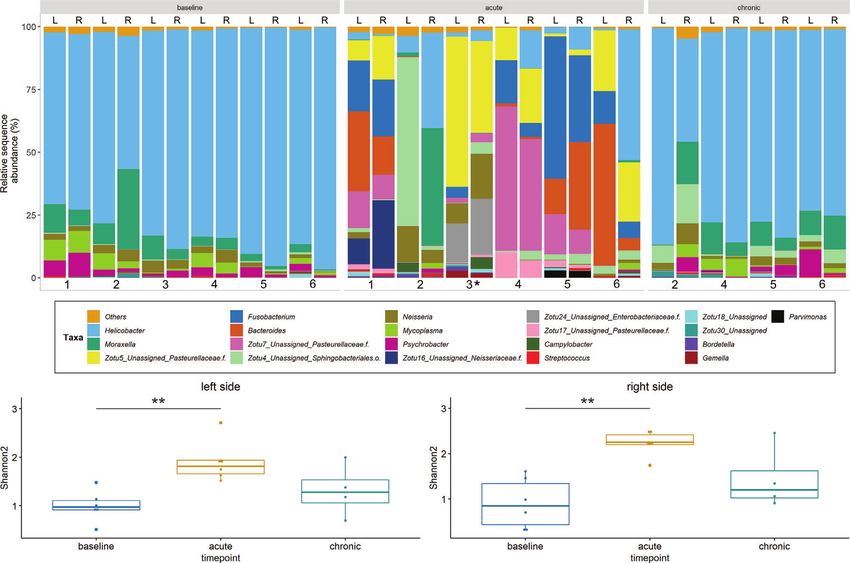
Lux et al. Bilateral Changes in a Unilateral Sinusitis Model
TABLE 1 | Endoscopic score grading for sinus inflammation. 27.8 +/- 15.7) compared to the right side (22.4 +/- 12.2) even
rabbit # Endoscopic scores
though the statistical significance was lacking (paired t-test, p =
0.28) (Figure 5). Rabbit #2, which had sinus opacification
baseline acute chronic confirmed by MRI, has a higher score for the right (control)
left right left right left right
sinus. Rabbit #4 showed similar scores for each side, while rabbits
#3, #5 and #6 had increased signs of inflammation in the left side,
1 0 0 3 2 NA NA although MRI images showed clear sinuses. Even though there
2 0 0 2 1 1 0
was only one sample (rabbit #3) from the acute stage, overall
3 0 0 NA NA NA NA
4 0 0 2 1 NA NA
scores were higher at week 14 (chronic stage) than those at week
5 0 0 3 1 1 0 4 (acute).
6 0 0 2 1 NA NA
0 = normal; 1 = mild inflammation; 2 = moderate inflammation; 3 = severe inflammation;
4 = severe inflammation with ulceration or polyps and luminal opacification.
Sinus Bacterial Community
Missing scores for animal #1 and #3 are due to premature death. Scores for animal #4 and Sequencing data from a total of 32 samples that were obtained
#6 at the chronic stage were not recorded. from the left and right sinuses of 6 rabbits at baseline (n = 12),
NA, not available. acute (n = 12) and chronic stage (n = 8) were evaluated. Data
were stratified by animals for PERMANOVA which revealed that
animals). There was no evidence of opacification in the time point (i.e. baseline vs. acute vs. chronic) accounts for 48% of
contralateral sinus at week 4 and week 14 in all animals. the observed variation (p < 0.001). Nostril side did not affect
bacterial community structure significantly (p = 0.6). The
Histology baseline sinus microbiota was heavily dominated by the genus
The entire sinus mucosa was scanned on both sides for signs of Helicobacter which, together with Moraxella and Neisseria (both
inflammation. Some evidence of mucosal and submucosal at much lower relative abundances), accounted for 85 – 90% of
inflammation was identified in all animals as shown in the assigned sequences (Figure 6A). A significant compositional
Figures 3 and 4: 1) hyperplasia of submucosal glands in the change was observed at the acute inflammatory stage compared
left sinuses (Figure 3A), 2) elevated number of goblet cells in the to the baseline bacterial community. The abundance of ZOTUs
left sinuses (Figure 3B), 3) infiltration of inflammatory cells assigned to several genera, including Helicobacter, Moraxella and
(Figure 4A), 4) loosening of the extracellular matrix or oedema Neisseria was significantly decreased while there was an apparent
(Figure 4B), 5) cellular exudate in the lumen (Figure 4C), and 6) rise in the abundance of ZOTUs classified as Bacteroides,
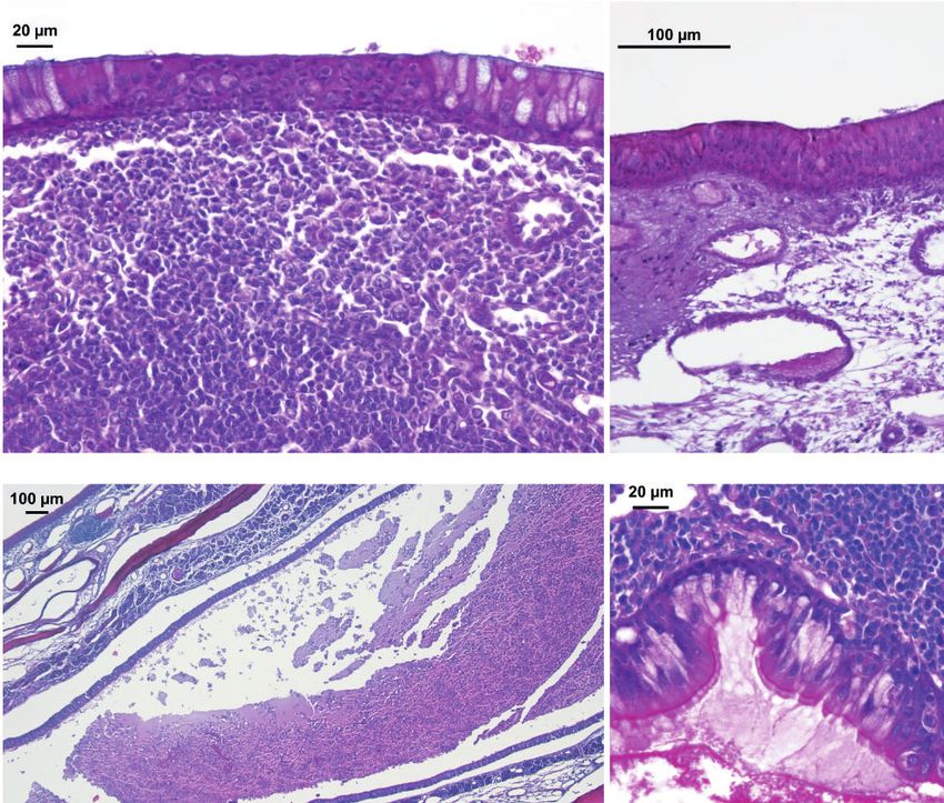
epithelial denudation (Figure 4D). The histologic evidence of Fusobacterium and the family Pasteurellaceae (Figure S1). At
inflammatory processes was observed in the left (blocked) side as the chronic stage, the sinus bacterial community returned to
well as in the right (control) side of the same rabbit in all studied baseline composition within all sinuses. The overall number of
animals. Rabbits’ left sinuses had a higher score (mean +/- SD, taxa found in the left sinuses was significantly elevated at the
FIGURE 2 | MRI scans of rabbit sinuses in coronal view. Severe and partial sinus opacification on the left side at the acute and chronic stage indicated by yellow arrows.
Frontiers in Cellular and Infection Microbiology | www.frontiersin.org 5 September 2021 | Volume 11 | Article 585625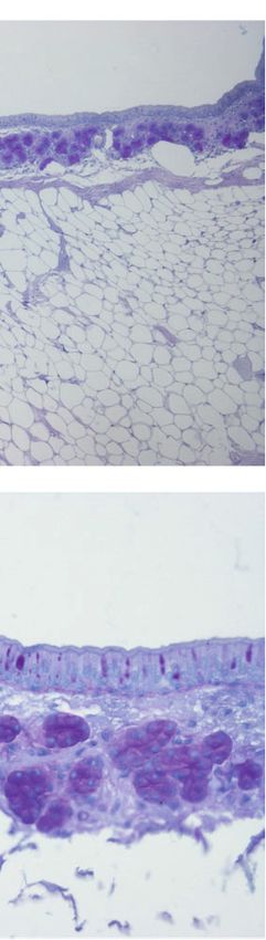
Lux et al.                                                                                                              Bilateral Changes in a Unilateral Sinusitis Model

                                 A

                                 B

 FIGURE 3 | PAS staining of sinus mucosa showing signs of inflammation in the left (blocked) side and right (control) side at week 14. (A) Increased number of
 submucosal glands. (B) Goblet cell hyperplasia. Images are chosen exemplary and do not represent left and right sinus of one animal.

                                  A                                                B

                                  C                                                D

 FIGURE 4 | H&E staining of sinus mucosa showing signs of inflammation at week 14. (A) Epithelial infiltration of inflammatory cells. (B) Loose subepithelial matrix.
 (C) Luminal exudate. (D) Epithelial denudation.

Frontiers in Cellular and Infection Microbiology | www.frontiersin.org            6                                    September 2021 | Volume 11 | Article 585625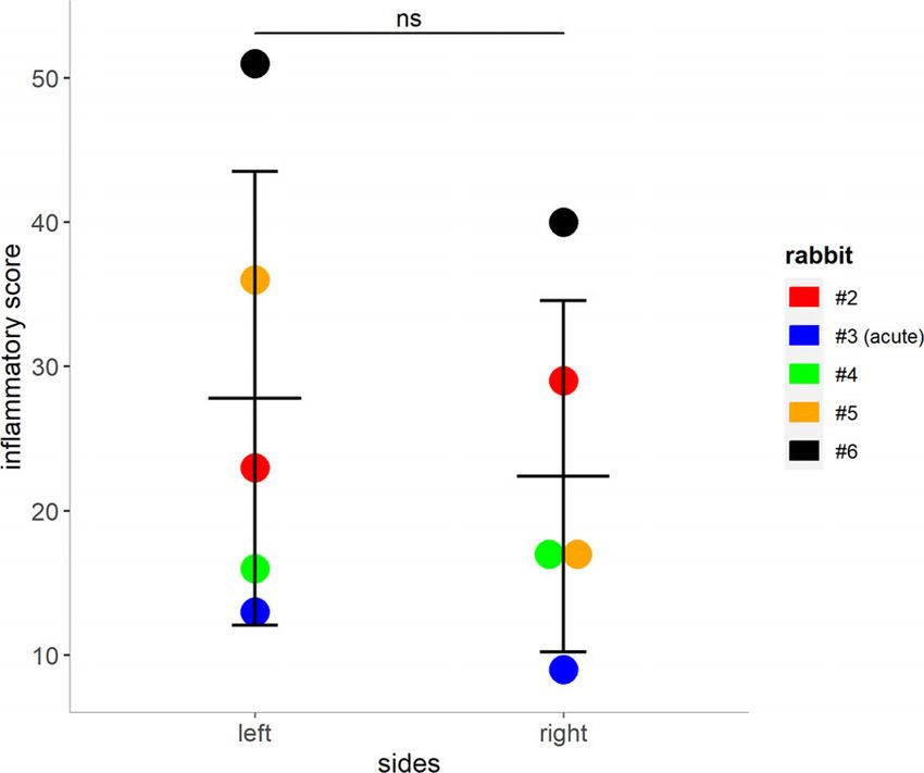
Lux et al.                                                                                                              Bilateral Changes in a Unilateral Sinusitis Model

                                                                                       acute inflammatory phase (Figure 6B). It is of note that alpha
                                                                                       diversity patterns from the right (control) side (Figure 6C)
                                                                                       closely mirrored those of the left side. Beta diversity analysis
                                                                                       showed no differences between the bacterial communities in the
                                                                                       left (blocked) and right (control) side at all time points (data
                                                                                       not shown).

                                                                                       Comparison With Bacterial Sequence Data
                                                                                       From a Previously Published Study
                                                                                       16S rRNA gene amplicon data from 10 samples pertaining to the
                                                                                       left sinuses of 10 rabbits (four from baseline, three from rabbits
                                                                                       killed at week 4 and three from rabbits killed at week 14) from a
                                                                                       previous study (Cho et al., 2018) were obtained to enable direct
                                                                                       comparisons of bacterial community profiles between studies.
                                                                                       Baseline bacterial communities in both studies were similar, with
                                                                                       dominance by members of the genus Helicobacter. In the current
                                                                                       study, Moraxella, Mycoplasma and Neisseria were the three most
                                                                                       abundant genera after Helicobacter. While Mycoplasma was
 FIGURE 5 | Mucosal score grading for rabbits 2 – 5. Higher scores indicate            found to be the second most abundant genus, Moraxella and
 an increase of inflammatory markers within the left and right (control) sinuses        Neisseria were not amongst the 20 most abundant ZOTUS in the
 of the rabbits (shown as mean + SD, paired t-test, p = 0.28). Tissue from             earlier study. At the acute stage, Lactobacillus and Streptococcus
 rabbit #3 was harvested during the acute phase (day 8 post blockage) due to
                                                                                       were the most abundant genera in the 2018 study, with a greater
 premature death. ns, not significant.
                                                                                       diversity of other, low-abundance taxa emerging in place of

             A

             B                                                                     C

 FIGURE 6 | Bacterial community profiles and alpha diversity based on 16S rRNA gene sequence data. (A) Profiles are shown at genus level for the left (L) and right
 (R) side of rabbits’ sinuses. Samples missing at the chronic stage are due to the premature death of two animals. ZOTUs that could not be confidently assigned to
 genus level are presented as single ZOTUs with the next higher taxonomic classification. (B) + (C) Alpha diversity for the left and right sides. Shannon2 indices are
 shown for samples from different time points of the study. Significance levels: p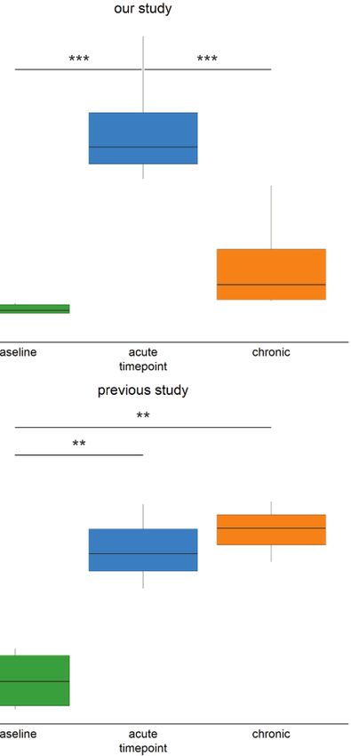
Lux et al.                                                                                                                Bilateral Changes in a Unilateral Sinusitis Model

Helicobacter. In contrast to our findings, the previous study                           develop bilateral sinonasal inflammation in an animal model. To
documented a persistently altered bacterial community at the                           our limited knowledge, this is the first study to demonstrate that
chronic stage (Figure S2).                                                             unilateral sinus blockage can develop inflammation in the
    Multidimensional scaling analysis reaffirmed our observation                        contralateral side in an animal model. Obstruction resulted in
that the baseline composition of the bacterial communities in                          acute sinusitis and three out of four rabbits did have increased
both studies were similar (i.e. close clustering of samples)                           signs of bilateral inflammation histologically at week 14.
(Figure 7A). By contrast, acute and chronic phase samples                              However, persistent radiologic inflammatory signs were only
differed markedly between the two studies. Accordingly,                                seen in one out of four rabbits that reached the endpoint of the
persistent changes in beta diversity, as indicated by the                              study (eight weeks after removal of obstruction).
distance of samples to the centroid, were observed for samples
of the previous study (Figure 7C). In our study, group dispersion                      Acute Versus Chronic Sinusitis
was significantly increased in samples from the acute phase                             We have found that the model is excellent for the study of acute
only (Figure 7B).                                                                      inflammation but challenging for the study of long-term
                                                                                       inflammatory changes that would be required to model CRS.
                                                                                       Endoscopic but not radiologic findings consistent with acute
                                                                                       sinusitis were observed in all study animals after four weeks of
DISCUSSION                                                                             blockage in the treated middle meatus. However, success in
                                                                                       establishing long term mucosal changes in these animals, all of
In this study, we have replicated a recently established animal                        which were New Zealand white rabbits, differed between the two
model that uses a period of middle meatal obstruction to cause                         studies. We hypothesise that this discrepancy may be attributed
unilateral sinusitis and assessed whether such obstruction could                       to any or some of the differences between the two studies:

       A                                                                                            B

 FIGURE 7 | Comparison of bacterial community diversity between the study by Cho et al. (2018), and our study. (A) Principal Coordinate Analysis (PCoA) of sinus
 bacterial community structures at different time points. A two-dimensional PCoA plot was constructed based on Bray-Curtis dissimilarity. The time point of collection
 referring to baseline, acute and chronic inflammation is illustrated by colour, the origin of the sample from the 2018 or the current study is indicated by shape. Only
 rabbits left sides are shown. (B, C) Box plots indicating the distances of samples to the centroid. Asterisks indicate that the multivariate homogeneity of group
 dispersions with Tukey’s honest significant difference resulted in a significant difference. Significance levels: p < 0.01 (**), p < 0.001 (***).

Frontiers in Cellular and Infection Microbiology | www.frontiersin.org             8                                     September 2021 | Volume 11 | Article 585625
Lux et al. Bilateral Changes in a Unilateral Sinusitis Model
anaesthetic procedure (injectable only in the previous study vs. more accurately represent a physiologic response of the sinus
injectable and gaseous in the current study), source of animals microbiome when compared to bacterial inoculation models.
(lab bred (Pasteurellaceae free) vs. farm raised), specimen
collecting sites/process, induction of acute sinusitis with full Bilateral Sinus Inflammation From
opacification and weight (2-4 kg vs. 4-6 kg) of the rabbits. Unilateral Sinus Outflow Obstruction
Hunter and colleagues investigated the role of airway mucus as We found in our pilot study that extending the period of sinus
a nutrient source (e.g., short chain fatty acids) in stimulating the outflow obstruction from two to four weeks resulted in increased
growth of putative pathogens (e.g., Pseudomonas spp.) in the inflammation. The more severe sinusitis on the obstructed side
cystic fibrosis airway (Flynn et al., 2016). This study appears to lead to inflammation on the contralateral (control)
demonstrated that potential pathogens (e.g., Staphylococcus, side which was associated with significant changes to the sinus
Pseudomonas), that cannot utilize the mucin, do not establish microbiota at week 4 (acute stage). In comparison, the previous
an airway infection until anaerobic, mucin-fermenting bacteria description of this model reported consistent inflammation and
have colonized. Therefore, it seems crucial to generate an opacification of all obstructed sinuses at two weeks which
anaerobic sinus cavity filled with pathogenic mucus during the persisted for another 12 weeks while contralateral sinuses
acute sinusitis period to induce chronic inflammatory features remained clear (Cho et al., 2018). However, the bacterial
later in this animal model. Furthermore, subtle differences could community profiles of the contralateral (control) side were not
have arisen due to the study being performed by another research investigated in that study.
team and at a different location, resulting in an altered The study by Perloff et al. clearly demonstrated an extensive
environment for the rabbits. inflammatory involvement extending from the infected sinus to
It is also of note that radiologic imaging in this study was the bone in rabbits infected with P. aeruginosa (Perloff et al.,
done by MRI while the previous study used micro-computed 2000). In this study, the changes in the microbiota during the
tomography (µCT). The latter is the preferred method to acute phase (an increase in anaerobic bacteria) could initiate the
measure sinusitis in rabbit models and could be more sensitive inflammatory process noted in the chronic phase, even though
in detecting sinus opacification (Kerschner et al., 2000). In the bacterial community composition had returned to baseline
addition, Cho et al. (2018) reported sinus opacifications in all by that time (week 14). CRS generally presents bilaterally and if
rabbits during the acute phase when assessed using high the potential for inflammation to spread locally is also present in
resolution µCT. There was a discrepancy between radiology human patients, it could explain some of the clinical
(MRI) and histology findings in our study and MRI results did observations. One of the major advantages of creating a
not reflect the subtle histologic changes in the sinus mucosa. unilateral model is that the contralateral side can be used as a
Similarly, when Ozan et al. inoculated Staphylococcus aureus in a control (Lux et al., 2019). Considering these findings, however,
rabbit model, the histopathological and CT findings in we advise careful monitoring of the mucosa and microbiota of
experimental rabbits were not correlated (Ozcan et al., 2011). the contralateral sinus and recommend that the contralateral
Histological findings as previously described (Cho et al., 2018) sinus may not be used as a true negative control.
could be reproduced in our model, however results were The interaction between both sinuses observed in this study
rather inconsistent and were not always concordant with may be triggered by neural and vascular reflexes and warrants
macroscopic observations. It is of note that highly variable further investigation. The described model provides the basis to
changes in the mucosa could be found within the same side study these inflammatory pathways on a molecular level. In
in a single histological section. This highlights the difficulty addition, the release of systemic inflammatory mediators as
with histopathological examination of the sinonasal mucosa result of the unilateral intervention may promote contralateral
for signs of chronic inflammation, and the requirement to sinus inflammation. Specifically, the examination of cytokine
examine multiple sections of several different sites within a sinus patterns of the tissue inflammation in the obstructed and
(Jacob et al., 2001). Local variation in inflammatory changes unobstructed side in future studies may help reveal potential
throughout the sinus cavities has been observed in CRS patients, implications for CRS.
in whom certain areas of the sinus mucosa can be highly inflamed
while others show little signs of inflammation (Suh and Host Response in Rabbits
Kennedy, 2011). Farm-raised or wild rodents have immune responses that are
The microbiome results are in accordance with the results of different to those of laboratory-bred animals (Abolins et al.,
the previously published report of this model, with an increase in 2017). While antibody responses are elevated in wild animals due
bacterial diversity at the acute sinusitis phase (Cho et al., 2018). to more extensive antigenic challenges, other immunologic
However, results in the two studies differed at the chronic processes such as proliferation and cytokine release are
stage. We suspect that the distinct bacterial community profiles depressed (Abolins et al., 2017; Viney and Riley, 2017). The
reflect the contrasting inflammatory states in the two studies in free-living rabbits used in our study appear to have a more
association with the different immunologic patterns of farm-raised competent immune system that can maintain immune
versus lab-bred animals. In this rhinogenic sinusitis model, homeostasis. Therefore, immunopathology is minimised, and
inflammation is initiated from the impairment of the natural acute inflammation is resolved rather than progressing to a
sinus function. Thus, the observed microbiological changes chronic inflammatory state. Reduced immune cell proliferation
Frontiers in Cellular and Infection Microbiology | www.frontiersin.org 9 September 2021 | Volume 11 | Article 585625Lux et al. Bilateral Changes in a Unilateral Sinusitis Model
and cytokine response may contribute to the variable histological AUTHOR CONTRIBUTIONS
findings in this study.
There are other limitations when drawing conclusions about CL, KB, RD and MT planned and conceived the study. CL
human pathophysiology from animal findings. The previous carried out the experiments, data analysis and took the lead in
study found that the dominant bacterial phyla in the rabbit writing the manuscript with support from KB, RD, MT and
model were the same as those in human sinuses. In the current D-YC. JJ and CD helped to perform the surgical procedures.
study, a genus-level taxonomic classification was applied which SW-T helped to carry out the histological experiments and
revealed that the most abundant bacterial genus (Helicobacter) is analysis. All authors contributed to the article and approved
not commonly found in human sinuses. However, putative the submitted version.
disease-associated genera (e.g. Streptococcus) remain the same
between hosts. Despite different sinonasal bacterial community FUNDING
profiles in rabbits and humans, observing shifts in the microbial
community throughout the inflammatory process can provide CL, RD and MT were supported by funding from the Garnett
helpful insights into the pathogenesis of CRS in human patients. Passe a nd Rodney Williams Me morial F oundation
(91073712790). D-YC received funding from the NIH/National
Institutes of Allergy and Infectious disease (K08AI146220), the
CONCLUSION Triological Society Career Development Award 2019 and the
Cystic Fibrosis Foundation K08 Boost Award (CHO20A0-KB).
This study reaffirmed the ability to develop sinusitis in a rabbit
model without inoculation of any pathogens as previously ACKNOWLEDGMENTS
described (Cho et al., 2018). Even though it is technically
challenging to generate a persistent sinus mucosal A special thanks to Jodi Salinsky and Linley Nisbet for their
inflammation, this rabbit model seems to be reproducible and excellent help with animal anaesthesia and monitoring during
provides a potential for studying host-microbe interactions surgical procedures and to Beau Pontré for helping with
during sinonasal inflammation under a high level of radiological imaging. The authors would also like to thank
experimental control. We further demonstrated that bilateral Brett Wagner Mackenzie and Joey Siu for their assistance
sinonasal inflammation can be caused by inducing unilateral during surgical procedures as well as the Vernon Jenson Unit
sinus blockage. Further research is necessary to investigate this for animal accommodation and care.
novel observation and its potential implication for the clinical
observations seen in CRS.
SUPPLEMENTARY MATERIAL
The Supplementary Material for this article can be found online
DATA AVAILABILITY STATEMENT at: https://www.frontiersin.org/articles/10.3389/fcimb.2021.
585625/full#supplementary-material
The data presented in the study are deposited in the SRA NCBI
Supplementary Figure 1 | Pairwise comparisons of relative sequence
repository, accession numbers SRR12012423 - SRR12012402.
abundances between timepoints. Selected single ZOTUs are shown. Significance
levels: pLux et al. Bilateral Changes in a Unilateral Sinusitis Model
Chiu, A. G., Antunes, M. B., Palmer, J. N., and Cohen, N. A. (2007). Evaluation of the Enterotoxin B in C57BL/6 Mice. Allergol. Immunopathol. (Madr) 44 (1), 66–
In Vivo Efficacy of Topical Tobramycin Against Pseudomonas Sinonasal 75. doi: 10.1016/j.aller.2015.04.004
Biofilms. J. Antimicrob. Chemother. 59 (6), 1130–1134. doi: 10.1093/jac/dkm087 Lee, S., and Lane, A. P. (2011). Chronic Rhinosinusitis as a Multifactorial
Cho, D. Y., Hoffman, K., Skinner, D., Mackey, C., Lim, D. J., Alexander, G. C., et al. Inflammatory Disorder. Curr. Infect. Dis. Rep. 13 (2), 159–168. doi: 10.1007/
(2017). Tolerance and Pharmacokinetics of a Ciprofloxacin-Coated Sinus Stent s11908-011-0166-z
in a Preclinical Model. Int. Forum Allergy Rhinol. 7 (4), 352–358. doi: 10.1002/ Liang, K. L., Jiang, R. S., Wang, J., Shiao, J. Y., Su, M. C., Hsin, C. H., et al. (2008).
alr.21892 Developing a Rabbit Model of Rhinogenic Chronic Rhinosinusitis.
Cho, D. Y., Hunter, R. C., and Ramakrishnan, V. R. (2020). The Microbiome and Laryngoscope 118 (6), 1076–1081. doi: 10.1097/MLG.0b013e3181671b74
Chronic Rhinosinusitis. Immunol. Allergy Clin. North Am. 40 (2), 251–263. London, N. R.Jr., and Lane, A. P. (2016). Innate Immunity and Chronic
doi: 10.1016/j.iac.2019.12.009 Rhinosinusitis: What We Have Learned From Animal Models. Laryngoscope
Cho, D.-Y., Mackey, C., van der Pol, W. J., Skinner, D., Morrow, C. D., Schoeb, T. Investig. Otolaryngol. 1 (3), 49–56. doi: 10.1002/lio2.21
R., et al. (2018). Sinus Microanatomy and Microbiota in a Rabbit Model of Lusk, R. P. (1998). The Surgical Management of Chronic Sinusitis in Children.
Rhinosinusitis. Front. Cell Infect. Microbiol. 7, 540. doi: 10.3389/fcimb. Pediatr. Ann. 27 (12), 820–827. doi: 10.3928/0090-4481-19981201-09
2017.00540 Lux, C. A., Douglas, R. G., Cho, D. Y., Taylor, M. W., and Biswas, K. (2019).
Chowdhury, N. I., Chandra, R. K., Li, P., Ely, K., and Turner, J. H. (2019). Animal Models for Inflammatory Mucosal Disease and Their Potential for
Investigating the Correlation Between Mucus Cytokine Levels, Inflammatory Studying the Microbiome in Chronic Rhinosinusitis. Rhinology Online 2 (2),
Cell Counts, and Baseline Quality-of-Life Measures in Chronic Rhinosinusitis. 69–80. doi: 10.4193/rhinol/19.015
Int. Forum Allergy Rhinol. 9 (5), 538–544. doi: 10.1002/alr.22287 Marks, S. C. (1997). Acute Sinusitis in the Rabbit: A New Rhinogenic Model.
Cole, J. R., Wang, Q., Fish, J. A., Chai, B., McGarrell, D. M., Sun, Y., et al. (2014). Laryngoscope 107 (12), 1579–1585. doi: 10.1097/00005537-199712000-00001
Ribosomal Database Project: Data and Tools for High Throughput rRNA Migliavacca, R., Piltcher, O. B., Kliemann, L. M., Cerski, M. R., Meyer, F.,
Analysis. Nucleic Acids Res. 42 (Database issue), 633–642. doi: 10.1093/nar/ Oppermann, P. D. O., et al. (2014). An Experimental Model of Chronic
gkt1244 Rhinosinusitis in Rabbits Without Bacterial Inoculation. Acta Cir. Bras. 29 (5),
Edgar, R. C. (2010). Search and Clustering Orders of Magnitude Faster Than BLAST. 313–319. doi: 10.1590/s0102-86502014000500005
Bioinformatics 26 (19), 2460–2461. doi: 10.1093/bioinformatics/btq461 Ozcan, K. M., Ozcan, I., Selcuk, A., Akdogan, O., Gurgen, S. G., Deren, T., et al.
Edgar, R. (2016a). SINTAX: A Simple non-Bayesian Taxonomy Classifier for 16s (2011). Comparison of Histopathological and CT Findings in Experimental
and ITS Sequences. bioRxiv 074161. doi: 10.1101/074161 Rabbit Sinusitis. Indian J. Otolaryngol. Head Neck Surg. 63 (1), 56–59.
Edgar, R. C. (2016b). UNOISE2: Improved Error-Correction for Illumina 16s and doi: 10.1007/s12070-011-0120-2
ITS Amplicon Sequencing. bioRxiv [Preprint]. doi: 10.1101/081257 Paramasivan, S., Bassiouni, A., Shiffer, A., Dillon, M. R., Cope, E. K., Cooksley, C.,
Flynn, J. M., Niccum, D., Dunitz, J. M., and Hunter, R. C. (2016). Evidence and et al. (2020). The International Sinonasal Microbiome Study: A Multicentre,
Role for Bacterial Mucin Degradation in Cystic Fibrosis Airway Disease. PloS Multinational Characterization of Sinonasal Bacterial Ecology. Allergy 75 (8),
Pathog. 12 (8), e1005846. doi: 10.1371/journal.ppat.1005846 2037–2049. doi: 10.1111/all.14276
Fokkens, W. J., Lund, V. J., Hopkins, C., Hellings, P. W., Kern, R., Reitsma, S., et al. Pereira, M. E., Macri, N. P., and Creasy, D. M. (2011). Evaluation of the Rabbit
(2020). European Position Paper on Rhinosinusitis and Nasal Polyps 2020. Nasal Cavity in Inhalation Studies and a Comparison With Other Common
Rhinology 58 (Suppl S29), 1–464. doi: 10.4193/Rhin20.600 Laboratory Species and Man. Toxicol. Pathol. 39 (5), 893–900. doi: 10.1177/
Gocea, A., Taulescu, M., Trombitas, V., and Albu, S. (2013). Effects of Cryotherapy 0192623311409594
on the Maxillary Antrostomy Patency in a Rabbit Model of Chronic Perloff, J. R., Gannon, F. H., Bolger, W. E., Montone, K. T., Orlandi, R., and
Rhinosinusitis. BioMed. Res. Int. 2013, 101534. doi: 10.1155/2013/101534 Kennedy, D. W. (2000). Bone Involvement in Sinusitis: An Apparent Pathway
Ha, K. R., Psaltis, A. J., Tan, L., and Wormald, P.-J. (2007). A Sheep Model for the for the Spread of Disease. Laryngoscope 110 (12), 2095–2099. doi: 10.1097/
Study of Biofilms in Rhinosinusitis. Am. J. Rhinol. 21 (3), 339–345. 00005537-200012000-00023
doi: 10.2500/ajr.2007.21.3032 Shin, H. W. (2016). Animal Models in CRS and Pathophysiologic Insights Gained:
Herlemann, D. P., Labrenz, M., Jurgens, K., Bertilsson, S., Waniek, J. J., and A Systematic Review. Laryngoscope Investig. Otolaryngol. 1 (5), 116–123.
Andersson, A. F. (2011). Transitions in Bacterial Communities Along the 2000 doi: 10.1002/lio2.29
Km Salinity Gradient of the Baltic Sea. ISME J. 5 (10), 1571–1579. doi: 10.1038/ Suh, J. D., and Kennedy, D. W. (2011). Treatment Options for Chronic
ismej.2011.41 Rhinosinusitis. AnnalsATS 8 (1), 132–140. doi: 10.1513/pats.201003-028RN
Hoggard, M., Biswas, K., Zoing, M., Wagner Mackenzie, B., Taylor, M. W., and Tamashiro, E., Banks, C. A., Chen, B., Gudis, D. A., Dogrhamji, L., Myntti, M.,
Douglas, R. G. (2017). Evidence of Microbiota Dysbiosis in Chronic et al. (2009). In Vivo Effects of Citric Acid/Zwitterionic Surfactant Cleansing
Rhinosinusitis. Int. Forum Allergy Rhinol. 7 (3), 230–239. doi: 10.1002/alr.21871 Solution on Rabbit Sinus Mucosa. Am. J. Rhinol. Allergy 23 (6), 597–601.
Jacob, A., Faddis, B. T., and Chole, R. A. (2001). Chronic Bacterial Rhinosinusitis: doi: 10.2500/ajra.2009.23.3398
Description of a Mouse Model. Arch. Otolaryngol. Head Neck Surg. 127 (6), Van Crombruggen, K., Zhang, N., Gevaert, P., Tomassen, P., and Bachert, C.
657–664. doi: 10.1001/archotol.127.6.657 (2011). Pathogenesis of Chronic Rhinosinusitis: Inflammation. J. Allergy Clin.
Jia, M., Chen, Z., Du, X., Guo, Y., Sun, T., and Zhao, X. (2014). A Simple Animal Immunol. 128 (4), 728–732. doi: 10.1016/j.jaci.2011.07.049
Model of Staphylococcus Aureus Biofilm in Sinusitis. Am. J. Rhinol. Allergy 28 Viney, M., and Riley, E. M. (2017). The Immunology of Wild Rodents: Current
(2), e115–e119. doi: 10.2500/ajra.2014.28.4030 Status and Future Prospects. Front. Immunol. 8, 1481. doi: 10.3389/
Jia, M., Chen, Z., Guo, Y., Chen, X., and Zhao, X. (2017). Efficacy of Silk Fibroin- fimmu.2017.01481
Nano Silver Against Staphylococcus Aureus Biofilms in a Rabbit Model of Yin, J., Xu, B., Zeng, X., and Shen, K. (2018). Broncho-Vaxom in Pediatric Recurrent
Sinusitis. Int. J. Nanomedicine 12, 2933–2939. doi: 10.2147/IJN.S130160 Respiratory Tract Infections: A Systematic Review and Meta-Analysis. Int.
Jiao, J., Duan, S., Meng, N., Li, Y., Fan, E., and Zhang, L. (2016). Role of Ifn-g, IL- Immunopharmacol. 54, 198–209. doi: 10.1016/j.intimp.2017.10.032
13, and IL-17 on Mucociliary Differentiation of Nasal Epithelial Cells in Yoruk, O., Tatar, A., Keles, O. N., and Cakir, A. (2017). The Value of Nigella Sativa
Chronic Rhinosinusitis With Nasal Polyps. Clin. Exp. Allergy 46 (3), 449– in the Treatment of Experimentally Induced Rhinosinusitis. Acta
460. doi: 10.1111/cea.12644 Otorhinolaryngol. Ital 37 (1), 32–37. doi: 10.14639/0392-100X-1143
Kato, A. (2015). Immunopathology of Chronic Rhinosinusitis. Allergol. Int. 64 (2),
121–130. doi: 10.1016/j.alit.2014.12.006 Conflict of Interest: The authors declare that the research was conducted in the
Kerschner, J. E., Cruz, M. J., Beste, D. J., Donahue, K. M., and Kehl, K. S. (2000). absence of any commercial or financial relationships that could be construed as a
Computed Tomography vs. Magnetic Resonance Imaging of Acute Bacterial potential conflict of interest.
Sinusitis: A Rabbit Model. Am. J. Otolaryngol. 21 (5), 298–305. doi: 10.1053/
ajot.2000.9874 Publisher’s Note: All claims expressed in this article are solely those of the authors
Khalmuratova, R., Lee, M., Kim, D. W., Park, J. W., and Shin, H. W. (2016). and do not necessarily represent those of their affiliated organizations, or those of
Induction of Nasal Polyps Using House Dust Mite and Staphylococcal the publisher, the editors and the reviewers. Any product that may be evaluated in
Frontiers in Cellular and Infection Microbiology | www.frontiersin.org 11 September 2021 | Volume 11 | Article 585625Lux et al. Bilateral Changes in a Unilateral Sinusitis Model
this article, or claim that may be made by its manufacturer, is not guaranteed or Commons Attribution License (CC BY). The use, distribution or reproduction in other
endorsed by the publisher. forums is permitted, provided the original author(s) and the copyright owner(s) are
credited and that the original publication in this journal is cited, in accordance with
Copyright © 2021 Lux, Johnston, Waldvogel-Thurlow, Dassi, Douglas, Cho, Taylor accepted academic practice. No use, distribution or reproduction is permitted which
and Biswas. This is an open-access article distributed under the terms of the Creative does not comply with these terms.
Frontiers in Cellular and Infection Microbiology | www.frontiersin.org 12 September 2021 | Volume 11 | Article 585625You can also read