Unique iris and pupil morphology in lamnid sharks - Research ...
←
→
Page content transcription
If your browser does not render page correctly, please read the page content below
Unique iris and pupil morphology in lamnid sharks
Megan Burt ( mburt@ethosvet.com )
Dr. Greg Skomal
Massachusetts Division of Fisheries
Dr. Richard Dubielzig
University of Wisconsin Madison COPLOW
Research Article
Keywords: Iris, Pupil, Ophthalmology, Lamnid Shark, Ocular, Anatomy, Vision
Posted Date: September 15th, 2021
DOI: https://doi.org/10.21203/rs.3.rs-904993/v1
License: This work is licensed under a Creative Commons Attribution 4.0 International License.
Read Full License
Page 1/16
Abstract
Documentation of the iris concentrating on color, pupillary shape, and orientation has been reported in a
number of elasmobranch species, but has not been documented in lamnid sharks. This study examined
the eyes of three lamnid sharks, white shark (Carcharodon carcharias), mako shark (Isurus oxyrinchus),
and porbeagle shark (Lamna nasus) to characterize the iris color and pupil shape. All three species
possess a brown color iris circling a horizontal slit pupil. A blue limbal ring of color circles the iris caused
by the sclera and cartilage from the limbus which extends into the anterior chamber of the eye. The
unique characteristics of the iris and pupil shape are described and implications of these findings on
future research are discussed.

Introduction
The family Lamnidae consists of the white shark (Carcharodon carcharias), two species of mako shark,
the longfin mako (Isurus paucus), and the shortfin mako, (Isurus oxyrinchus), two species of mackerel,
the porbeagle shark (Lamna nasus), and the salmon shark (Lamna ditropis). Lamnid sharks inhabit
tropical to cold temperate waters worldwide and share many of the same physical and anatomical traits.

Sharks of the family Lamnidae are active, efficient predators convergent with tuna for “thunniform” body
shapes (Bernal et al. 2003a; Donley at al. 2004; Shadwick, 2005; Gemballa et al. 2006; Perry at al. 2007),
and posses vascular counter-current heat exchangers giving them the ability to maintain the temperature
of specific tissues elevated above ambient water temperature (reviewed in Brill et al. 1994; Dickson and
Graham 2004; Goldman et al. 2004). They rely heavily on their sensory system, including vision, for
survival and are generally described as having large eyes with round, black, or dark colored pupils
(Species At Risk Public Registry, Canada; Dewar, Eguchi, Hyde, Kinzey, Kohin, Moore, Taylor and Vetter,
2013; Helfman and Burgess, 2012, Alexis L. Morris, Elisa J. Livengood, Frank A. Chapman, 2018; Pelagic
Shark Research Foundation, www.pelagic.org). Various web-related ecotourism and anatomy sites often
refer to the iris of the white shark (Carcharodon carcharias) as being the color blue
(www.sharkcagediving.net, www.adventuresportsnetwork.com, www.quora.com, nautiluslivaboards.com).
These theories generally stem from photographs and videos of these sharks, as physiological and
anatomical research is limited due to worldwide protective status (Carcharodon carcharias), the inability
to attain tissue, and the overall difficulties of being able to safely examine an apex predator in the wild.

Among species, including elasmobranchs, a variety of pupil shapes occur, including circular, slit shaped
in a horizontal, vertical, or oblique slit, and multiple pinhole pupillary apertures (Hart, Lisney, and Collin,
2006).

Each pupil shape has evolved to provide certain benefits; for example, an advantage of a slit pupil over a
circular pupil is the more complete closure that can occur under very bright illumination (Walls, 1942). A
disadvantage to slit pupils however, is asymmetry in the transfer of spatial details to the retina, such that
linear detail perpendicular to the slit is blurred (Hart, Lisney, and Collin, 2006;Charman, 1991). A shark

                                                    Page 2/16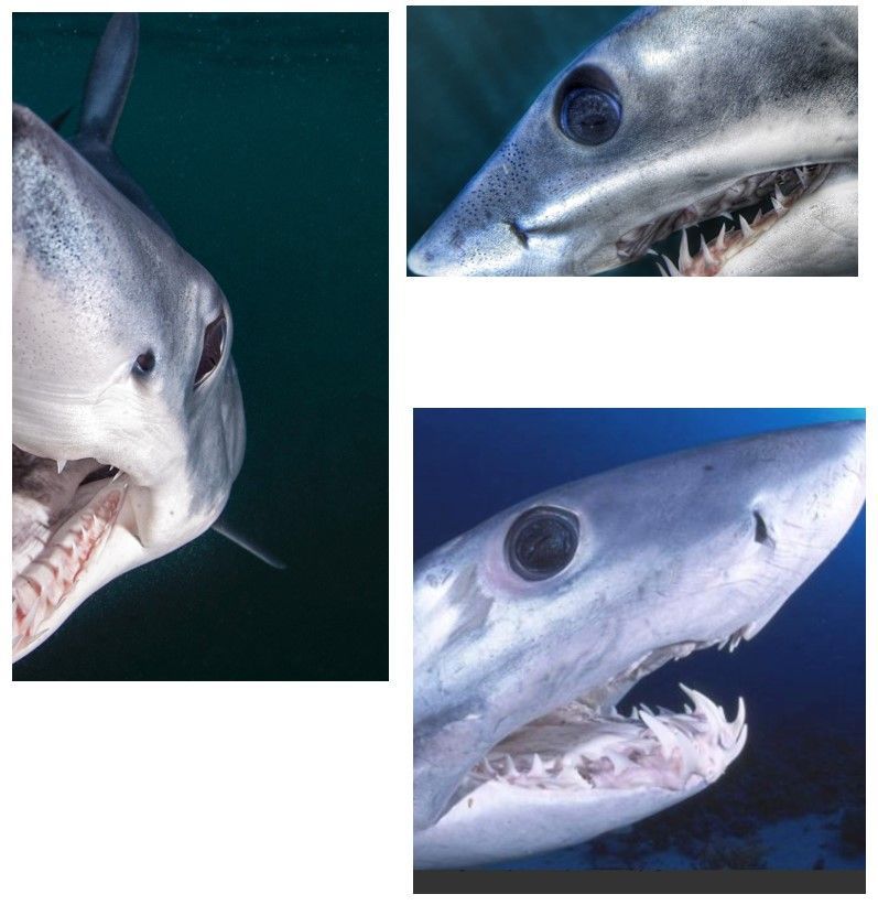
with a vertical slit pupil will be able to resolve detail in the vertical plane much better than in the
horizontal plane depending on the spatial resolving power of the retina and the amount of other
aberrations of the eye (Collin, 2018). Horizontal slit pupils are likely to sacrifice sharpness with the
advantage of having a wide field of vision for spotting vertical moving prey. In terrestrial animals, there
has been a correlation made between pupil shape and the environment/time of day in which the animal
hunts determining that ambush predators (snakes, dogs, cats) tend to have vertical slit pupils, and prey
animals (goats, horses, deer) tend to have horizontal slit pupils (Banks, Sprague, Schmoll, Parnell, Love,
2015). Establishing different retinal specializations in conjunction with lens structure and pupil shape
can better clarify how a shark may process an image. Retinal mapping has only been performed on one
Lamnid species (Carcharodon carcharias) prior to its protective status, which identified diurnal vision
based on identification, counts, and measurements of retinal neurons. The presence of a retinal ventro-
temporally located area centralis was also identified (Gruber and Cohen, 1985). Overall, pupil shape plays
several important roles in functional vision.

Studies on visual behavior has offered us glimpses into a shark’s visual environment and is readily
observed and researched in Carcharodon carcharias. Visual shape discrimination tests have been
performed (Strong, 1996), along with predator-prey interactions between seals or seal decoys (Fallows,
Aidan Hammerschlag 2011). One study determined that, during low-light conditions, Carcharodon
carcharias success rate of capture exceeds 55%, but drops below 40% during bright light conditions.
(Martin and Hammerschlag, 2012; Vermeij, 1982). Carcharodon carcharias have been known to attack
with the sun at their backs and find ways to use it to their advantage when seeking prey. (Huveneers,
Holman, and Robbins, 2015). Many of these field investigations have proved that the visual system in
Carcharodon carcharias is highly developed and they have learned to adapt to predator avoidance, prey
capture, and environmental changes.

Pupil shape and iris color have not been officially recorded in lamnid sharks. Determining pupil shape
could provide more insight on convergent evolution and may play a significant role in determining visual
capabilities and behaviors.

Materials And Methods
Subjects:
Six sets of white shark globes were harvested by one of the authors (GS) from sharks that had either
washed ashore deceased or had died after beaching themselves in the shallow water off Cape Cod,
Massachusetts (MA). Additionally, eyes from a mako and a porbeagle shark were also examined,
obtained from fisherman in Rhode Island.

The first set of white shark eyes were from a beached adult male white shark that died on shore in
Wellfleet, MA, in 2015. The head of the shark was placed in a freezer shortly after the time of death. In

                                                    Page 3/16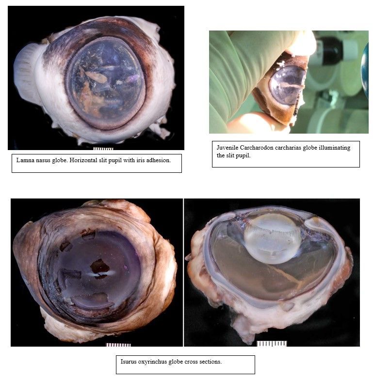
early 2018, the shark was thawed and both eyes were enucleated and placed in 10% Buffered
Formaldehyde.

Four juvenile white shark eyes were obtained from sharks that washed ashore in New York (NY) in June,
2018. The sharks were initially fixed in an ethanol bath for 6 days. Subsequently, the eyes were
enucleated and placed in 10% Buffered Formaldehyde.

Two white shark globes were obtained from a juvenile female in Scituate, MA in July 2018 and 2 more
globes from a from a juvenile male white shark were obtained in Truro, MA in August 2018. The eyes on
these sharks were enucleated immediately on site and placed in 10% Buffered Formaldehyde.

Two globes were obtained from an adult porbeagle shark and an adult mako shark in Rhode Island, in
August, 2018 after being caught by fisherman in a net. All globes were fixed in 10% Buffered
Formaldehyde.

Two white shark globes were obtained from a juvenile white shark that beached itself in Cape Cod,
Massachusetts (MA) in July of 2019.

Methods of Evaluation:
One set of white shark globes was dissected, photographed, and examined under a Zeiss optics operating
microscope in a surgical suite at Port City Veterinary Referral Hospital.

Underwater GoPro photographs and videos of white sharks in their natural environment were taken by
one of the authors (GS) in conjunction with his white shark population study with the Massachusetts
Division of Fisheries. Additionally, photographs of white sharks were obtained with permission from
George Probst, underwater photographer. These photos were taken with a Nikon camera in Guadalupe,
Mexico.

Photographs of salmon sharks were obtained with permission from Ron Watkins, underwater
photographer. These photos were taken with a Nikon camera in Alaska.

Photographs of mako sharks were obtained with permission from underwater photographer Joe Romerio.
These photos were taken with a Nikon camera in Rhode Island.

A total of 10 white shark, 2 porbeagle, and 2 mako shark globes were submitted to Dr. Richard Dubielzig,
DVM, DACVP, DACVO at the Comparative Ocular Pathology Laboratory of Wisconsin (COPLOW) for
histopathological evaluation. Gross photographs were obtained of all globes using a Canon DSLR. An
MRI was performed on one of the juvenile white shark eyes that was placed in 10% buffered
formaldehyde immediately following enucleation. All globes were sectioned in the vertical plane based on
the horizontal position of the pupil and hematoxylin & eosin and alcian blue PAS stains were performed.

Results
                                                 Page 4/16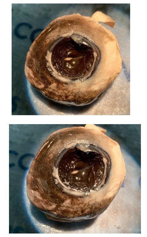
On initial exam of the first set of eyes received from the previously frozen adult white shark eyes,
cataracts were immediately noticeable likely caused by freezing. The pupil in both eyes was in a
horizontal slit position with iris adhesions (due to freezing), and the iris color was noticeably brown with a
slight blue hue in the limbus. These findings were confirmed with the use of a Zeiss ophthalmic surgical
microscope at Port City Veterinary Referral Hospital in Portsmouth, NH. Globe measurement on both
globes were 4 x 4.5in with one of the pupils in a horizontal slit position broken up by an iris adhesion.
Pupil diameter on one side of the adhesion was irregular and measured from 1–2 x 4mm and the
opposite pupil margin measured 1 x 3mm. The adhesion was 2mm long. The other pupil was also in a
horizontal slit position with irregularities measuring 1–2 x 8mm. The corneas measured 22 x 25mm.
Two of the juvenile white shark eyes from New York in June, 2018 were measured. Globes were 1 x 1.5in,
pupils were in a horizontal slit position at 1 x 10mm, and corneas measured 20 x 20mm.
Only one set of white shark globes, the juvenile female caught in Scituate, MA displayed bilaterally dilated
pupils. This shark was discovered deceased by fisherman in less than 24 hours after they deployed the
net. The globes on this shark measured 2 x 3in and the corneas were 20 x 23mm. Full dilation of the
pupils were present with iris irregularities. The amount of visible iris measured anywhere from 1-3mm.
One of the globes from the juvenile white shark in 2019 measured 2.5 x 3in, pupil was in a horizontal slit
position at 1 x 10mm, and cornea was 20 x 22mm.
The adult Porbeagle globes measured 4.5 x 5in. One of the pupils was slightly irregular in a horizontal
position measuring anywhere from 2–3 x 7mm. The other pupil was also in a horizontal slit position
measuring 2 x 5mm on one side of an iris adhesion and 2 x 3mm on the other side. The adhesion
measured 2mm long.
The Mako shark globes measured 2 x 3in with the pupils in a horizontal slit position measuring 1 x
10mm. The corneas measured 20 x 22mm.
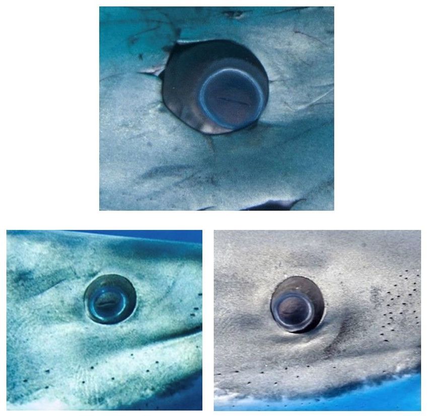
All online and personal photographs of white sharks examined after magnification confirmed a small,
elongated horizontal slit pupil and dark brown iris with the ring of blue around the limbus of the iris. No
visible cataracts, operculum, or iris adhesions are visibly noted in any of the photographs.
Even though we were not able to secure globes of the salmon shark (Lamna ditropis), ocular photographs
confirmed a horizontal slit pupil.
Pathology report confirmed a horizontal slit pupil in the white shark, mako, and porbeagle shark along
with a brown iris (Fig. 2, 5, 6, 7, 8). The blue ring mistakenly believed to be the iris, is in fact not the iris,
but a section of sclera and cartilage from the limbus that extends into the anterior chamber of the eye
(Fig. 4). The white shark globes that were thought to be the best preserved was evaluated by MRI prior to
cutting. That showed that the inner structure was uninterpretable, and when the globe was sectioned, it
was clear there was advanced autolysis making it impossible to recognize the internal structures of the
globe (Fig. 1). The sphincter muscle of the iris was best sampled histologically in the porbeagle shark. A
Page 5/16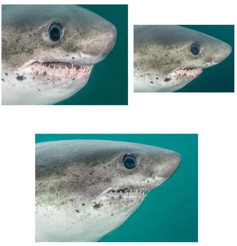
robust smooth muscle sphincter of the iris circles the horizontal pupil. A dilator muscle was hard to make
out (Fig. 3).

Discussion
In conclusion, this research reveals that the lamnid sharks, more specifically Carcharodon carcharias,
Isurus oxyrinchus, and Lamna nasus, have a horizontal slit pupil in the light adapted state. The one
female white shark with dilated pupils could potentially be the result of a “fight or flight” situation getting
caught in a gill net. None of the photographs and video examined showed any evidence of pupils in a
dilated state. Due to the robust smooth muscle found within the iris, it is believed that either the incoming
light from the sun at the surface of the ocean, and/or the camera and video lights are enough to
stimulate the pupil to quickly constrict. Further investigation of pupillary dilation and constriction in these
species is warranted.

With Carcharodon carcharias having a specialized retinal area centralis, it therefore requires a greater
degree of eye movement to locate prey vs other species of shark that have a dorsal or ventral streak
(Collin, 2018). The retinal variation of Carcharodon carcharias could very well be related to the habitat
and hunting methods it possesses, or it could simply be a part of how this species has evolved. Knowing
that Carcharodon carcharias possesses a horizontal slit pupil in conjunction with an area centralis, there
is also reason to believe that they must experience chromatic blurring based on the direction of how the
light hits the axis of the pupil. Knowing that this family of shark have a horizontal slit pupil, further
studies and tests could potentially lead to a better understanding of certain behaviors, and evolution
within this species. Determining the retinal specializations of Isurus oxyrinchus, Lamna nasus, and
Lamna ditropis will be helpful in further studies of convergent evolution. Because the Lamnid sharks rely
so heavily on vision for survival, understanding their complete visual anatomy will help paint a better
picture of migration, navigation, and habitat techniques, and could also help in understanding how
Climate Change may be altering these habits.

Declarations
Competing interests:

The authors declare no competing interests.

References
Bernal D, Sepulveda C, Mathieu-Costello O, Graham JB. 2003a. Comparative studies of high performance
swimming in sharks. I. Red muscle morphometrics, vascularization and ultrastructure. J Exp Biol
206:2831–2843.

Donley JM, Sepulveda CA, Konstantinidis P, Gemballa S, Shadwick RE. 2004. More than skin deep:
Convergent evolution in mechanical design of lamnid sharks and tunas. Nature 429:61–65.
                                                   Page 6/16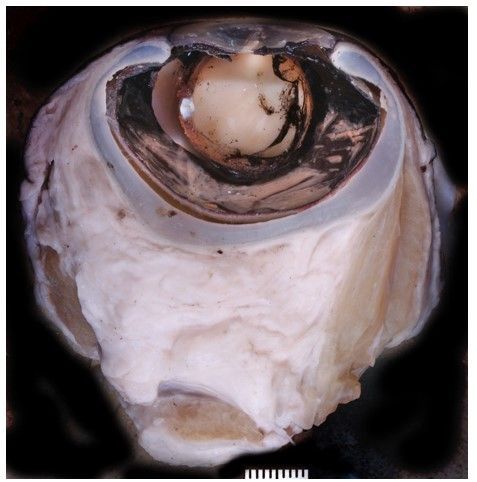
Shadwick RE. 2005. How tunas and lamnid sharks swim: An evolutionary convergence. Am Sci 93:524–
531.
Perry CN, Cartamil DP, Bernal D, Sepulveda CA, Theilmann RJ, Graham JB, Frank LR. 2007. Quantification
of red myotomal muscle volume and geometry in the shortfin mako shark (Isurus oxyrinchus) and the
salmon shark (Lamna ditropis) using T-1-weighted magnetic resonance imaging. J Morphol 268:284–
292.
Brill RW, Dewar H, Graham JB (1994) Basic concepts relevant to heat transfer in fishes, and their use in
measuring the physiological thermoregulatory abilities of tunas. Environ Biol Fish 40:109–124.
Dickson KA, Graham JB (2004) Evolution and consequences of endothermy in fishes. Physiol Biochem
Zool 77:998–1018.
Goldman KJ, Anderson SD, Latour RJ, Musick JA (2004) Homeothermy in adult salmon sharks, Lamna
ditropis. Environ Biol Fish 71:403–411
Species At Risk Public Registry, Canada COSEWIC Assessment and Status Report on the White Shark
Carcharodon carcharias in Canada, 2018. www.sararegistry.gc.ca/
Dewar, Eguchi, Hyde, Kinzey, Kohin, Moore, Taylor and Vetter. Status Review of the Northeastern Pacific
Population of White Sharks under the Endangered Species Act December, 2013. Pg 9.
Helfman, Burgess. Sharks: The Animal Answer Guide. Johns Hopkins University Press. Pg 64. May 15,
2014.
Alexis L. Morris, Elisa J. Livengood, and Frank A. Chapman. Sharks for the Aquarium and Considerations
for Their Selection. Reviewed November 2018, University of Florida Fisheries and Aquatic Sciences
Program, UF/IFAS Extension.Publication #FA179
www.sharkcagediving.net, www.adventuresportsnetwork.com, www.quora.com, nautiluslivaboards.com
Gruber SH, Cohen JL. Visual system of the white shark, Carcharodon carcharias, with emphasis on retinal
structure. Mem South Calif Acad Sci 1985; 9:61–72
Strong WR. Shape discrimination and visual predatory tactics in white sharks. In: Klimley P, Ainley DG,eds.
Great White Sharks: The Biology of Carcharodon carcharias. New York, New York: Academic Press, 1996
Fallows Chris, Martin R. Aidan, Hammerschlag Neil (2011) Comparisons between White S hark–Pinniped
Interactions at Seal Island (South Africa) with Other Sites in California. Global Perspectives on the
Biology and Life History of the Great White Shark, Chapter: 9: Publisher: CRC Press
Martin R. Aidan, Hammerschlag Neil (2011) Marine predator prey contests: Ambush and speed versus
vigilance and agility. Marine Biology Research, 2012; 8: 90-94
Page 7/16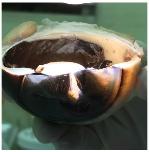
Vermeij GV. 1982. Unsuccessful predation and evolution. American Naturalist 120:701-20.

Huveneers C, Holman D, Robbins R et al. White sharks exploit the sun during predatory approaches. Am
Nat 2015; 185: 562–570.

Figures

Figure 1

Cross section of the adult white shark globe.

                                                Page 8/16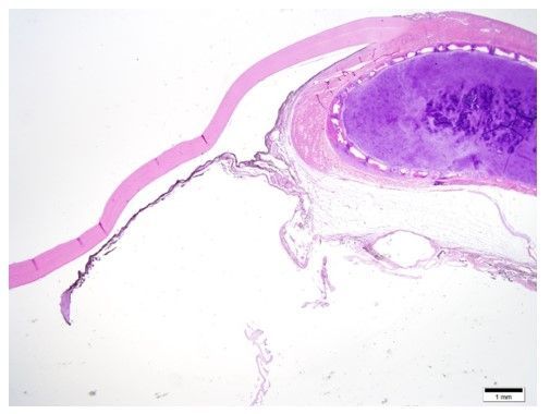
Figure 2

Illuminating the horizontal slit pupil in the adult white shark globe.

                                                   Page 9/16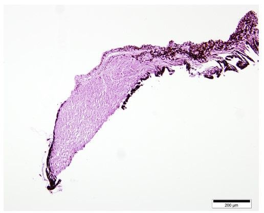
Figure 3

Magnification of the pupillary margin of the iris comprised of smooth muscle.

Figure 4

                                               Page 10/16
Magnification of the sclera and cartilage extending into the anterior chamber of the eye causing the
apparent “blue ring”.
Figure 5
GoPro Footage of Carcharodon carcharias from Dr. Greg Skomal and the Massachusetts Division of
Fisheries.
Page 11/16Figure 6
Carcharodon carcharias globe dissection 3/11/2020. Photos taken by Megan Burt
Page 12/16Figure 7
Carcharodon carcharias from underwater photographer George Probst.
Page 13/16Figure 8
Photos of Isurus oxyrinchus obtained from underwater photographer, Joe Romerio.
Page 14/16Figure 9
Photos of Lamna ditropis obtained via underwater photographer, Ron Watkins.
Page 15/16Figure 10
Globes
Page 16/16You can also read