UNIVERSITÄTSKLINIKUM HAMBURG-EPPENDORF
←
→
Page content transcription
If your browser does not render page correctly, please read the page content below
UNIVERSITÄTSKLINIKUM HAMBURG-EPPENDORF

           Institut für Klinische Pharmakologie und Toxikologie

                      Prof. Dr. med. Rainer H. Böger

Kinetik und Dynamik der oralen Supplementation mit Homoarginin
                    in gesunden Probanden

                              Dissertation

           zur Erlangung des Grades eines Doktors der Medizin
          an der Medizinischen Fakultät der Universität Hamburg.

                              vorgelegt von:

                            Mirjam Schönhoff
                      geboren in Georgsmarienhütte

                             Hamburg 2019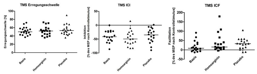
Angenommen von der
Medizinischen Fakultät der Universität Hamburg am: 20.05.2019

Veröffentlicht mit Genehmigung der
Medizinischen Fakultät der Universität Hamburg.

Prüfungsausschuss, der/die Vorsitzende: Prof. Dr. Edzard Schwedhelm

Prüfungsausschuss, zweite/r Gutachter/in: PD Dr. Chi-un Choe

                                                                      2
Inhaltsverzeichnis

1    Publikation ................................................................................................... 4
2    Darstellung der Publikation ........................................................................ 13
    2.1    Einleitung ............................................................................................ 13
    2.2    Methoden ............................................................................................ 15
     2.2.1       Probanden .................................................................................... 15
     2.2.2       Studiendesign ............................................................................... 16
     2.2.3       Pulswellengeschwindigkeit (PWV) und Augmentations-Index (AIx)..
                 ...................................................................................................... 17
     2.2.4       Flow-mediated vasodilation (FMD) ............................................... 18
     2.2.5       Transkranielle Magnetstimulation (TMS) ...................................... 19
     2.2.6       Studienprodukt.............................................................................. 19
     2.2.7       Messung und Analyse der kinetischen Parameter ........................ 20
     2.2.8       Statistische Auswertung ............................................................... 20
    2.3    Ergebnisse .......................................................................................... 21
     2.3.1       Charakteristika der Probanden ..................................................... 21
     2.3.2       Kinetikanalysen............................................................................. 22
     2.3.3       Dynamische Endpunkte der Studie............................................... 22
    2.4    Diskussion ........................................................................................... 25
    2.5    Zusammenfassung .............................................................................. 28
    2.6    Summary ............................................................................................. 29
    2.7    Abkürzungsverzeichnis ....................................................................... 30
    2.8    Literaturverzeichnis ............................................................................. 31
3    Erklärung des Eigenanteils ........................................................................ 34
4    Danksagung............................................................................................... 35
5    Lebenslauf ................................................................................................. 36
6    Eidesstattliche Erklärung ........................................................................... 37

                                                                                                                        3
British Journal of Clinical                                                  Br J Clin Pharmacol (2016) 82 1477–1485      1477
              Pharmacology

CLINICAL TRIALS
Oral supplementation with L-homoarginine in
young volunteers
Correspondence Edzard Schwedhelm, PhD, Department of Clinical Pharmacology and Toxicology, University Medical Centre Hamburg-
Eppendorf, Martinistr. 52, 20246 Hamburg, Germany. Tel.: +49 40 7410 54891; Fax: +49 40 7410 59757; E-mail: schwedhelm@uke.de

Received 13 April 2016; Revised 14 July 2016; Accepted 17 July 2016

Dorothee Atzler1,2,3, Mirjam Schönhoff1, Kathrin Cordts1,2, Imke Ortland4, Julia Hoppe5,
Friedhelm C. Hummel5, Christian Gerloff5, Ulrich Jaehde4, Annika Jagodzinski2,6, Rainer H. Böger1,2,
Chi-un Choe5 and Edzard Schwedhelm1,2
1
    Department of Clinical Pharmacology and Toxicology, University Medical Centre Hamburg-Eppendorf, Hamburg, Germany, 2DZHK (Deutsches
Zentrum für Herz-Kreislauf-Forschung e.V.), partner site Hamburg/Kiel/Lübeck, Germany, 3Vascular Biology, Institute for Stroke and Dementia
Research, Klinikum der Universität München Ludwig Maximilians-University of Munich, Munich, Germany, 4Institute of Pharmacy Department of
Clinical Pharmacy, University of Bonn, Bonn, Germany, 5Department of Neurology, University Medical Center Hamburg-Eppendorf, Hamburg,
Germany, and 6Department of General and Interventional Cardiology, University Heart Center Hamburg-Eppendorf, Hamburg, Germany

Keywords asymmetric dimethylarginine, L-arginine, L-homoarginine, nitric oxide, vascular function

AIMS
Low blood concentrations of the naturally occurring amino acid L-homoarginine (L-hArg) are related to impaired cardiovascular
outcome and mortality in humans and animals. L-hArg is a weak substrate of nitric oxide synthase and an inhibitor of arginases
in vitro. The aim of our study was to obtain kinetic and dynamic data after oral L-hArg supplementation.

METHODS
In a double-blind, randomized, placebo-controlled crossover study, 20 young volunteers received 125 mg L-hArg once daily for
4 weeks. Kinetic parameters (Cmax, Tmax and AUC0-24h) were calculated after ingestion of single and multiple doses of oral
supplementation as primary endpoint. Secondary endpoints that were evaluated were routine laboratory, L-arginine, asymmetric
dimethylarginine (ADMA), pulse wave velocity (PWV), augmentation index (AIx), flow-mediated vasodilatation (FMD),
corticospinal excitability, i.e. motor threshold (MT), and cortical excitability, i.e. intracortical inhibition (ICI) and facilitation (ICF).

RESULTS
One hour after ingestion (Tmax), L-hArg increased the baseline L-hArg plasma concentration (2.87 ± 0.91 μmol l 1, mean ± SD) by
8.74 ± 4.46 [95% confidence intervals 6.65; 10.9] and 17.3 ± 4.97 [14.9; 19.6] μmol l 1 (Cmax), after single and multiple doses,
respectively. Once-only and 4 weeks of supplementation resulted in AUCs0-24h of 63.5 ± 28.8 [50.0; 76.9] and 225 ± 78.5 [188;
2624] μmol l 1*h, for single and multiple doses, respectively. Routine laboratory parameters, L-arginine, ADMA, PWV, AIx, FMD,
MT, ICI and ICF did not change by L-hArg supplementation compared to baseline.
CONCLUSION
Once daily orally applied 125 mg L-hArg raises plasma L-hArg four- and sevenfold after single dose and 4 weeks of supplemen-
tation, respectively, and is safe and well tolerated in young volunteers.

© 2016 The British Pharmacological Society                                                                          DOI:10.1111/bcp.13068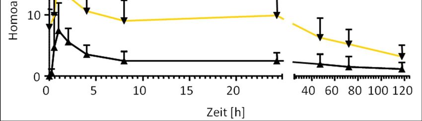
D. Atzler et al.

WHAT IS ALREADY KNOWN ABOUT THIS SUBJECT
• L-Homoarginine (L-hArg) is an amino acid found in pea pulses (Lathyrus sativus and cicera). It is a weak substrate for nitric
  oxide (NO) synthases (NOS) and an inhibitor of arginases.
• In clinical and epidemiological studies, low circulating L-hArg is associated with impaired cerebrovascular and cardiovas-
  cular outcome.
• Orally administered L-hArg is readily absorbed in rats and pigs with a recovery of >95% of unmetabolized L-hArg in urine.
WHAT THIS STUDY ADDS
• Our data show that oral supplementation with 125 mg L-hArg raises plasma L-hArg concentrations four- and sevenfold
  after single and multiple dosing in humans, respectively.
• Four weeks of supplementation did not change vascular and neuronal function in young volunteers, nor did any toxic
  side-effects occur.

Tables of Links

  TARGETS                                                                         LIGANDS
                                                                                                                           G   G
  Enzymes [1]                                 Nitric oxide synthases              L-arginine                              N ,N -dimethyl-L-arginine
  Arginase                                    Transporters [2]                    creatine

  Dimethylarginine                            SLC7 family
  dimethylaminohydrolases
  Arginine:glycine
  amidinotransferase

  These Tables list key protein targets and ligands in this article that are hyperlinked to corresponding entries in http://www.guidetopharmacology.
  org, the common portal for data from the IUPHAR/BPS Guide to PHARMACOLOGY [3], and are permanently archived in the Concise Guide to
  PHARMACOLOGY 2015/16 [1, 2].

Introduction                                                                   from guanidated lysine residues in casein and soya-bean pro-
                                                                               teins is readily absorbed in the jejunum and ileum of
The L-arginine/nitric oxide (NO) pathway plays an important                    Göttingen minipigs and broiler chickens [16, 17]. In line with
role in vascular haemostasis and L-arginine has been supple-                   these in vitro and in vivo data, oral supplementation of 1 and
mented to optimize health and welfare [4]. However, after                      10 mg kg 1 body weight L-hArg were almost quantitatively
oral administration, L-arginine is subject to extensive pre-                   (>95%) excreted unmetabolized in urine in pigs and rats, re-
systemic and systemic elimination by arginases and oral                        spectively [18]. The aim of this study was to investigate the ki-
doses required to increase plasma concentrations range from                    netic and dynamic properties of single and multiple oral
3 to 8 g/day [5, 6]. The non-proteinogenic amino acid                          doses of 125 mg L-hArg in young humans and the effect of
L-homoarginine (L-hArg) interferes with the L-arginine/NO                      multiple doses on the endothelial and vascular function and
pathway and has been identified as a risk marker for cardio-                    cortical excitability.
vascular, cerebrovascular and kidney diseases as well as for
cardiovascular and all-cause mortality (reviewed in [7, 8]).
L-hArg is a weak substrate of NO synthase (NOS) and arginase.
The maximal activity (Vmax) for murine NOS-dependent NO                        Methods
formation is similar for L-hArg and L-arginine, but the
Michaelis–Menten constant (Km) is 10–20 times higher for                       Subjects
L-hArg [9]. The Km value for L-hArg of rat liver arginase is                   Twenty-four healthy volunteers (15 female, 9 male) without
7.2 mmol l 1 which is about 70-fold above its actual concen-                   evidence of disease were found eligible for this study
tration in rat liver [10, 11]. The Vmax of rat liver arginase is 130           (Figure 1). They were recruited from the participating depart-
times faster for L-arginine compared with L-hArg [10]. These                   ments and from students at the University Medical Centre
properties make L-hArg a competitive inhibitor of rat liver ar-                Hamburg-Eppendorf. Exclusion criteria were sitting blood
ginase and render unlikely a strong catabolism in mammals.                     pressure ≥ 160/100 or ≤90/60 mmHg, sitting heart rate-
L-hArg is a substrate for the y+-transporter system which is re-                99 bpm or ≤50 beats per minute (bpm), a history of clinically
sponsible for stereoselective uptake and secretion of cationic                 significant hypotensive episodes or symptoms of fainting,
amino acids in many cells [12]. Furthermore, in vitro data                     dizziness, or light-headedness, a body mass index (BMI) ≥32
identified high L-hArg as a non-competitive inhibitor of alka-                  or ≤16 at screening, a history or symptoms of cardiovascular
line phosphatases (ALP, [13–15]). Of note, L-hArg originating                  disease, particularly coronary artery disease, arrhythmias, or

1478   Br J Clin Pharmacol (2016) 82 1477–1485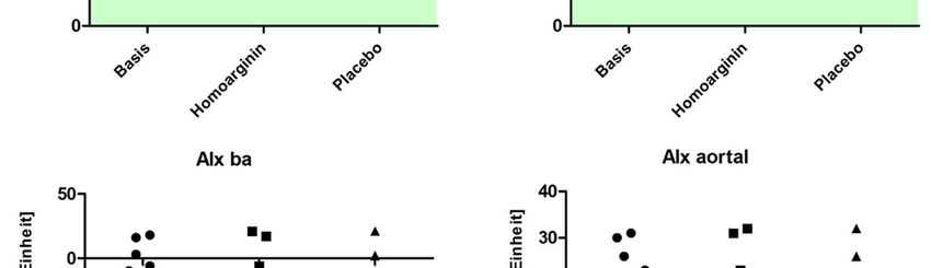
L-hArg in humans

Figure 1
CONSORT diagram

congestive heart failure, a history of significant central        dimethylarginine (ADMA) determinations were performed
nervous system disease, including transient ischemic attack,     and adverse events were evaluated. At baseline and after
stroke, seizure disorder, or behavioural disturbances, the use   each supplementation period (L-hArg and placebo), dynamic
of any drugs, a history of hepatitis B or C, and/or human        analyses applying plethysmography [i.e. pulse wave velocity
immunodeficiency virus (HIV 1 + 2), participation in an           (PWV) and augmentation index (AIx)], ultrasound [i.e. flow-
investigational drug or medical device study within 30 days      mediated vasodilatation (FMD)], and transcranial magnetic
of first dosing, donation of blood or blood products within       stimulation [TMS, i.e. motor threshold (MT), intracortical in-
the last 2 months (male) or 3 months (female) prior to study,    hibition (ICI), intracortical facilitation (ICF)] were recorded.
and pregnancy (female). Written informed consent was             Two individuals abandoned study participation without
obtained from all participants. The study was planned as a       statement of reasons.
non-drug study and the study protocol was approved by the
Ethics Committee of the Hamburg Board of Physicians
(PV4038) accordingly. The investigation was conducted in         Biochemical analyses
accordance with the Declaration of Helsinki and registered       Plasma L-hArg, L-arginine and ADMA concentrations were
at clinicaltrials.gov (NCT02675660).                             determined in 20 participants by liquid chromatography
                                                                 (LC)-tandem mass spectrometry (MS) analysis as described
Study design                                                     previously [19, 20]. Briefly, 25 μL aliquots of plasma were
In a double-blind, placebo-controlled crossover design, 22       spiked with stable isotope-labelled L-hArg, L-arginine and
participants were randomized to receive either 125 mg L-hArg     ADMA, which served as internal standards. Proteins were
or placebo once daily in the morning for 4 weeks each            precipitated with 100 μL of methanol, filtrated through a
(Figure S1). Placebo and L-hArg capsules were provided by        0.22 μm hydrophilic membrane (Multiscreen HTS™,
Wellnest International Ltd. (West Sussex, UK), the latter        Millipore, Molsheim, France), derivatized with butanolic
being marketed as a dietary supplement. Cellulose capsules       1 N HCl, and analysed by LC-tandem MS (Varian 1200 MS,
contained lactose or 119 ± 13 mg L-hArg (mean ± SD, n = 7)       Agilent Technologies, Santa Clara, USA). Quantification was
and cornstarch as excipient. The study periods were separated    performed by calculation of peak area ratios and calibration
by a washout phase of 4 weeks, and the sequence of the           with known concentrations of analytes in dialysed EDTA
medications was randomly chosen in each participant. The         plasma. Limits of quantification were 0.01 μmol l 1 for
study was preceded by a run-in phase, where all participants     L-hArg, 0.25 μmol l 1 for L-arginine and 0.005 μmol l 1 for
received a single dose of 125 mg L-hArg. Blood samples           ADMA. For all arginine metabolites, coefficients of variation
(2.7 ml EDTA vacutainer) for plasma L-hArg determinations        were ≤7.5% [19, 20]. Blood counts, blood glucose, serum cre-
were drawn at time points 0, 15, 30 min, 1, 2, 4, 8, 24, 48,     atinine, glutamic oxaloacetic transaminase (GOT), glutamic
72 and 120 h after single and multiple doses of L-hArg and       pyruvic transaminase (GPT), ALP, and high sensitive
placebo, respectively. At baseline, after each supplementation   C-reactive protein (hsCRP) were determined with routine
period (L-hArg and placebo) and after 4 weeks of follow-up,      laboratory assays. Estimated glomerular filtration rate (eGFR)
biochemical analyses including L-arginine and asymmetric         was calculated with the CKD-EPI formula [21].

                                                                                 Br J Clin Pharmacol (2016) 82 1477–1485    1479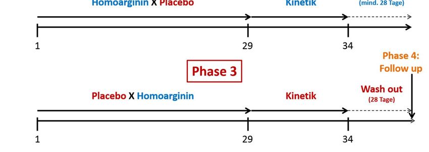
D. Atzler et al.

Kinetic analyses                                                 Results
Kinetic parameters, i.e. maximum plasma concentration
(Cmax), time to maximum plasma concentration (Tmax)              Baseline characteristics of investigated subjects are listed in
and area under the plasma concentration–time curve               Table 1. All participants were healthy Asian-Caucasian with
(AUC0-24h), were calculated for L-hArg after single dose         no history or symptoms of cardiovascular disease, particu-
and multiple doses. AUCs were calculated for up to 24 h.         larly coronary artery disease, arrhythmias, congestive heart
To account for possible circadian rhythms of endogenous          failure, transient ischemic attack, stroke, seizure disorder or
L-hArg, plasma concentrations following L-hArg adminis-          behavioural disturbances. Baseline L-hArg concentration
tration at each time point were corrected for individual         was 2.87 ± 0.91 μmol l 1, mean ± SD, with no difference
baseline (time point zero of single and multiple measure-        between women and men, i.e. 2.66 and 3.13 μmol l 1, respec-
ment) and placebo data prior to calculation of Cmax, Tmax,       tively (P = 0.26, Student's t-test for unpaired data). Oral
and AUC0-24h values. Even for corrected data, the                supplementation with 125 mg L-hArg increased the plasma
calculation of half-life was still not possible. All kinetic     concentrations of L-hArg (Cmax) after single and multiple
calculations were performed using Excel (version 2010,           doses by 8.74 ± 4.46 [95% confidence intervals 6.65; 10.9]
Microsoft Corporation, Redmont, USA).                            and 17.3 ± 4.97 [14.9; 19.6] μmol l 1, respectively (Table 2).
                                                                 The AUC0-24h was 3.5-fold higher after multiple dosing com-
                                                                 pared with a single dose of L-hArg. Cmax was reached after 1 h
                                                                 irrespectively of the dosing regimen.
Dynamic analyses
Systolic (SBP) and diastolic blood pressure (DBP) was mea-
sured at baseline in three independent examinations at
supine position after 5 min of resting, and results were
                                                                 Table 1
averaged. PWV and AIx were obtained in supine position
by plethysmography, applying the vascular explorer system        Baseline characteristics of participantsa
(Enverdis, Jena, Germany). Augmented pressure was calcu-
lated as the difference between the second systolic peak                                                                              Standard
and the first systolic peak, and AIx was calculated as the ra-                                                      Mean               deviation
tio between augmented pressure and pulse pressure. Values
                                                                   Age (years)                                     35                 14
were normalized to a heart rate of 75 bpm. Central PWV
was assessed recording waveforms at the femoral and ca-            Gender (n, %)                                   11 females (55%)
rotid site, sequentially. FMD was assessed in the volunteers'      Smoker (n, %)                                   5 (25%)
right arm by high resolution ultrasound (12 MHz linear                             2
array transducer, Sienna, Siemens, Munich, Germany) as             BMI (kg m           )                           24                 2.9
                                                                                                               b
described previously [22]. In brief, longitudinal echo scans       Blood pressure (mmHg)
of the brachial artery were obtained before and after reac-            Systolic                                    119                9.3
tive hyperaemia. FMD was calculated as the percent in
                                                                       Diastolic                                   75                 6.8
artery diameter 1 min after cuff release relative to the
diameter before cuff release. Corticospinal excitability, i.e.     Blood counts/clinical chemistry
MT, and cortical excitability, i.e. ICI and ICF, were evalu-           Leukocytes (c/nL)                           6.0                1.3
ated during rest with well-established single and paired-
                                                                       Thrombocytes (c/nL)                         255                62
pulse TMS protocols using a 7 cm diameter figure of 8
shaped coil and two Magstim 200 stimulators (Magstim                   GOT (U/L)                                   25.8               9.8
Co., Whitland, Carmarthenshire, UK) and Signal software                GPT (U/L)                                   21.3               8.9
4.05 and a CED1902-amplifier (both Cambridge Electronic
                                                                       ALP (U/L)                                   46                 18
Design, Cambridge, UK) for TMS data recording and pro-
cessing [23, 24]. Two subjects were excluded from ICI and          Blood glucose (mg/dL)                           76                 18
ICF measurement; due to high MT and low recruitment,                                       c
                                                                   hsCRP (mg/dL)                                   0.9                [0.9; 2.2]
no stable test stimulus motor evoked potential >0.2 mA                                     1
                                                                   eGFR (ml min                )                   100                15
could be achieved (and with low amplitude test stimuli
no reliable ICI nor ICF can be elicited [25]).                     L-hArg (μmol l
                                                                                               1
                                                                                                   )               2.87               0.91
                                                                                                       1
                                                                   L-Arginine (μmol l                      )       80                 20
                                                                                           1
                                                                   ADMA (μmol l                )                   0.60               0.08
Statistical analyses                                               a
All data are given as mean ± standard deviation (SD) [95%            Data are given as mean ± standard deviation unless otherwise
                                                                   indicated.
confidence intervals, if appropriate] or median [25th; 75th         b
                                                                     Average of three independent measurements.
percentile]. Statistical comparisons were made by Student's        c
                                                                     Median [25th; 75th percentile].
t-test (two-tailed) for unpaired or paired data of two groups      ADMA, asymmetric dimethylarginine; ALP, alkaline phosphatase;
and repeated measures ANOVA with Newman–Keuls post hoc             BMI, body mass index; eGFR, estimated glomerular filtration rate
test for paired data of four groups. Statistical analysis was      computed using the CKD-EPI formula, GOT, glutamic oxaloacetic
performed with GraphPad Prism (version 5 for Windows,              transaminase; GPT, glutamic pyruvic transaminase; L-hArg,
La Jolla, USA).                                                    L-homoarginine, hsCRP, high-sensitivity C-reactive protein.

1480   Br J Clin Pharmacol (2016) 82 1477–1485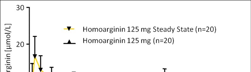
L-hArg in humans

Table 2
Kinetic characteristics of L-homoarginine in human plasma after a single dose and four weeks of 125 mg oral supplementationa

                                               Single dose                                  Multiple dose                                P valueb
                 1
  Cmax [μmol l       ]                         8.74 ± 4.46 [6.65; 10.9]                     17.3 ± 4.97 [14.9; 19.6]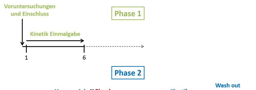
D. Atzler et al.
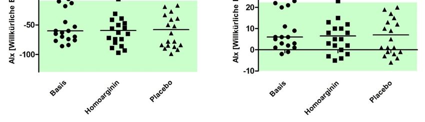
Figure 3
Transcranial magnetic stimulation (TMS) and flow-mediated vasodilatation (FMD) were performed at baseline and after 4 weeks of supplemen-
tation with L-homoarginine and placebo (n = 20 for FMD, n = 18–20 for TMS). Corticospinal excitability, i.e. motor threshold (MT), cortical
excitability, i.e. intracortical inhibition (ICI) and facilitation (ICF), were evaluated with single and paired-pulse TMS protocols. *P < 0.05 vs. base-
line, repeated measures ANOVA with Newman–Keuls post hoc test
difference in L-hArg concentrations between women and latter might not sufficiently be inhibited by a single dose once
men, 125 mg L-hArg tended to increase Cmax more in women daily of a weak arginase inhibitor [5]. Experimental data
compared with men, i.e. by 18.6 and 15.7 μmol l 1, respec- showed that L-hArg is an inhibitor of ALP [13–15] and clinical
tively (P = 0.20, Student's t-test for unpaired data). The sex observations revealed a negative correlation between circu-
difference was most likely due to the lower body weight in lating L-hArg and ALP levels in patients undergoing coronary
women, i.e. 63 vs. 82 kg. angiography [28]. However, physiological concentrations of
Multiple doses of 125 mg L-hArg increased the Cmax L-hArg, i.e. 0.2–3 μmol l 1 do not inhibit ALP, but induce
sevenfold over baseline as compared with a fourfold in- osteogenic transformation of vascular smooth muscle cells,
crease after a single dose. We did not perform serial blood augmenting vascular calcification in experimental athero-
collections to evaluate Cmax, Tmax, and AUC0-24h at differ- sclerosis [29]. In the present investigation, the applied dosing
ent time-points after multiple dosing regimens and thus regime of L-hArg did not alter ALP activity or vascular
do not know if the tripling of AUC0-24h represents the phenotypes.
steady state kinetics. As a substrate for the y+-transporter Lower plasma concentrations of L-hArg have been re-
system, L-hArg is likely to be taken up by several organs ported in a variety of clinical conditions, among them
[12]. Even though we corrected our data for baseline and coronary artery disease, congestive heart failure, ischemic
placebo concentrations to account for diurnal variation in stroke and myocardial infarction [28, 30–35]. Low L-hArg
L-hArg plasma concentrations, we were not able to calcu- has been linked to a worsened prognosis in patients
late the terminal half-life in our study. with renal and cardiovascular disease as well as to cardio-
In line with previous observations made for the oral vascular and all-cause mortality in the general population
supplementation with L-arginine, L-hArg did not follow [28, 36–38]. These retrospective and prognostic cohort
first-order elimination kinetics in a single compartment studies provide no experimental evidence in favour of a
model [22]. L-hArg is a competitive inhibitor of rat liver supplementation with L-hArg.
arginase [10]. We therefore investigated whether 4 weeks of To date, L-hArg has only been supplemented in several
125 mg L-hArg supplementation increases L-arginine plasma animal models [16–18, 32, 39, 40]. In all studied species (i.e.
concentrations or its endogenous methylation product chickens, pigs, rats and mice) orally supplemented L-hArg
ADMA [27]. Even though we did not find increased L-arginine was readily absorbed in the intestine, it increased L-hArg
nor ADMA concentrations in our investigation, we cannot plasma concentrations and was excreted almost quantita-
rule out that alternative dosing regimens, i.e. higher doses tively and unmetabolized into the urine. Oral supplementa-
or shorter dosing intervals, might influence L-arginine me- tion of C57BL/6 mice for 4 weeks with 14 mg l 1 L-hArg in
tabolism. L-Arginine is not only subject to systemic but also drinking water (approx. 2 mg kg 1 body weight) resulted in
to extensive pre-systemic elimination by arginases, and the a threefold increase in L-hArg plasma concentration from
1482 Br J Clin Pharmacol (2016) 82 1477–1485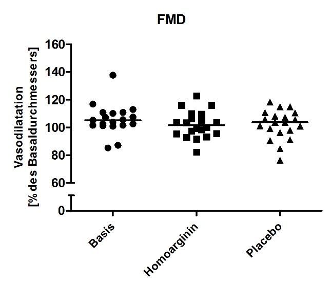
L-hArg in humans

0.14 to 0.46 μmol l 1 [32]. Supplementation of 125 mg L-hArg         studies, and L-hArg plasma concentrations decline with
once daily (approx. 2 mg kg 1 body weight) for 4 weeks in            progression of chronic kidney disease [38, 46]. At baseline
humans resulted in a sevenfold increase (Cmax) in L-hArg             we observed relatively high L-hArg plasma concentrations
plasma concentration. These data clearly indicate that me-           possibly attributed to the young age and normal kidney
tabolism of L-hArg is different between mice and men and             function in our study population (Table 1). We did not in-
needs further investigation. Although the increase in L-hArg         vestigate urinary excretion of L-hArg or AGAT expression,
observed in mice was rather moderate, the applied dose               which might be altered upon L-hArg supplementation. Pre-
significantly improved neurological outcome in an experi-             viously it was shown that L-arginine is extensively metabo-
mental model of stroke [32]. So far, murine and human data           lized by arginase in the gut wall and liver [5, 47]. This
indicate that genetic alterations of L-arginine:glycine              limits its oral bioavailability as a substrate for NOS and
amidinotransferase (AGAT) are responsible for changes in             subsequent effect on vascular function. L-hArg is an alter-
L-hArg levels [32, 41]. Therefore, AGAT itself might represent       native substrate for NOS not subjected to elimination by
a possible target for future interventions to regulate L-hArg        arginase [9, 10]. However, to date it is still speculative
levels. Furthermore, it is still unknown whether the therapeu-       whether the beneficial effects of L-hArg are solely due to
tic potential of L-hArg supplementation is translatable to           interactions with L-arginine/NOS metabolism. The ratio of
humans. However, our data show that L-hArg supplementa-              L-arginine over the endogenous NOS inhibitor ADMA is
tion in humans is feasible.                                          one predictor for the substrate availability for NOS [48].
    In experimental and clinical studies L-hArg was associated       In our study, supplementation with L-hArg did not change
with endothelial function, e.g. FMD [42], kidney function,           L-arginine, ADMA or the L-arginine/ADMA ratio. In line
e.g. eGFR [38], neurotoxicity, e.g. altered neuronal excitabil-      with this, we did not observe any improvement of endo-
ity [43], blood pressure [44] and glucose metabolism [39].           thelial function; neither FMD, nor PWI or AIx were
Supplementing 20 young individuals with 125 mg L-hArg                changed. This does not render changes in endothelial
once daily did not change FMD, eGFR, ICI, ICF, SBP or DBP.           function impossible after longer supplementation period
We observed a moderate increase in blood glucose after               or in subjects with pre-existing cardiovascular diseases.
L-hArg supplementation (Table S1). This could be an adverse          Nevertheless, the primary endpoint of our study was the
reaction to the supplement, but experimental findings in              determination of kinetic parameters and our study was
obese mice have shown an opposite effect of L-hArg supple-           sufficiently powered for this purpose.
mentation on blood glucose. Oral supplementation of                      In conclusion, the results of the present study provide a
C57BL/6 mice for 16 weeks with 14 and 28 mg l 1 L-hArg in            rationale for larger, prospective clinical studies with longer
drinking water blunted a metabolic phenotype induced by a            treatment periods to investigate the effects of oral supple-
high-fat diet; i.e. L-hArg fostered insulin secretion and ame-       mentation with 125 mg L-hArg in patients with cardiovascu-
liorated blood glucose levels [39]. MT seemed significantly           lar or metabolic disease.
increased after placebo compared with baseline, but one
outlier contributed to this effect. Furthermore, MT of placebo
and L-hArg supplementation groups did not differ signifi-
cantly from each other. Despite the clinical studies showing         Competing Interests
associations between L-hArg and clinical phenotypes, no evi-
dence for a direct effect is given. At least for the dosing period   All authors have completed the Unified Competing Interest
applied to healthy individuals in the current study, no im-          form at http://www.icmje.org/coi_disclosure.pdf (available
pact, neither harm nor benefit, was observed.                         on request from the corresponding author) and declare: DA
    Na+/K+-ATPase is a crucial enzyme responsible for the            had a grant from the European Community in the previous
active transport of sodium and potassium ions in the ner-            3 years; CG had grants and personal fees with Bayer
vous system necessary to maintain the ionic gradient for             Healthcare, Boehringer Ingelheim, GlaxoSmithKline,
neuronal excitability. In vitro studies showed an inhibitory         Lundbeck, Pfizer, Sanofi Aventis, UCB, Merck Serono, EBS
effect of L-hArg on Na+/K+-ATPase in the synaptic plasma             technologies, Silk Road Medical, German Research Council,
membrane from cerebral cortex of young rats at concentra-            German Ministry of Science and Education and the
tions of 5–20 μmol l 1 [43]. However, in our study we did            European Community in the previous 3 years; CUC had a
not observe any alterations of the cortical excitability by          grant with the Else Kröner-Fresenius Stiftung in the previous
hArg supplementation, i.e. neither ICI nor ICF were                  3 years; there are no other relationships or activities that
changed. Given its polarity, transport of L-hArg across the          could appear to have influenced the submitted work.
blood–brain barrier is likely to require an active transport             The excellent medical and technical assistance of A. Dehn,
system. L-hArg is a substrate for the y+-transporter system          S. Griesbach, M. Kastner, J. Lockowandt A. Steenpass and
[12] and has been reported to act as a competitive inhibitor         J. Wiener is appreciated. Dr Atzler acknowledges the support of
of L-arginine uptake by porcine endothelial cells [45]. How-         the European Community under a Marie Curie Intra-European
ever, in mice, L-hArg was reported to be taken up into the           Fellowship for Career Development and Dr Choe was funded
brain [32]. Thus, it can only be concluded from the present          by an Else Kröner Memorial Stipendium from the Else
data that supplementation of 125 mg L-hArg for 4 weeks               Kröner-Fresenius Stiftung. This publication was funded by
does not seem to interfere with cortical excitability in             LMU Munich's Institutional Strategy LMUexcellent within the
healthy individuals.                                                 framework of the German Excellence Initiative (DA). The contri-
    AGAT is expressed predominantly in the kidney and                butions to sample and data collection made by volunteers are
liver, L-hArg and GFR are positively associated in cohort            gratefully acknowledged.

                                                                                     Br J Clin Pharmacol (2016) 82 1477–1485   1483
D. Atzler et al.
Contributors regulation by subunit–subunit interaction. Jpn J Pharmacol 1994;
64: 97–102.
Conception and design of the work: DA, CUC, ES. Analysis 16 Schmitz M, Hagemeister H, Erbersdobler HF. Homoarginine
and interpretation of data: DA, MS, KC, IO, JH, CUC, ES. labeling is suitable for determination of protein absorption in
Drafting or revising the manuscript: DA, MS, KC, IO, UJ, miniature pigs. J Nutr 1991; 121: 1575–80.
CUC, AJ, ES. Final approval of the manuscript: DA, MS, KC, 17 Siriwan P, Bryden WL, Annison EF. Use of guanidinated dietary
IO, JH, FCH, CG, UJ, AJ, RHB, CUC, ES. DA, MS, ES, and protein to measure losses of endogenous amino acids in poultry.
CUC contributed equally. Br J Nutr 1994; 71: 515–29.
18 Hou Y, Hu S, Jia S, Nawaratna G, Che D, Wang F, et al. Whole-
body synthesis of L-homoarginine in pigs and rats supplemented
with L-arginine. Amino Acids 2016; 48: 993–1001.
19 Atzler D, Mieth M, Maas R, Böger RH, Schwedhelm E. Stable
References isotope dilution assay for liquid chromatography-tandem mass
1 Alexander SPH, Fabbro D, Kelly E, Marrion N, Peters JA, Benson spectrometric determination of L-homoarginine in human
HE, et al. The Concise Guide to PHARMACOLOGY 2015/16: plasma. J Chromatogr B Analyt Technol Biomed Life Sci 2011;
Enzymes. Br J Pharmacol 2015; 172: 6024–109. 879: 2294–8.
2 Alexander SPH, Kelly E, Marrion N, Peters JA, Benson HE, 20 Schwedhelm E, Maas R, Tan-Andresen J, Schulze F, Riederer U,
Faccenda E, et al. The Concise Guide to PHARMACOLOGY Böger RH. High-throughput liquid chromatographic-tandem
2015/16: Transporters. Br J Pharmacol 2015; 172: 6110–202. mass spectrometric determination of arginine and dimethylated
arginine derivatives in human and mouse plasma. J Chromatogr B
3 Southan C, Sharman JL, Benson HE, Faccenda E, Pawson AJ,
Analyt Technol Biomed Life Sci 2007; 851: 211–19.
Alexander SP, et al. The IUPHAR/BPS Guide to PHARMACOLOGY
in 2016: towards curated quantitative interactions between 1300 21 Levey AS, Stevens LA, Schmid CH, Zhang YL, Castro AF 3rd,
protein targets and 6000 ligands. Nucl Acids Res 2016; 44: Feldman HI, et al. A new equation to estimate glomerular
D1054–68. filtration rate. Ann Intern Med 2009; 150: 604–12.
4 Wu G, Meininger CJ. Arginine nutrition and cardiovascular 22 Schwedhelm E, Maas R, Freese R, Jung D, Lukacs Z, Jambrecina A,
function. J Nutr 2000; 130: 2626–9. et al. Pharmacokinetic and pharmacodynamic properties of oral
L-citrulline and L-arginine: impact on nitric oxide metabolism. Br
5 Morris SM Jr. Enzymes of arginine metabolism. J Nutr 2004;
J Clin Pharmacol 2008; 65: 51–9.
134 (10 Suppl): 2743S–2747S ;discussion 65S–7S.
23 Zimerman M, Heise KF, Hoppe J, Cohen LG, Gerloff C,
6 Böger RH. The pharmacodynamics of L-arginine. Altern Ther
Hummel FC. Modulation of training by single-session
Health Med 2014; 20: 48–54.
transcranial direct current stimulation to the intact motor
7 Atzler D, Schwedhelm E, Choe CU. L-homoarginine and cortex enhances motor skill acquisition of the paretic hand.
cardiovascular disease. Curr Opin Clin Nutr Metab Care 2015; 18: Stroke 2012; 43: 2185–91.
83–8.
24 Ziemann U, Reis J, Schwenkreis P, Rosanova M, Strafella A,
8 Pilz S, Meinitzer A, Gaksch M, Grübler M, Verheyen N, Drechsler Badawy R, et al. TMS and drugs revisited 2014. Clin Neurophysiol
C, et al. Homoarginine in the renal and cardiovascular systems. 2015; 126: 1847–68.
Amino Acids 2015; 47: 1703–13.
25 Kujirai T, Caramia MD, Rothwell JC, Day BL, Thompson PD,
9 Moali C, Boucher JL, Sari MA, Stuehr DJ, Mansuy D. Substrate Ferbert A, et al. Corticocortical inhibition in human motor cortex.
specificity of NO synthases: detailed comparison of L-arginine, J Physiol 1993; 471: 501–19.
homo-L-arginine, their N omega-hydroxy derivatives, and N
26 Atzler D, Appelbaum S, Cordts K, Ojeda FM, Wild PS, Münzel T,
omega-hydroxynor-L-arginine. Biochemistry 1998; 37: 10453–60.
et al. Reference intervals of plasma homoarginine from the
10 Reczkowski RS, Ash DE. Rat liver arginase: kinetic mechanism, German Gutenberg Health Study. Clin Chem Lab Med 2016; 54:
alternate substrates, and inhibitors. Arch Biochem Biophys 1994; 1231–7.
312: 31–7.
27 Tsikas D, Wu G. Homoarginine, arginine, and relatives: analysis,
11 Yang Y, Wu Z, Jia S, Dahanayaka S, Feng S, Meininger CJ, et al. metabolism, transport, physiology, and pathology. Amino Acids
Safety of long-term dietary supplementation with L-arginine in 2015; 47: 1697–702.
rats. Amino Acids 2015; 47: 1909–20.
28 März W, Meinitzer A, Drechsler C, Pilz S, Krane V, Kleber ME, et al.
12 White MF, Gazzola GC, Christensen HN. Cationic amino acid Homoarginine, cardiovascular risk, and mortality. Circulation
transport into cultured animal cells. I. Influx into cultured human 2010; 122: 967–75.
fibroblasts. J Biol Chem 1982; 257: 4443–9.
29 Alesutan I, Feger M, Tuffaha R, Castor T, Musculus K, Buehling SS,
13 Lin CW, Fishman WH. L-Homoarginine. An organ-specific, et al. Augmentation of phosphate-induced osteo-/chondrogenic
uncompetitive inhibitor of human liver and bone alkaline transformation of vascular smooth muscle cells by homoarginine.
phosphohydrolases. J Biol Chem 1972; 247: 3082–7. Cardiovasc Res 2016; 110: 408–18.
14 Rufo MB, Fishman WH. L-homoarginine, a specific inhibitor of 30 Atzler D, Baum C, Ojeda F, Keller T, Cordts K, Schnabel RB, et al.
liver-type alkaline phosphatase, applied to the recognition of Low homoarginine levels in the prognosis of patients with acute
liver-type enzyme activity in rat intestine. J Histochem Cytochem chest pain. J Am Heart Assoc 2016; 5: e002565.
1972; 20: 336–43.
31 Atzler D, Rosenberg M, Anderssohn M, Choe CU, Lutz M, Zugck
15 Suzuki K, Yoshimura Y, Hisada Y, Matsumoto A. Sensitivity of C, et al. Homoarginine – an independent marker of mortality in
intestinal alkaline phosphatase to L-homoarginine and its heart failure. Int J Cardiol 2013; 168: 4907–9.
1484 Br J Clin Pharmacol (2016) 82 1477–1485L-hArg in humans
32 Choe CU, Atzler D, Wild PS, Carter AM, Böger RH, Ojeda F, et al. cerebral cortex by guanidino compounds accumulating in
Homoarginine levels are regulated by L-arginine:glycine hyperargininemia. Brain Res 1999; 838: 78–84.
amidinotransferase and affect stroke outcome: results from
44 van der Zwan LP, Davids M, Scheffer PG, Dekker JM, Stehouwer
human and murine studies. Circulation 2013; 128: 1451–61.
CD, Teerlink T. L-Homoarginine and L-arginine are
33 Drechsler C, Meinitzer A, Pilz S, Krane V, Tomaschitz A, Ritz E, antagonistically related to blood pressure in an elderly
et al. Homoarginine, heart failure, and sudden cardiac death in population: the Hoorn study. J Hypertens 2013; 31: 1114–23.
haemodialysis patients. Eur J Heart Fail 2011; 13: 852–9.
45 Bogle RG, Moncada S, Pearson JD, Mann GE. Identification of
34 Pilz S, Meinitzer A, Tomaschitz A, Drechsler C, Ritz E, Krane V, inhibitors of nitric oxide synthase that do not interact with the
et al. Low homoarginine concentration is a novel risk factor for endothelial cell L-arginine transporter. Br J Pharmacol 1992; 105:
heart disease. Heart 2011; 97: 1222–7. 768–70.
35 Pilz S, Tomaschitz A, Meinitzer A, Drechsler C, Ritz E, Krane V, 46 Drechsler C, Kollerits B, Meinitzer A, März W, Ritz E, König P, et al.
et al. Low serum homoarginine is a novel risk factor for fatal Homoarginine and progression of chronic kidney disease: results
strokes in patients undergoing coronary angiography. Stroke from the Mild to Moderate Kidney Disease Study. PLoS One 2013;
2011; 42: 1132–4. 8: e63560.
36 Atzler D, Gore MO, Ayers CR, Choe CU, Böger RH, de Lemos JA, 47 Castillo L, deRojas TC, Chapman TE, Vogt J, Burke JF,
et al. Homoarginine and cardiovascular outcome in the Tannenbaum SR, et al. Splanchnic metabolism of dietary arginine
population-based Dallas Heart Study. Arterioscler Thromb Vasc in relation to nitric oxide synthesis in normal adult man. Proc
Biol 2014; 34: 2501–7. Natl Acad Sci U S A 1993; 90: 193–7.
37 Pilz S, Teerlink T, Scheffer PG, Meinitzer A, Rutters F, Tomaschitz 48 Böger RH, Vallance P, Cooke JP. Asymmetric dimethylarginine
A, et al. Homoarginine and mortality in an older population: the (ADMA): a key regulator of nitric oxide synthase. Atheroscler
Hoorn study. Eur J Clin Invest 2014; 44: 200–8. Suppl 2003; 4: 1–3.
38 Tomaschitz A, Meinitzer A, Pilz S, Rus-Machan J, Genser B,
Drechsler C, et al. Homoarginine, kidney function and
cardiovascular mortality risk. Nephrol Dial Transplant 2014; 29: Supporting Information
663–71.
39 Stockebrand M, Hornig S, Neu A, Atzler D, Cordts K, Böger RH, Additional Supporting Information may be found in the
et al. Homoarginine supplementation improves blood glucose in online version of this article at the publisher’s web-site:
diet-induced obese mice. Amino Acids 2015; 47: 1921–7.
40 Pentyala J, Rao SLN. Sustained nitric oxide generation with http://onlinelibrary.wiley.com/doi/10.1111/bcp.13068/suppinfo.
L-homoarginine. Res Commun Biochem Cell Mol Biol 1999; 3:
223–32. Figure S1 Study design. Time points indicated are days.
41 Davids M, Ndika JD, Salomons GS, Blom HJ, Teerlink T.
Kinetic and dynamic parameters were evaluated after single
Promiscuous activity of arginine:glycine amidinotransferase is dose and at the end of multiple doses (L-homoarginine and
responsible for the synthesis of the novel cardiovascular risk placebo).
factor homoarginine. FEBS Lett 2012; 586: 3653–7. Table S1 Laboratory and anthropometric phenotypes at
baseline, after 4 weeks of supplementation (L-homoarginine
42 Valtonen P, Laitinen T, Lyyra-Laitinen T, Raitakari OT, Juonala M,
Viikari JS, et al. Serum L-homoarginine concentration is elevated
and placebo), and after four weeks of follow-up.
during normal pregnancy and is related to flow-mediated Table S2 Treatment-emergent adverse events experienced
vasodilatation. Circ J 2008; 72: 1879–84. by one or more participants during the treatment period.
43 da Silva CG, Parolo E, Streck EL, Wajner M, Wannmacher CM,
Wyse AT. In vitro inhibition of Na+,K(+)-ATPase activity from rat
Br J Clin Pharmacol (2016) 82 1477–1485 1485Inhaltsverzeichnis
Inhaltsverzeichnis ............................................................................................... 3
2 Darstellung der Publikation ........................................................................ 13
2.1 Einleitung ............................................................................................ 13
2.2 Methoden ............................................................................................ 15
2.2.1 Probanden .................................................................................... 15
2.2.2 Studiendesign ............................................................................... 16
2.2.3 Pulswellengeschwindigkeit (PWV) und Augmentations-Index (AIx)
17
2.2.4 Flow-mediated vasodilation (FMD) ............................................... 18
2.2.5 Transkranielle Magnetstimulation (TMS) ...................................... 19
2.2.6 Studienprodukt.............................................................................. 19
2.2.7 Messung und Analyse der kinetischen Parameter ........................ 20
2.2.8 Statistische Auswertung ............................................................... 20
2.3 Ergebnisse .......................................................................................... 21
2.3.1 Charakteristika der Probanden ..................................................... 21
2.3.2 Kinetikanalysen............................................................................. 22
2.3.3 Dynamische Endpunkte der Studie............................................... 22
2.4 Diskussion ........................................................................................... 25
2.5 Zusammenfassung .............................................................................. 28
2.6 Summary ............................................................................................. 29
2.7 Abkürzungsverzeichnis ....................................................................... 30
2.8 Literaturverzeichnis ............................................................................. 31
3 Erklärung des Eigenanteils ........................................................................ 34
4 Danksagung............................................................................................... 35
5 Lebenslauf ................................................................................................. 36
6 Eidesstattliche Erklärung ........................................................................... 37
32 Darstellung der Publikation
2.1 Einleitung
In dieser Studie soll die Kinetik und Dynamik von Homoarginin bei seiner
Supplementation in gesunden Probanden untersucht werden.
Homoarginin ist eine nicht-proteinogene Aminosäure, die sich besonders durch
ihr natürliches Vorkommen in Pflanzen als auch im Menschen als
Nahrungsergänzungsmittel eignen könnte. Man findet Homoarginin im Menschen
in geringen Konzentrationen von 2-3 µmol/l (Atzler et al. 2015). Zum einen
produziert der menschliche Körper selbst in geringen Mengen Homoarginin,
indem die Arginin-Glycin-Amidinotransferase (AGAT) die Guanodinogruppe des
Arginins auf Lysin überträgt (Choe et al. 2013, Davids et al. 2012). Zum anderen
kann die Aminosäure über die Nahrung aufgenommen werden, so wurde sie in
Spezies der Platterbse (Lathyrus cicera und Lathyrus sativus) sowie der Linse
(Lens culinaris) gefunden (Rao 2011). In der menschlichen Ernährung spielen
die Platterbsen vor allem in Afrika und Asien, aber auch im südlichen Europa eine
Rolle, während sie diesbezüglich in Mitteleuropa kaum Verwendung finden.
Für ein geeignetes Nahrungsergänzungsmittel ist natürlich von Relevanz, ob und
wie es überhaupt vom Körper aufgenommen wird, hierzu existieren bislang nur
wenige Untersuchungen. Eine Studie von 1991, bei der Göttinger Minischweinen
Homoarginin über die Nahrung zugeführt wurde, hat ergeben, dass die
Aminosäure fast vollständig im Jejunum und Ileum des Dünndarms resorbiert
wurde (Schmitz et al. 1991). In einer weiteren Studie wurde Schweinen und
Ratten oral 1 oder 10 mg/kg Körpergewicht Homoarginin supplementiert. Diese
Mengen wurden von den Tieren zu 95% unmetabolisiert im Urin wieder
ausgeschieden. Dies zeigt zum einen, dass eine Resorption im Intestinaltrakt
stattgefunden haben muss, zum anderen, dass die Metabolisierung des
Homoarginins im Körper der Schweine und Ratten limitiert zu sein scheint (Hou
et al. 2016).
Die Bedeutung des Homoarginins für den menschlichen Organismus ist noch
nicht abschließend geklärt, es wurden aber verschiedene Zusammenhänge
gefunden, in denen die Aminosäure eine Rolle spielt. So fungiert Homoarginin
als alternatives Substrat der Stickstoffmonoxid (NO)-Synthase (NOS) (Hrabak et
al. 1994, Moali et al. 1998). Dieses Enzym findet sich unter anderem im
13Gefäßendothel und dient dort als Signalmolekül und führt zur Vasodilatation der
Gefäße. Die NOS zeigt im Vergleich zu Homoarginin eine deutlich höhere Affinität
zu ihrem Hauptsubstrat Arginin (Moali et al. 1998). Außerdem dient Homoarginin
auch als ein Inhibitor der Arginase, einem Enzym, das Arginin abbaut, sodass
hohe Homoarginin-Konzentrationen auch zu erhöhten Arginin-Spiegeln und
somit zu einem insgesamt erhöhten Substratangebot für die NOS führen können
(Hrabak et al. 1994, Reczkowski und Ash 1994).
Wenn auch noch nicht ganz klar ist, auf welchem Weg Homoarginin im Körper
wirkt, ist eine hohe Homoarginin-Plasmakonzentrationen in klinischen und
epidemiologischen Studien mit verringerter kardiovaskulärer Morbidität und
Mortalität assoziiert worden. Sowohl die Ludwigshafen Risk and Cardiovascular
Health Study (LURIC), bei der Patienten teilnahmen, die sich aufgrund von
Herzkreislauferkrankungen einer Koronarangiographie unterzogen, als auch die
deutsche Diabetes Dialyse (4D)-Studie, die dialysepflichtige Diabetiker
einschloss, als auch die HOORN-Studie, die Frauen und Männer zwischen 50
und 75 Jahren untersuchte, zeigten einen Zusammenhang zwischen niedrigen
Homoargininspiegeln im Blut und erhöhter Sterblichkeit insgesamt sowie
erhöhter Sterblichkeit nach Herz-Kreislauferkrankungen. Daten dieser Studien
ergaben außerdem eine Korrelation zwischen niedrigen Homoargininwerten und
einem erhöhten Risiko für schwere Schlaganfälle und einem erhöhten Risiko für
eine schlechtere Nierenfunktion (März et al. 2010, Pilz et al. 2011, Pilz et al.
2014).
Die Zusammenschau der Erkenntnisse, die bereits zu Homoarginin erhoben
wurden, lässt es sowohl spannend als auch sinnvoll erscheinen, Homoarginin als
mögliches Nahrungsergänzungsmittel näher zu untersuchen. Seine positiven
Effekte auf das Herz-Kreislauf-System sind relevant, wenn man bedenkt, dass
Erkrankungen dieses Systems wie Herzinfarkte oder Schlaganfälle zu den
häufigsten Erkrankungsgruppen und Todesursachen in den Industrienationen
gehören. Diese Erkrankungen erfordern zumeist eine lebenslange Behandlung,
die eine enorme Belastung für die Patienten als auch für das Gesundheitssystem
darstellt. So sollte in der Forschung ein besonderes Augenmerk auf die
Prävention von Erkrankungen des Herz-Kreislaufsystems gelegt werden. Zudem
geben zum einen die spezifischen Eigenschaften von Homoarginin als
körpereigene Aminosäure und zum anderen Forschungsergebnisse an anderen
14Säugetieren, die belegen, dass Homoarginin intestinal resorbiert wurde,
Anhaltspunkte für die Annahme, dass eine Supplementation mit Homoarginin
zunächst mal möglich und voraussichtlich auch sicher ist. Vor diesem
Hintergrund entwickelte sich das Ziel der Studie, die Kinetik und Dynamik von
Homoarginin nach der Einmalgabe bzw. einer vierwöchigen Einnahme zu
beobachten. Dabei lag mein Schwerpunkt in dieser Arbeit insbesondere auf den
dynamischen Endpunkten der Studie, nämlich dem Effekt des Homoarginins auf
die Gefäßfunktion und die kortikale Erregbarkeit.
2.2 Methoden
2.2.1 Probanden
Es wurden 24 freiwillige und gesunde Probanden in die Studie aufgenommen,
davon waren 15 weiblich und 9 männlich. Die Probanden waren zum Zeitpunkt
des Einschlusses zwischen 21 und 60 Jahren alt. Grundvoraussetzung für die
Teilnahme an der Studie war die unterschriebene Einverständniserklärung, die
im Rahmen der Aufklärung über die Studie ausgefüllt wurde. Es bestand eine
Reihe weiterer Ausschlusskriterien für mögliche Probanden dieser Studie. So
durfte ein Proband bei der Erstuntersuchung keine Blutdruckwerte über
160/100 mmHg oder unter 90/60 mmHg, sowie keine Herzfrequenz größer 99
Schlägen pro Minute oder unter 50 Schlägen pro Minute aufweisen. Ein Body
Mass Index über 32 kg/m2 oder unter 16 kg/m2 führte ebenfalls zum Ausschluss.
Wenn der Proband in der Vorgeschichte entweder kardiologische
Vorerkrankungen (z.B. Koronare Herzkrankheit, Arrhythmien oder
Herzinsuffizienz) oder neurologische Vorerkrankungen (z.B. Transiente
ischämische Attacken, Apoplex, Anfallserkrankungen oder psychische
Verhaltensstörungen) aufwies, war die Teilnahme an der Studie nicht möglich.
Die Probanden sollten nicht an Infektionskrankheiten, insbesondere nicht an
Hepatitis B, C oder HIV leiden. Falls Grund zu der Annahme bestand, dass der
Proband allergisch auf einen Inhaltsstoff des Supplements reagieren könnte oder
ein Medikament einnimmt, das mit dem Supplement reagieren könnte, wurde
ebenfalls von einer Teilnahme abgesehen. Zudem durften die Probanden keine
Metallimplantate haben, um auch an allen Untersuchungen der Studie
teilnehmen zu können. Für die Zeit der Teilnahme in der Studie wurden die
Probanden gebeten, einige Empfehlungen zu berücksichtigen. Sie sollten 21
15Tage vor Beginn sowie während der Studie keine Medikamente einnehmen, die
sich auf die Untersuchungsergebnisse auswirken könnten, insbesondere
nichtsteroidale Antiphlogistika, antioxidative Vitamine wie Vitamin C und E,
Folsäure, Fischölkapseln, Knoblauchextrakt sowie allgemein vasoaktive
Medikamente. Zudem wurde auf eine möglichst ausgewogene Ernährung
inklusive Fleisch und Fisch hingewiesen. Auf die Teilnahme an weiteren Studien
zu Medizinprodukten oder Medikamenten und auf eine Blutspende sollte
während der Dauer der Studie verzichtet werden.
2.2.2 Studiendesign
Es handelt sich um eine doppelblinde, Placebo-kontrollierte, randomisierte Studie
im Crossover-Design. Die Studie lässt sich in vier Phasen aufteilen, wobei Phase
1 aus den Voruntersuchungen sowie der Einmalgabe von 125 mg L-Homoarginin
besteht. In den Phasen 2 und 3 findet jeweils eine vierwöchige Einnahmephase
von L-Homoarginin oder Placebo mit den dazugehörenden Untersuchungen
statt. Die beiden Phasen trennt eine vierwöchige Auswaschphase. Die letzte
Phase folgt ebenfalls auf eine vierwöchige Auswaschphase und beinhaltet die
Follow-up-Untersuchungen.
Zu Beginn fand bei jedem Probanden eine Basisuntersuchung statt, die so auch
zu allen weiteren Messzeitpunkten durchgeführt wurde. Diese bestand aus der
Messung des Blutdrucks und Pulses, der Messung verschiedener
Laborparameter (Erythrozyten, Leukozyten, Thrombozyten, Hämoglobin, GOT,
GPT, Glukose, Kreatinin, CRP, Homoarginin, L-Arginin, Asymmetrisches
Dimethylarginin (ADMA)) sowie die Erhebung von Größe, Gewicht,
Medikamentenanamnese, Vorerkrankungen sowie Sport- und
Ernährungsgewohnheiten anhand eines Fragebogens. Außerdem wurden die
dynamischen Endpunkte der Studie erhoben durch eine
Plethysmographiemessung, eine Ultraschalluntersuchung der Arteria brachialis
und durch Transkranielle Magnetstimulation. Die erste Phase endete mit einer
Einmalgabe von 125 mg L-Homoarginin gefolgt von 11 Blutentnahmen zur
Bestimmung der Single-dose-Kinetik. Die Einnahme der Kapsel entsprach dem
Zeitpunkt 0 (t0), hier erfolgte die erste Blutentnahme (EDTA-Monovette, 2,7 ml),
weitere Blutentnahmen folgten nach 15 Minuten (t1/4), 30 Minuten (t1/2), 1 Stunde
(t1), 2 Stunden (t2), 4 Stunden (t4), 8 Stunden (t8), 24 Stunden (t24), 48 Stunden
(t48), 72 Stunden (t72) und 120 Stunden (t120). Die Proben wurden bei 4 °C in der
16Zentrifuge (Labofuge 400R, Heraeus Instruments, Hanau, Deutschland) bei
2000 rpm für 20 Minuten zentrifugiert. Das überstehende
Plasma wurde bei 20 °C eingefroren. Mindestens eine Woche nach der
Einmalgabe begann die vierwöchige Einnahme von Homoarginin oder Placebo
einmal täglich morgens. Die Probanden wurden zu Beginn der Studie per Zufall
einer Gruppe zugeordnet. In der letzten Einnahmewoche fand erneut die
Messung der dynamischen Parameter statt. Am Ende der Einnahmephase
erfolgten die Blutentnahmen wie oben beschrieben sowie die Erhebung der
Sicherheitsparameter (Laborparameter, Blutdruck und Puls). Vom Zeitpunkt der
letzten Einnahme an folgte nun eine mindestens vierwöchige Auswaschphase.
Die dritte Phase ähnelte der zweiten Phase, es erfolgte die vierwöchige
Einnahme des Homoarginins oder Placebos, je nachdem welche Kapsel noch
nicht eingenommen wurde. Erneut fanden die Messungen für die dynamischen
Endpunkte und die Blutentnahmen für die kinetischen Endpunkte sowie die
Erhebung der Sicherheitsparameter statt. Darauf folgte ebenfalls eine
Auswaschphase von vier Wochen, am Ende dieser Zeit wurden erneut die
Sicherheitsparameter in einer Follow-up-Untersuchung erhoben.
Abb.1: Ablauf der Studie in ihren einzelnen Phasen
2.2.3 Pulswellengeschwindigkeit (PWV) und Augmentations-
Index (AIx)
Die Messung der PWV und des AIx erfolgte mit dem Vascular Explorer (enverdis
GmbH, Jena, Deutschland). Die PWV wurde ermittelt über die gleichzeitige
17Manschettenmessung am Oberarm und Knöchel auf diastolischem Druckniveau,
so wurde die Zeitdifferenz der Pulswelle gemessen und daraus die
Pulswellengeschwindigkeit brachial-ankle (PWV ba) errechnet. Über diese
Methode sowie die Messung der reflektierten Pulswelle war es der Software
außerdem möglich, über die aortale Pulswellengeschwindigkeit (PWV ao) die
Pulswellengeschwindigkeit carotid-femoral (PWV cf) zu errechnen. Für die
Messung des AIx nutzte die Software einen Schwingungssensor in der
brachialen Manschette, dieser nahm die Oszillationen auf diastolischem und
suprasystolischem Druckniveau wahr. Aus dem Verhältnis der Augmentation
zum Pulsdruck errechnete die Software dann sowohl einen aortalen
Augmentationsindex (AIx ao) als auch einen Augmenationsindex brachial-ankle
(AIx ba). Die Werte wurden standardisiert auf eine Herzfrequenz von 75 Schlägen
pro Minute angegeben.
2.2.4 Flow-mediated vasodilation (FMD)
Die Untersuchung erfolgte am liegenden Probanden bei leicht erhöhtem
Oberkörper. Der Proband lag vor Messbeginn fünf Minuten in Ruhe. Zuerst wurde
der Blutdruck nach Riva-Rocci gemessen. Zur Darstellung der Arteria brachialis
in der rechten Ellenbeuge wurde ein 12 MHz-Linearschallkopf im B-Mode eines
Ultraschallgeräts (Sonoline G50, Siemens, München, Deutschland) genutzt. Zu
Beginn wurde eine basale Messung des Durchmessers der Arterie durchgeführt.
Hierfür wurde die Arterie im Longitudinalschnitt dargestellt. Um sicher zu gehen,
dass die Messung auch zu späteren Messzeitpunkten an derselben Lokalisation
durchgeführt wird, wurde der Messpunkt etwa 2 cm oberhalb der Bifurkation der
Arteria brachialis in die Arteria radialis und die Arteria ulnaris festgelegt. Nach der
Messung des basalen Durchmessers wurde eine Blutdruckmanschette am
Oberarm proximal des Messpunktes angelegt, auf suprasystolische Werte
aufgepumpt und für fünf Minuten belassen, um eine Ischämie zu erzeugen.
Daraufhin wurde die Luft aus der Manschette abgelassen und es stellte sich eine
reaktive Hyperämie mit nachfolgender Vasodilatation im rechten Arm ein. Im
Zeitraum von 45-60 Sekunden nach Ablassen der Luft wurde erneut der
Durchmesser der Arterie gemessen, da zu diesem Zeitpunkt die maximale
Vasodilatation erwartet wird (Coretti et al. 2002). Die Auswertung der Aufnahmen
erfolgte mit Hilfe des Brachial Analyzer for Research (Version 6, Medical Imaging
18You can also read