Unusual echocardiographic evidence of hypercoagulation in usual left atrial appendage as the first and only sign of COVID-19
←
→
Page content transcription
If your browser does not render page correctly, please read the page content below
Folia Morphol.
                                                                                                                                        Vol. 80, No. 3, pp. 714–717
                                                                                                                                      DOI: 10.5603/FM.a2021.0059
                                                                     CASE REPORT                                                      Copyright © 2021 Via Medica
                                                                                                                                                   ISSN 0015–5659
                                                                                                                                                  eISSN 1644–3284
                                                                                                                                               journals.viamedica.pl

Unusual echocardiographic evidence of
hypercoagulation in usual left atrial appendage
as the first and only sign of COVID-19
M. Świątczak , R. Nowak, A. Faran, E. Wabich, G. Raczak, M. Klimkiewicz,
L. Daniłowicz-Szymanowicz
Department of Cardiology and Electrotherapy, Medical University of Gdansk, Poland

[Received: 14 April 2021; Accepted: 20 May 2021; Early publication date: 25 May 2021]

                                                    Coronavirus disease 2019 (COVID-19) is a condition caused by a novel virus,
                                                    severe acute respiratory syndrome coronavirus 2 (SARS-CoV-2). The disease’s
                                                    course ranges from entirely asymptomatic to severely ill patients. Hypercoagu-
                                                    lation is often a complication of this disease, worsening the prognosis, which is
                                                    extremely important in patients at higher risk of thromboembolic events, such
                                                    as atrial fibrillation (AF), where thrombus formation in the left atrial appendage
                                                    (LAA) is frequent. LAA could be of various sizes, volumes, and shapes, distinguish
                                                    several morphologies, from which the WindSock LAA is the most frequent. In
                                                    contrast, thromboembolic complications occur most frequently in patients with
                                                    AF and the Cactus LAA. We present a clinical case of a 70-year-old woman with
                                                    an initial negative real-time polymerase chain reaction (RT-PCR) test for SARS-
                                                    -CoV-2, suspicion of device-related infection after dual pacemaker implantation,
                                                    AF, and LAA without thrombus in the initial transoesophageal echocardiography
                                                    (TEE). Despite apixaban treatment, spontaneous restoration of sinus rhythm, and
                                                    WindSock LAA morphology, the sludge in LAA was diagnosed in control TEE. The
                                                    patient did not present any typical clinical COVID-19 symptoms but re-checked
                                                    the RT-PCR test for SARS-CoV-2 infection was positive. The described case presents
                                                    echocardiographic evidence of hypercoagulation as the first and only feature
                                                    of SARS-CoV-2 condition besides the usual morphological presentation of the
                                                    WindSock LAA. (Folia Morphol 2021; 80, 3: 714–717)

                                                    Key words: COVID-19, SARS-CoV-2, coronavirus, atrial fibrillation, left
                                                    atrial appendage

                        INTRODUCTION                                                    to severely ill patients. Hypercoagulation is often
    Coronavirus disease 2019 (COVID-19) is a condi-                                     a complication of this disease, worsening the prog-
tion caused by a novel virus, severe acute respirato-                                   nosis [3]. This complication seems to be aggravated
ry syndrome coronavirus 2 (SARS-CoV-2) [10]. The                                        in patients at higher risk of thromboembolic events,
real-time polymerase chain reaction (RT-PCR) test                                       such as patients with atrial fibrillation (AF), in whom
is the most reliable in diagnosing COVID-19. The                                        a thrombus formation in the left atrial appendage
disease’s course ranges from entirely asymptomatic                                      (LAA) is frequent. Additional factors that increase

Address for correspondence: Ludmiła Daniłowicz-Szymanowicz, MD, PhD, Department of Cardiology and Electrotherapy, Medical University
of Gdansk, ul. Dębinki 7, 80–211 Gdańsk, Poland, tel: +58 349 39 10, fax: +58 349 39 20, e-mail: ludwik@gumed.edu.pl
This article is available in open access under Creative Common Attribution-Non-Commercial-No Derivatives 4.0 International (CC BY-NC-ND 4.0) license, allowing to down-
load articles and share them with others as long as they credit the authors and the publisher, but without permission to change them in any way or use them commercially.

                                                                                 714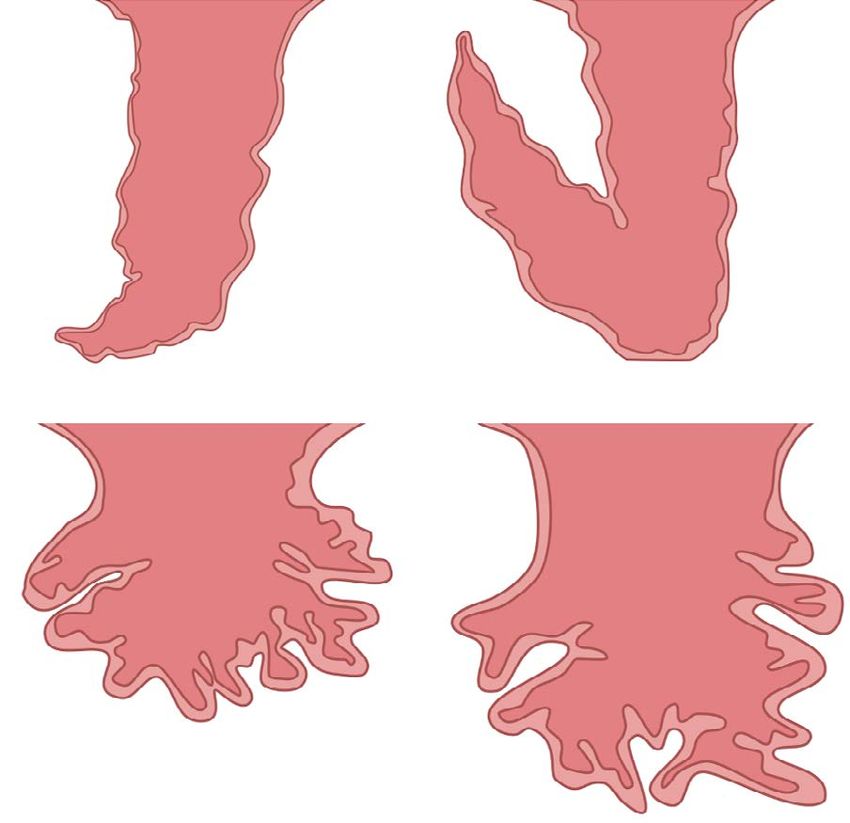
M. Świątczak et al., Unusual echocardiographic evidence of hypercoagulation in COVID-19
A B
C D
E F
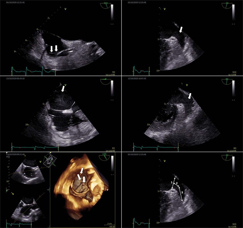
Figure 1. The figures visualise the image in transoesophageal echocardiography performed twice in the described patient. On admission to
the Cardiology Department, the patient had linear echoes on the atrial electrode (panel A — arrows) and a clean left atrial appendage (LAA)
(panel B — arrow). In a follow-up study after the spontaneous return of sinus rhythm and a week of apixaban (5 mg BID) therapy, the linear
echoes on the atrial lead disappeared, while the left atrium filled with highly hyperechogenic blood (panel C — arrow) and LAA with the
sludge (panel D — arrow) were visualised. Additionally, the long persistence of the bubbles of midazolam in the right atrium was observed
(panel E — arrows). Panel F presents the dimensions of the LAA in the described patient; 1 — 1.7 cm, 2 — 0.8 cm, 3 — 2.4 cm.
the risk of formation of thrombus in the LAA are the maker implantation, performed 1 week before the
specific anatomy of the LAA and the low left ventric- admission. Additionally, the urinary tract infection
ular ejection fraction. We present a clinical case that was diagnosed, and empirical antimicrobial therapy
presents SARS-CoV-2 infection as an additional cause was initiated, leading to a quick reduction of inflam-
of LAA thrombus formation. matory markers. There were no pathologies connect-
ed with ventricle lead; however, in transoesophageal
CASE PRESENTATION echocardiography (TEE), using two- and three-di-
A 70-year-old woman with AF and negative RT- mensional techniques, linear echoes associated with
-PCR test for SARS-CoV-2 was transferred from the the atrial electrode were visualised (Fig. 1A). Due to
district hospital to the Cardiology Department with the short time after the implantation and negative
suspicion of device-related infection after dual pace- blood culture results, the thrombus was anticipated,
715Folia Morphol., 2021, Vol. 80, No. 3
A B of 2 (54%) lobes, the second most frequent are an LAA
with 3 (23%) lobes, then with 1 (20%) lobe and the least
frequent is the presence of 4 (3%) lobes. The orifice of
the LAA leads through the neck to the central append-
age cavity, which can have a different shape depending
on the type of an LAA. According to Wang et al. [12], the
most frequent LAA anatomy is the WindSock LAA (Fig.
2A), which has no obvious bands, and one dominant
C D lobe of sufficient length is the basic structure. Variations
of this type of LAA appear with the different locations
and number of secondary lobes descending downward
from the dominant lobe. The second most frequent
anatomy is the Cauliflower LAA (Fig. 2C), which, like
the previous type, does not have obvious bands; an LAA
is characterized by a limited overall length and more
complex internal features. Its varieties are described by
a more irregular shape of the orifice of the LAA, the
Figure 2. General scheme showing the four most common
types of the left atrial appendage (LAA); A. The WindSock number of significant lobes, and the lack of the dom-
LAA; B. The ChickenWing LAA; C. The Cauliflower LAA; inant lobe. The ChickenWing LAA (Fig. 2B) is the third
D. The Cactus LAA.
most common type; the main feature of that type of
LAA is an obvious band in the proximal or middle part
not bacterial vegetation. The LAA had typical mor- of the dominant lobe or a backward fold of the LAA at
phology and sizes, as presented in Figure 1. Despite some distance from the orifice of the LAA. This type may
AF, there was no sludge or thrombus in the LAA differ in the presence or absence of additional lobes or
(Fig. 1B). The spontaneous restoration of sinus rhythm twigs, with a different measured distance to this bend,
was observed. The treatment with apixaban (5 mg and with different orientations of the bends to the main
BID) was initiated, and empirical antibiotic therapy lobe. The last most common type is the Cactus LAA
was continued. After 1 week of the treatment, TEE (Fig. 2D); its main feature is the dominant lobe with
was repeated with no linear echoes within the atri- secondary lobes extending from the dominant lobe to
al electrode. Interestingly, despite permanent sinus the superior and inferior directions [12].
rhythm and anticoagulation therapy with apixaban, According to the literature, thromboembolic com-
the left atrium was filled with highly hyperechogenic plications occur most frequently in patients with AF
blood (Fig. 1C) and the LAA with a sludge (Fig. 1D). and the Cactus LAA [4]. Additionally, the small size
Moreover, the long persistence of the bubbles of of LAA, the presence of secondary lobes, the narrow
midazolam in the right atrium has been observed orifice of the LAA, and excessive trabeculations result
(Fig. 1E). The patient did not present any typical clin- in low LAA peak flow velocities that could significantly
ical COVID-19 symptoms [2], but had neutropenia, increase the risk of thrombus formation LAA [1]. It
lymphopenia, low procalcitonin, hypoalbuminaemia, should also be emphasized that the large size of the
and increased C-reactive protein. We re-checked the left atrium and the reduced left ventricular ejection
RT-PCR test for SARS-CoV-2 infection, and the result fraction are additional risk factors for the develop-
was positive. ment of thromboembolic complications.
In the described case, we presented echocardi-
DISCUSSION ographic evidence of hypercoagulation as the first
The LAA lies within the pericardium, in the left and only feature of SARS-CoV-2 infection in the usual
atrioventricular sulcus atop the left circumflex artery’s morphological presentation of the WindSock LAA
proximal part and extends between the anterior and (Fig. 1F). Despite the use of apixaban treatment, the
the lateral walls of the LA near the left pulmonary veins lack of features promoting thrombus formation in
[11]. The LAA could be of various sizes, volumes, and the LAA (Fig. 1D), such as excessive trabeculations,
shapes and often has several lobes [5, 8, 11]. Veinot et a narrow junction of the proximal lobe of the LAA,
al. [11] found that the most common is the presence a narrow junction between the distal lobe of the LAA
716M. Świątczak et al., Unusual echocardiographic evidence of hypercoagulation in COVID-19
and its proximal lobe, or the presence of additional 3. Connors J, Levy J. Thromboinflammation and the hyperco-
LAA lobes, there has been a sludge in the LAA. The agulability of COVID-19. J Thromb Haemost. 2020; 18(7):
1559–1561, doi: 10.1111/jth.14849.
coagulation abnormalities in COVID-19 are postulated
4. Di Biase L, Santangeli P, Anselmino M, et al. Does the
to result from acute inflammation in the organism left atrial appendage morphology correlate with the
and increased activity of inflammatory mediators [7]. risk of stroke in patients with atrial fibrillation? Results
The urinary tract infection could additionally attenu- from a multicenter study. J Am Coll Cardiol. 2012; 60(6):
531–538, doi: 10.1016/j.jacc.2012.04.032, indexed in
ate the hypercoagulation status in our patient.
Pubmed: 22858289.
5. Kamiński R, Kosiński A, Brala M, et al. Variability of the
CONCLUSIONS left atrial appendage in human hearts. PLoS One. 2015;
The connection between hypercoagulation 10(11): e0141901, doi: 10.1371/journal.pone.0141901,
indexed in Pubmed: 26544191.
features and COVID-19 in a patient without other
6. Marietta M, Vandelli P, Mighali P, et al. COVID-19 HD
typical infection indicators seems to be particular- Study Group. Randomised controlled trial comparing
ly difficult, as in the presented case. Based on the efficacy and safety of high versus low Low-Molecular
available literature about COVID-19 management, Weight Heparin dosages in hospitalized patients with
low-molecular-weight heparin should be considered severe COVID-19 pneumonia and coagulopathy not re-
quiring invasive mechanical ventilation (COVID-19 HD):
for thromboembolic complications prophylaxis [6].
a structured summary of a study protocol. Trials. 2020;
In contrast, oral anticoagulant therapy is not recom- 21(1): 574, doi: 10.1186/s13063-020-04475-z, indexed in
mended due to its limited effectiveness, confirmed Pubmed: 32586394.
in our patient. The LAA in the case presented above 7. Panigada M, Bottino N, Tagliabue P, et al. Hypercoag-
ulability of COVID-19 patients in intensive care unit:
did not show any features predisposing to thrombus
A report of thromboelastography findings and other
formation; the LAA was of standard size and not parameters of hemostasis. J Thromb Haemost. 2020;
narrow. Besides, a thrombus developed despite the 18(7): 1738–1742, doi: 10.1111/jth.14850, indexed in
patient’s persistent sinus rhythm and anticoagu- Pubmed: 32302438.
lant treatment, which suggests a hypercoagulability 8. Panyawongkhanti M, Fuktongphan P, Chentanez V. Mor-
phometric study of the left atrial appendage related to
state in the course of COVID-19. The presented case closure device deployment: a cadaveric study in Thai pop-
additionally shows that a negative test for SARS- ulation. Folia Morphol. 2020; 79(1): 79–85, doi: 10.5603/
CoV-2 infection does not always give a full guarantee FM.a2019.0066, indexed in Pubmed: 31162625.
that the patient is not infected, and the patient’s 9. Rozwadowska K, Raczak G, Sikorska K, et al. Influence
of hereditary haemochromatosis on left ventricular wall
clinical manifestation should be taken into account
thickness: does iron overload exacerbate cardiac hypertro-
in further clinical decisions. It is worth emphasizing phy? Folia Morphol. 2019; 78(4): 746–753, doi: 10.5603/
that modern echocardiography, including three-di- FM.a2019.0025, indexed in Pubmed: 30835340.
mensional techniques, can be recognised as a part 10. Sławiński G, Lewicka E. What should a cardiologist know
about coronavirus disease 2019? Kardiol Pol. 2020; 78(4):
of comprehensive imaging technology that could be
278–283, doi: 10.33963/KP.15302, indexed in Pubmed:
helpful in COVID-19 diagnosis [9]. 32336069.
11. Veinot JP, Harrity PJ, Gentile F, et al. Anatomy of the
Conflict of interest: None declared normal left atrial appendage: a quantitative study of
age-related changes in 500 autopsy hearts: implications
for echocardiographic examination. Circulation. 1997;
REFERENCES 96(9): 3112–3115, doi: 10.1161/01.cir.96.9.3112, indexed
1. Al-Saady NM, Obel OA, Camm AJ. Left atrial appendage: in Pubmed: 9386182.
structure, function, and role in thromboembolism. Heart. 12. Wang Y, Di Biase L, Horton RP, et al. Left atrial ap-
1999; 82(5): 547–554, doi: 10.1136/hrt.82.5.547, indexed pendage studied by computed tomography to help
in Pubmed: 10525506. planning for appendage closure device placement.
2. Carfì A, Bernabei R, Landi F, et al. Persistent symptoms in J Cardiovasc Electrophysiol. 2010; 21(9): 973–982,
patients after acute COVID-19. JAMA. 2020; 324(6): 603, doi: 10.1111/j.1540-8167.2010.01814.x, indexed in
doi: 10.1001/jama.2020.12603. Pubmed: 20550614.
717You can also read