Usefulness of 3D Digital Subtraction Angiography in the Endovascular Approach of Cerebral Aneurysms
←
→
Page content transcription
If your browser does not render page correctly, please read the page content below
case series

                                  Usefulness of 3D Digital Subtraction
                                  Angiography in the Endovascular
                                  Approach of Cerebral Aneurysms
                                  Utilidad de la angiografía por sustracción digital 3D en el abordaje
                                  endovascular de aneurismas cerebrales
                                                                                                                       Nelson Oswaldo Lobelo García1
                                                                                                                        Alejandra Navarrete Sánchez2
                                                                                                                      Mauricio Enrique Moreno Mejía3
                                                                                                                                Cristian Camilo Páez4
                                                                                                                     Cihara Valessa Avendaño Padilla5
    Key words (MeSH)
    Digital subtraction
      angiography
                                  Summary
    Intracranial aneurysm         Introduction: The trend in management of intracranial aneurysms has shifted during the last decades
    Embolization, therapeutic     to minimally invasive endovascular procedures. The usefulness of new imaging tools such as digital
    Endovascular procedures       subtraction angiography in 3D (3D DSA), added to the experience of neurointerventional radiologists,
                                  have led to greater definition and accuracy in the study of intracranial aneurysms. Objective: To
                                  describe the usefulness of three-dimensional digital subtraction angiography for pre and post
    Palabras clave (DeCS)         embolization approach of intracranial aneurysms. Methodology: A cross-sectional study between
    Angiografía de                January 2016 and April 2017 in patients diagnosed with arterial cerebral aneurysms at the Hospital
      sustracción digital         Infantil Universitario San José in Bogota, Colombia. Results: 32 patients were included, 71.8% (n = 23)
    Aneurisma intracraneal
                                  were women. Among the risk factors for aneurysm rupture, the most frequent was age above 40
                                  years (81.8%). The most frequent location was in the Right Middle Cerebral Artery (MCA) (30.3%).
    Embolización terapéutica
                                  All cases corresponded to saccular aneurysms. In the immediate post-embolization angiographic
    Procedimientos                control it was evidence that 16 cases (48.5%) presented residual sac. Conclusions: The realization
      endovasculares              of multiplanar projections with 3D angiographic reconstruction allows for a better characterization
                                  of the aneurysm and evaluation of the adjacent anatomical structures, being very useful for the
                                  planning of the procedure and in the follow-up.

                                  Resumen
                                  Introducción: En el manejo de los aneurismas intracraneales la tendencia ha sido realizar
                                  procedimientos endovasculares mínimamente invasivos. Nuevas herramientas en imágenes, como
                                  la angiografía por sustracción digital en 3D (ASD 3D), sumadas a la experiencia de los radiólogos
1
 Neuro-interventionist radio-     neurointervencionistas, han llevado a una mayor definición y precisión en el estudio del aneurisma
logist. Assistant professor.      intracraneal. Objetivo: Describir la utilidad de la técnica de angiografía por sustracción digital
Fundación Universitaria de        tridimensional para el abordaje pre y postembolización de los aneurismas intracraneales. Metodología:
Ciencias de la Salud. Hospital
                                  Estudio de corte transversal entre enero de 2016 y abril de 2017 en pacientes diagnosticados con
Infantil Universitario de San
José.. Bogotá, Colombia.          aneurisma de arterias cerebrales, en el Hospital Infantil Universitario San José, en Bogotá, Colombia.
                                  Resultados: Se incluyeron 32 pacientes, de los cuales 71,8 % (n = 23) fueron mujeres. Entre los
2
 Radiologist. Fundación Uni-      factores de riesgo para ruptura del aneurisma, el más frecuente fue edad mayor a 40 años (81,8 %).
versitaria de Ciencias de la
Salud. Bogotá, Colombia.          La localización más usual fue en la arteria cerebral media (ACM) derecha (30,3 %). Todos los casos
                                  correspondieron a aneurismas saculares. En el control angiográfico postembolización inmediato
3
 Radiologist. Fundación Uni-      se evidenció que 16 casos (48,5 %) presentaron saco residual. Conclusiones: La realización de
versitaria de Ciencias de la
Salud. Pontificia Universidad
                                  proyecciones multiplanares con reconstrucción angiográfica 3D brinda información adicional para
Javeriana. Bogotá, Colombia.      una mejor caracterización del aneurisma y evaluación de las estructuras anatómicas adyacentes,
                                  por lo que es de gran utilidad para planear el procedimiento y para el seguimiento.
4
 Resident of Radiology and
Diagnostic Imaging, Funda-
ción Universitaria de Cien-
cias de la Salud. Bogotá,
Colombia.
                                  Introduction                                                imaging tools, such as 3D digital subtraction angiography
5
 Epidemiologist. Department
of Diagnostic Imaging, Hos-            In recent decades, the trend in the management of      (3D ASD) and the experience of neurointerventional
pital Infantil Universitario de   intracranial aneurysms is based on minimally invasive       radiologists, which facilitates greater definition and
San José. Bogotá, Colombia.
                                  endovascular procedures, which shows that intravascular     accuracy in the characterization of the intracranial
- Institution: Diaimage, De-
                                  treatment compared to surgical treatment reduces the risk   aneurysm, the physiological state of the communicating
partment of Diagnostic Ima-
ges. Hospital Infantil Univer-    of death or long-term disability (1).                       vessels (vasospasm), as well as the anatomical structures.
sitario de San José. Bogotá,           The factors involved in performing an appropriate      This decreases the time of procedures and, in turn, the
Colombia.
                                  procedure increasingly depend on the usefulness of new      frequency of complications (2-4).

Rev. Colomb. Radiol. 2020; 31(1): 5283-8                                                                                                       5283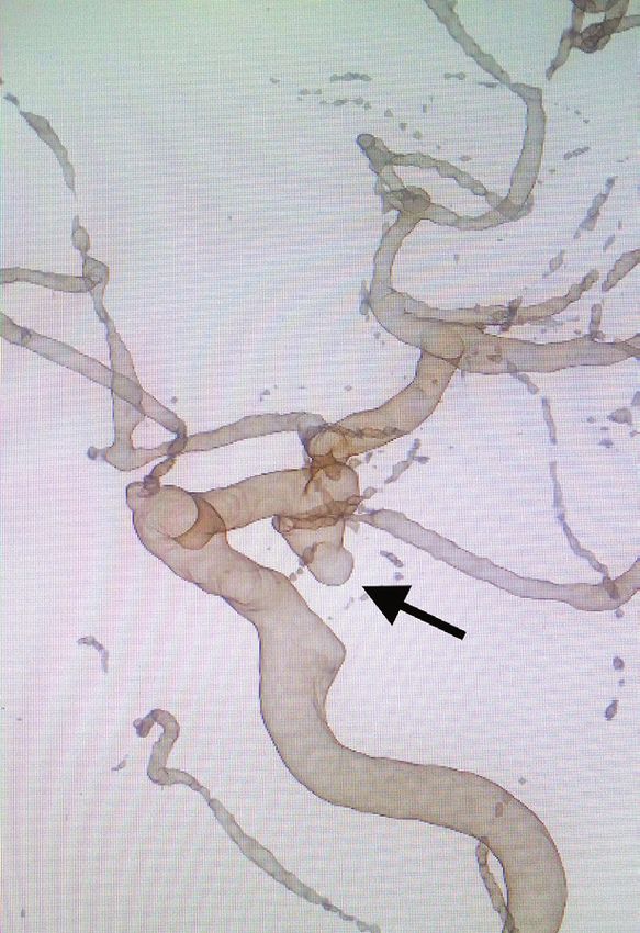
case series
ASD with 2D projections was used as the modality of choice for all of them were treated by means of coil embolization. As indepen-
the study of intracranial vascular structures for several years, with dent variables were taken into account sex, age, risk factors for the
imaging in the anteroposterior, lateral, and oblique planes. ASD 3D formation of cerebral aneurysms in each patient; location, size and
is a technological evolution of ASD 2D that allows the visualization shape of the aneurysm, characteristics of the afferent vessel and ves-
of intracranial vessels from any possible projection. The ASD 3D sels originating in the aneurysm sac. Also recorded were: the duration
requires a flat detector angiograph with a rotating C-arm. Two rotatio- of the procedure, the post-treatment results, and the characteristics
nal image acquisitions are performed, the first to obtain a subtraction of the afferent vessel in the immediate post-treatment control 3D
mask and the second to acquire the images during the injection of angiography.
the contrast medium. In the second acquisition, the arc rotates 180 The data were obtained from the workstation of a TOSHIBA
degrees in approximately 4-8 seconds, while 13 ml of contrast medium angiograph model INFX-8000V/W6, Series W6C12Y2019, using
is administered through an injector at a rate of 2.5 mL/s. Subsequent the VÍTREA FX application (Version 6.3). The demographics and
reconstructions with specialized software allow for suppression of clinical records of each patient.
unnecessary vessels, image rotation and magnification, among other For the realization of the angiography, the institutional protocol
applications (2). was followed in each one of the patients and later on, 3D reconstruc-
The purpose of this article is to illustrate with clinical cases the tion was performed with the mentioned software.
usefulness of 3D digital subtraction angiography in the endovascular A univariate analysis of the variables included was made to es-
approach to brain aneurysms and to compare the results found before tablish absolute and relative frequencies by means of the statistical
the intervention and in the immediate postoperative period with what package SPSS Statistics V24.0. No missing data were available.
has been published in the literature. A literature search was carried out in the PubMed, OVID and
ClinicalKey databases, with the keywords digital subtraction an-
giography, intracranial aneurysms, therapeutic embolization and
Methodology endovascular procedures, to compare the results obtained with those
A cross-sectional study was conducted between January 2016 described in the literature.
and April 2017 with patients diagnosed with brain artery aneurysm. We had the approval of the hospital’s medical ethics committee
Thirty-two patients were included who underwent digital subtraction and complied with the regulations in force in Colombia.
angiography with 3D reconstruction; 33 aneurysms were diagnosed;
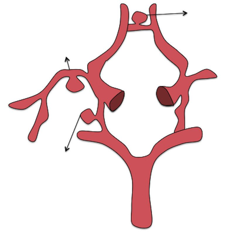
- Anterior communicating artery 27.3%
Right middle
cerebral artery
30.3%
- Posterior communicating artery
21.2% Other locations: Internal carotid artery
- Communicating segment 3%
- Ophthalmic segment 6.1%
- Supraclinoid 3%
- Right bifurcation 3%
- Median left cerebral artery 6.1%.
Figure 1. Graphic representation of the most frequent location of aneurysms in the population studied.
5284 Usefulness of 3D Digital Subtraction Angiography in the Endovascular Approach of Cerebral Aneurysms.
Lobelo N., Navarrete A., Moreno M., Páez C., Avendaño C.
case series
a b
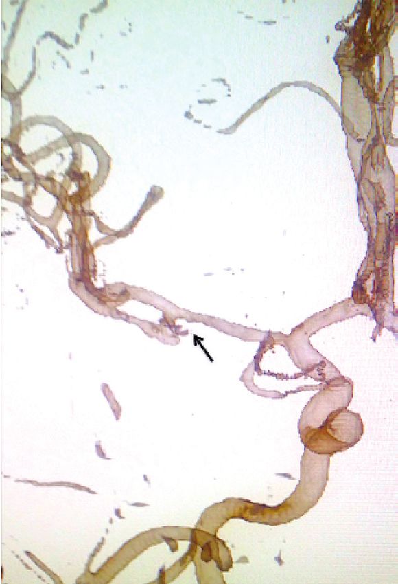
Figure 2. Right middle cerebral artery
saccular aneurysm. a). 3D pre-embolization
reconstruction (arrow). b) 3D post-embolization
reconstruction. Note the neck portion of the
remaining aneurysmal sac in the right middle
cerebral artery (arrow).
a b
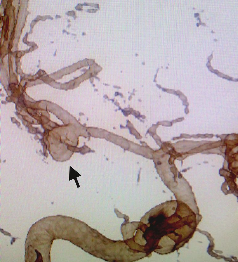
Figure 3. Saccular aneurysm of the anterior communicating
artery. a) 3D pre-embolization reconstruction (arrow). b).
3D post-embolization reconstruction. Note the complete
occlusion of the aneurysmal sac (arrow).
a b
Figure 4. Saccular aneurysm of the posterior
communicating artery. a) 3D pre-embolization
reconstruction (arrow) b) 3D post-embolization
reconstruction. Note the remaining aneurysmal sac
portion in the posterior communicating artery (arrow).
Rev. Colomb. Radiol. 2020; 31(1): 5283-8 5285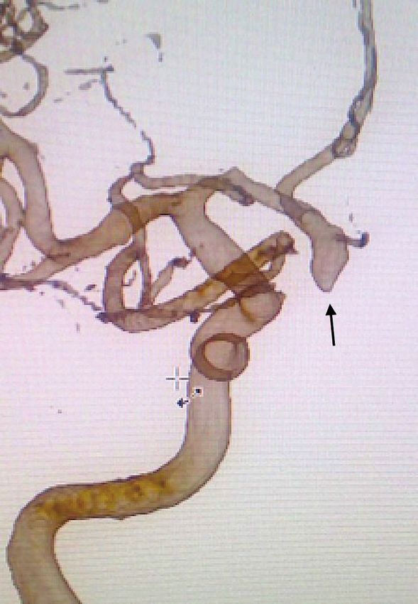
case series

Results                                                                     ture, probably influenced by other factors such as: most of the patients
    Of the 32 patients included in the study, 71.8% (n = 23) were women     presented small aneurysms and complete occlusion or only residual
and 29.2% were men (n = 9). The minimum age was 3 years and the             neck was obtained in more than half of the cases.
maximum was 90 years, with a mean of 55.5 years (Standard Devia-                 Some authors have considered 3D ASD as the reference diagnostic
tion [SD] of 17.9 years). Among the risk factors for aneurysm rupture,      image for the evaluation of intracranial circulation in general and the
age over 40 years was found in 27 patients (81.8%), 17 patients were        planning of treatment of brain aneurysms, and complications as low
hypertensive (53.1%) and 5 were smokers (15.6%). Table 1 shows the          as 0.3% have been found in expert hands (9).
characteristics of the population studied. The most frequent locations           Previously published studies have demonstrated the superior
were: right middle cerebral artery (MCA) with 10 cases (30.3%),             capability of 3D ASD when compared to 2D ASD and rotational
anterior communicating artery (ACoA) with nine cases (27.3%) and            angiography, evidencing the possibility of acquiring images in the
posterior communicating artery (ACoP) with seven cases (21.2%)              cranio-flow axis in high resolution, the elimination of structures made
(Figure 1). The size of the aneurysms ranged from 3.6 mm to 26 mm,          of metallic material prostheses and spirals (stents, coils) or overlapping
with an average of 8.44 mm; there was a greater frequency of small          structures and the performance of simulation processes. In addition, it
aneurysms (less than 10 mm), corresponding to 93.9% (n = 31). As for        is an intracranial hemodynamic technique, which achieves in real time
afferent vessel characteristics, it was documented that in most cases it    the assessment of the physiological state of the collateral vessels, the
was normal (60.6%). In 21 aneurysms 63.6% of the cases there were           afferent vessel of the aneurysm and the vessels within the aneurysm
vessels originating from the aneurysm sac. All cases corresponded to        sac (10, 11).
saccular aneurysms (Figure 2). In the post-embolization angiographic             In our research, regarding the size of the aneurysm with the 3D
control it was shown that 16 cases (48.5%) presented residual sac (fi-      ASD technique, we were able to detect small aneurysms from 3.6 mm.
gures 3 and 4) and that the characteristics of the afferent vessel were     Previous research has documented that 3D angiography can detect
normal in most of these, 23 cases (69.7%) (figure 5). The duration of       aneurysms up to 0.5 mm.
the procedure was in the range of 14 to 106 minutes with an average              2D angiography has been attributed a sensitivity and specificity
of 39 minutes (table 2).                                                    greater than 90% for detecting aneurysms measuring 3 mm; however,
                                                                            it has marked limitations for visualizing aneurysms smaller than 2
                                                                            mm (12, 13).
Tabla 1. Características de la población estudiada
                                                                                 In the 33 aneurysms in our study, their shape could be adequately
                                                                            classified as saccular and images of sufficient quality were obtained to
                                                        Porcentaje          plan endovascular treatment. The literature describes that the ability
                Variable                         n
                                                           (%)              of 2D angiography to accurately show the shape of the aneurysm has
 Sexo                                                                       a lower performance compared to 3D ASD images (3).
                                                                                 Regarding the definition of the afferent vessel characteristics and the
 Mujer                                          23           69,7
                                                                            presence of vessels originating from the aneurysm sac, this technique
 Hombre                                          9            27,3
                                                                            manages to eliminate the overlapping of vascular structures, thanks to
 Edad
                                                                            the post-processing in the working console and the 360-degree view
 Menores de 40 años                              5            18,2          of the aneurysm. In this way it was possible to detect that in 18.2% of
 Mayores de 40 años                             27            81,8          the cases the afferent vessel was dysplastic and that 63.6% of the cases
 Factores de riesgo de ruptura de aneurisma                                 there were vessels in the aneurysm sac. Although the role of 3D ASD
 Hipertensión arterial                          17            53,1          to avoid the overlapping of vascular structures was not specifically
 Fumadores                                       5            15,6
                                                                            described in the present research, several studies have been published
                                                                            that support the superiority of this characteristic of 3D ASD over 2D
 Malformación arteriovenosa asociada             1            3,1
                                                                            when characterizing these findings (11).
 Ninguno                                         4            12,5
                                                                                 In some studies, 3D ASD is recommended as the standard study for
                                                                            angiographic follow-up of patients with coiled embolized aneurysms,
Discussion                                                                  to increase the detection rate of remaining aneurysms and to identify
     Intracranial aneurysms are abnormal external dilations in the wall     afferent vessel stenosis (6, 8, 9, 12). In our study, the remaining aneu-
of intracranial blood vessels; the saccular variant is the most common.     rysm sac (most of it small size) was identified as the main immediate
They are frequently located in the anterior circulation (90%) and           post-treatment result by 48.5%.
commonly affect women in the 5th decade of life (5-7), all of which
are frequent in the population of this study. The etiology of saccular
aneurysms is still unknown (8); however, predisposing risk factors for
their development and rupture have been studied, such as systemic
arterial hypertension and smoking, present in 53.1% and 15.6% of our
patients, respectively. Another risk factor per se is age over 40 years,
which corresponds to 81.8% of the population. In this research with
more than one year of follow-up there were no cases of aneurysm rup-

5286                                           Usefulness of 3D Digital Subtraction Angiography in the Endovascular Approach of Cerebral Aneurysms.
                                                                                             Lobelo N., Navarrete A., Moreno M., Páez C., Avendaño C.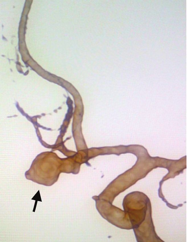
case series

 a                                               b

                                                                                           Figure 5. Saccular aneurysm of the anterior communicating
                                                                                           artery. a) 3D pre-embolization reconstruction (arrow) b)
                                                                                           3D post-embolization reconstruction. Note the normal
                                                                                           state of the afferent vessel (arrow).

                                                                     stabilization, since, depending on the size and configuration of the
                                                                     aneurysm, subsequent re-interventions may be necessary to complete
Table 2. Characteristics evaluated in 3D angiography
                                                                     treatment. This medium- and long-term follow-up was not possible,
of the aneurysm and afferent vessel before and after                 partly due to administrative factors derived from the health system,
embolization                                                         since some patients were referred for follow-up and control in other
                                                      Percentaje     institutions. However, the vast majority of patients were assessed by the
                 Variable                   n                        treating physician, with evidence of complete closure of the aneurysms.
                                                         (%)
                                                                     Additionally, it is considered that a classification should be applied to
                                 Size
                                                                     determine the percentage of remaining sac, with that it would be easier
 Small ( < 10 mm)                           31            94
                                                                     and more objective the way to compare in the follow-up studies the
 Large ( > 10 mm)                           1             3
                                                                     significant variations that the aneurysms present.
 Giant ( > 25 mm)                           1             3

                    Afferent vessel characteristics
 Normal                                    20            60,6
                                                                     Conclusion
                                                                         This observational study shows that multiplanar projections with 3D
 Dysplastic                                 6            18,2
                                                                     reconstruction allow a complete characterization of the aneurysm and
 With vasospasm                             7            21,2        evaluation of the adjacent anatomical structures, which is very useful
                     Vessels in the aneurysm sac                     for planning the procedure and in the post-embolization follow-up,
 Yes                                        21           63,6        especially to determine the degree of occlusion of the treated aneurysms.
 No                                         12           36,4

                        Post-treatment result                        References
 Residual bag                               16           48,5        1.   Molyneux AJ, Kerr RSC, Yu L-M, et al. International subarachnoid aneurysm trial
                                                                          (ISAT) of neurosurgical clipping versus endovascular coiling in 2143 patients with
 Residual neck                              10           30,3             ruptured intracranial aneurysms: a randomised comparison of effects on survival,
                                                                          dependency, seizures, rebleeding, subgroups, and aneurysm occlusion. Lancet (London,
 Complete occlusion                         7            21,2
                                                                          England). 2005;366(9488):809-17.
          Characteristics of post-embossing afferent cup             2.   Cieściński J, Serafin Z, Strześniewski P, et al. DSA volumetric 3D reconstructions of
                                                                          intracranial aneurysms: A pictorial essay. Polish J Radiol. 2012;77(2):47-53.
 Normal                                    23            69,7        3.   Anxionnat R, Bracard S, Ducrocq X, et al. Intracranial aneurysms: Clinical Value
 With vasoespasm                            6            18,2             of 3D digital subtraction angiography in the therapeutic decision and endovascular
                                                                          treatment. Radiology. 2001;218(3):799-808.
 Dysplastic                                 3             9,1        4.   Tanoue S, Kiyosue H, Kenai H, et al. Three-dimensional reconstructed images after
                                                                          rotational angiography in the evaluation of intracranial aneurysms: Surgical correlation.
 With stenosis                              1            3,0              Neurosurgery. 2000;47(4):866-71.
                                                                     5.   Robbins SL, Kumar V, Cotran RS. Robbins and Cotran pathologic basis of disease.
                                                                          8th ed. Philadelphia PA: Saunders/Elsevier; 2010.
                                                                     6.   Zhou B, Li M-H, Wang W, et al. Three-dimensional volume-rendering technique in the
                                                                          angiographic follow-up of intracranial aneurysms embolized with coils. J Neurosurg.
Study limitations and recommendations for                                 2010;112(3):674-80.
                                                                     7.   Hacein-Bey L, Provenzale JM. Current imaging assessment and treatment of intra-
further studies                                                           cranial aneurysms. Am J Roentgenol. 2011;196(1):32-44.
    Our findings should be replicated with medium- and long-term     8.   Grobelny TJ. Brain aneurysms: Epidemiology, treatment options, and milestones of
                                                                          endovascular treatment evolution. Disease-a-Month. 2011;57(10):647-55.
follow-up controls to determine aneurysm recurrence, shrinkage, or

Rev. Colomb. Radiol. 2020; 31(1): 5283-8                                                                                                                5287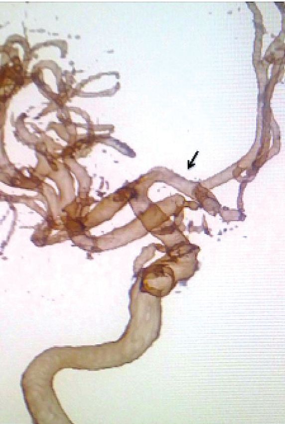
case series

9.    Fifi JT, Meyers PM, Lavine SD, et al. Complications of modern diagnostic cerebral
      angiography in an Academic Medical Center. JVIR. 2009;20:442-7.
10.   Bau Alegría J. Reconstrucción 3D angiográfica en el diagnóstico y el tratamiento de
      aneurismas cerebrales. Imagen Diagnóstica. 2010;1(2):51-5.
11.   Sugahara T, Korogi Y, Nakashima K, et al. Comparison of 2D and 3D digital subtraction
      angiography in evaluation of intracranial aneurysms. AJNR Am J Neuroradiol.
      2002;23(9):1545-52.
12.   Van Rooij WJ, Sprengers ME, de Gast AN, et al. 3D Rotational angiography: The new
      gold standard in the detection of additional intracranial aneurysms. Am J Neuroradiol.
      2008;29(5):976-9.
13.   Hochmuth A, Spetzger U, Schumacher M. Comparison of three-dimensional rotational
      angiography with digital subtraction angiography in the assessment of ruptured cerebral
      aneurysms. Am J Neuroradiol. 2002;23(7):1199-205.

Correspondence
      Mauricio Enrique Moreno Mejía
      Hospital Infantil Universitario de San José
      Carrera 52 # 67A -71
      Bogotá, Colombia
      mmorenomejia@hotmail.com

Received for evaluation: December 1, 2019
Accepted for publication: March 8, 2020

5288                                                         Usefulness of 3D Digital Subtraction Angiography in the Endovascular Approach of Cerebral Aneurysms.
                                                                                                           Lobelo N., Navarrete A., Moreno M., Páez C., Avendaño C.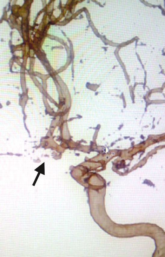
You can also read