Validation of Horizontal Genes Transfer Using Selected Clinical Strain of P. Mirabsilis for Swarming Activity - Open Journal Systems
←
→
Page content transcription
If your browser does not render page correctly, please read the page content below
3418     Indian Journal of Forensic Medicine & Toxicology, October-December 2020, Vol. 14, No. 4

          Validation of Horizontal Genes Transfer Using Selected
           Clinical Strain of P. Mirabsilis for Swarming Activity

                          Rusul Idan Mohsin Ali1,3, Muslim Idan Mohsin2, Abbas Al-Muhna3
  1
   Assistant Lecturer in Department of Medical-laboratory-techniques, Al-Toosi University College, Najaf, Iraq,
  2
   Lecturer in Department of Medical Pathological Analyses, Faculty of Science, University of Kufa, Najaf, Iraq,
              3Professor in Department of biology, Faculty of Science, University of Kufa, Najaf, Iraq

                                                              Abstract
       Introduction: Proteus mirabilis expresses several virulence factor which enable them to colonize, survive
       and grow in the host organism. This study was to investigate whether P. mirabilis could deliver a spontaneous
       plasmids to competent strains, and to investigate whether P. mirabilis can be as a donor strains to delivery
       their virulent such as adhesion and swarming activity.

       Methodology: PCR was used to detect the motility genes that encoded in P. mirabilis. Using also
       transformation assay to transfer these genes of Proteus to standard strain. PCR technique used again to
       determine the whether genes were transfer or not after transformation.

       The results: the findings show that 9 (filD, flhC, flhD, motA, motB, fliL, flaA, flgM,) genes were encoded
       on chromosomal DNA of P. mirabilis. Interestingly data, only 3 (flaA, flgM and filD ) genes were carried
       on different plasmids of P. mirabilis. The results also show that spontaneous plasmids of P. mirabilis can
       transfer to standard strains of E. coli. We found one gene transfer to E. coli S17λ filD. The gene(filD) has a
       role in the P. mirabilis swarming motility.

       Conclusions: we demonstrated that these genes can carry not only on chromosomal DNA but also in
       Plasmidal DNA and transferring genes associated-characteristics, swarming motility

       Key words: filD ,flhC, flhD, motA, motB, flaA, Proteus mirabilis, swarming motility.

                        Introduction                                  is well known for its ability to form biofilms, and thereby
                                                                      resists antibiotic treatment [5].
       One of opportunistic pathogen is Proteus mirabilis
which is can cause severe invasive diseases, in critically                 Swarming motility of the Proteus is made up
ill and patients who have immunocompromised [1]. It                   of circular waves on solid media [6]. These waves
is an important source of hospital-acquired infections                include consolidation and swarming rings, during the
[2]
    . P. mirabilis is the main cause of urinary tract,                consolidation phase, the cells are in short form, and in
respiratory tract and wounds infections, burns, digestive             the swarming phase, they are in long form [6].
tract infections, ear infection and otitis [3]. P. mirabilis
has also a wide virulence factors which enable them to                    P. mirabilis have many virulence factors. Some
colonize, survive and grow in the host organism such                  of these virulence factors encoded in plasmids could
as swarming, fimbriae, urease, hemolysin, protease and                be acquired from hospitals or environments. We try to
Lipopolysaccharides (LPS) [4]. Furthermore, P. mirabilis              demonstrate whether clinical strains have acquired some
                                                                      genes related with swarming activity and can transfer
                                                                      these genes to standard bacteria strains, which is E. coli
Corresponding author:                                                 S17λ.
Rusul Idan Mohsin,
Rusul@altoosi.edu.iq M.no: 009647813634994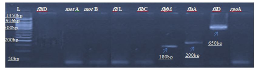
Indian Journal of Forensic Medicine & Toxicology, October-December 2020, Vol. 14, No. 4   3419

                   Methodology                                      Isolation of total Plasmid
    Bacterial strains:                                              DNA plasmid were extracted from all bacteria
                                                               isolates using QIAprep spin miniprep kit (Qiagen)
     We isolated 40 samples of P. mirabilis from
                                                               according to the manufacturer’s instructions.
different site of infection, and we selected (P23) which
has plasmids and a strongly swarming. P. mirabilis                  Gel electrophoresis
(P23) was grown in lysogeny agar. It was also grown
an overnight culture and were cultured at 37 °C to reach             PCR product was separated based on their size,
OD600 0.4. It was equal 1x10⁸. E. coli S17λ was provided       using gel electrophoresis. 2g/100mL of agarose from
from Life technologies New England Biolabs®.                   Fisher Scientific was dissolved in tris base, acetic
                                                               acid and EDTA (TAE) buffer by boiling. The agarose
    PCR detection of genes encoded in chromosomal or           solution was left in 25˚C to allow cooling around 55 °C,
plasmidal DNA                                                  and then 5µL of 0.5 mg/ml of ethidium bromide to stain
                                                               the DNA fragments. Typically, 1 μL of 5X DNA gel
    Isolation of total DNA
                                                               loading dye (Thermo) and 5μL of samples were mixed
    The Genomic DNA Extraction Kit was used for                before loading in the gel. The UV transilluminator was
DNA extraction by manufacturer’s protocol performed            used and images was captured using an EDAS 290
according to (Intron\ Korea).                                  imaging system (Kodak) to visualise the DNA pieces.

    Table 1 primers quotation

                                                                                        Amplicon
 Gene type                        Primer Sequence (5’-3’)                                                     Reference
                                                                                        size (bp)

              F               AAGGCTTCCGCAATGTTTAGAC
    flhD                                                                                   190                    [7]
              R               GTTGCAAATCATCCACTCTGGA
              F                GATGGTGACGGGGAATATGAA
   mot A                                                                                   215                    [8]
              R                 CCATTTCCCCAGCAGGTCTA
              F                 GCGTTACGTCCACATCTCAA
   mot B                                                                                   200                    [9]
              R                 ATGTCGCGCATATAGGGTTC

              F                 GGTGATCGCCATTATTGCAG
    fliL                                                                                   220                    [8]
              R                 AGCGTAACGTGATCCCTATG
              F              GCCAGTGAAAAAAGCATTGTTCA
    flhC                                                                                   500           GenBank: O34202
              R               CCAACAGCCTGTACTCTCTGTTC

              F                 CGCACAAATCCACTTATCCC
   flgM                                                                                    180                    [8]
              R                 GGGCAACTTTTTCGACATTG

              F                 TGCTGGTGCAACTTCATACG
    flaA                                                                                   200                    [9]
              R                 TTTGTCAGCACCTTCCAGTG
              F               GCCGCAAGTATTTCATCGCTGGG
    fliD                                                                                   650           GenBank: P42274
              R               CCTTTGGAATTACTGTTGTTTTC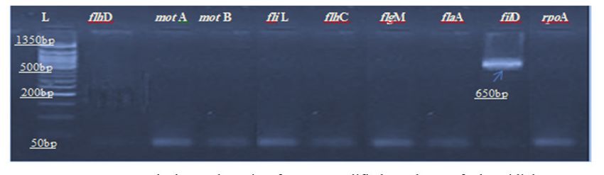
3420 Indian Journal of Forensic Medicine & Toxicology, October-December 2020, Vol. 14, No. 4
Statistical Analyses are required for swimming and twitching motility of
Proteus mirabilis. The stator complexes are not only
Statistical analyses are performed using GraphPad
encoded in P. mirabilis but also in most pathogenic
Prism.8. Significance was determined using ozone
bacteria that need to swarm in over surfaces [12]. All
correlation test. Data are presented as mean ± standard
isolates 40 (100%) have motA and fliL genes ,while
error of mean.
motB was detected in 37 isolates (92.5%), Fig1. These
results were in consistent with result of [13] [14]. This
The Results and Discussion
findings were similar with other researchers [15] [16]. As
Detection of swarming genes shown in Fig 2, From 8 genes, which were encoded in
DNA chromosome, only 3 genes were carrying on DNA
Nine virulence genes rpoA, flhC, flhD, motA,
plasmid of P. mirabilis. The percentage of genes filD ,
motB, fliL, flaA, flgM,filD that carried on P. mirabilis
flaA and flgM, were accounted in 3(60%),3(60%) and
DNA were studied. These genes have a role to induce
2(40%) isolates respectively. These results in regards
many infections [8] [9] [10]. The aim of this section was
to swarming activity genes were similar with findings
to quantify P. mirabilis genes whether were encoded in
obtained by [17, 18]. [15] [16]. They demonstrated that the
chromosomal and/or plasmid.
virulence genes are encoded in DNA plasmodial or
The motA, motB and fliL genes play a role in the chromosomal by using plasmid curing, which is clean
rotary motor of flagella [11]. Two motA and motB genes the bacteria from any plasmid.
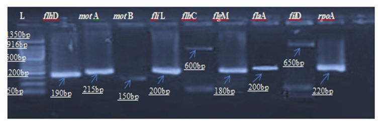
Figure (1) Agarose gel electrophoresis of PCR amplified products of chromosomal DNA genes using species-
specific PCR primer sets interested in this study. Lanes of genes are examined P. mirabilis isolate NO.23 .
Lane L, 1350 bp (ladder).
Figure (2) Agarose gel electrophoresis of PCR amplified products of plasmidial DNA genes using species-
specific PCR primer sets interested in this study. Lanes of genes are examined P. mirabilis isolate NO.23 .
Lane L, 1350 bp (ladder).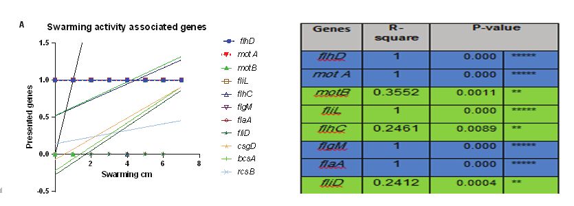
Indian Journal of Forensic Medicine & Toxicology, October-December 2020, Vol. 14, No. 4   3421

    Correlation of P. mirabilis swarming activity and 8        swarming activity. as demonstrated in Figs (3A and B).
genes                                                          Many studies are investigated the correlations between
                                                               virulence genes and swarming. For example, study
    The relation between swarming activity and flhC,           has been shown that fliF, motA, motB, flhC, fliG, and
flhD, motA, motB, fliL, flgM, flaA& fliD genes were            fliM genes of Salmonella enterica were related with
conducted in different isolates of P. mirabilis. The           swarming [19]. However, not only these genes have a role
results showed there are a good correlation between            in swarming, but also wosA , flaA, flhD, flgM and fliL
flhC, flhD, motA, motB, fliL, flgM, flaAa&fliD with            genes have a role in this phenomenon [20]

              Figure (3 A and B) Relationship between swarming activity and associated genes.

    Swarming is totally positively correlated with fihD,       transformation. It was accomplished using E. coli S17λ
motA, flgM and flaA (R-square 1, Kruskal–Wallis).              competent cells and isolates P.mirabilis donor cells. E.
Swarming is also positively correlated with motB, flhC         coli S17λ competent cells because one of them only
and fliD genes ( P values were between 0.001-0.01) . The       used in transformation based on the gene profile of these
significant differences between all genes were tested          competent bacterial cells. Briefly, for transformation, we
by using ozone correlation test in Graphpad prism-8            used heat shock method based on antibiotics resistance
program. Bule means direct role in swarming, green             marker. It was purified the E. coli S17λ based on whether
means indirect role in swarming activity.                      successfully resisted to antibiotics and received the
                                                               plasmid from P. mirabilis. The results appear that both
    Genes transferring
                                                               methods were shown that only 1 gene are transferred to
     This step was established to detect which gene            E. coli S17λ from the DNA plasmid extracted from P.
can transfer from P. mirabilis to competent bacterial          mirabilis. In addition P. mirabilis gene can also transfer
cells using the horizontal gene transfer method,               to E. coli S17λ during transformation process, which
                                                               were included: filD. Fig (4).

 Figure (4) Agarose gel electrophoresis of PCR amplified products of plasmidial DNA gene using species-
specific PCR primer sets interested in this study. Lanes of genes are examined E.coli S17λ competent strain
                              after transformation . Lane L, 1350 bp (ladder).
3422 Indian Journal of Forensic Medicine & Toxicology, October-December 2020, Vol. 14, No. 4
Conclusions flhDC operon of Escherichia coli K-12. Journal of
Microbiology, 2013. 51(1): p. 140-144.
P. mirabilis can form swarming and attach to
Uroepithelial cells strongly. From 8 different genes, 8. Cusick, K., et al., Perturbation of FliL interferes
which have a role in swarming, only 1 gene could be with Proteus mirabilis swarmer cell gene expression
expressed in plasmid profile, and transfer to standard and differentiation. Journal of bacteriology, 2012.
strain. 194(2): p. 437-447.
9. Maisuria, V.B., Z. Hosseinidoust, and N. Tufenkji,
Declaration: The authors have no conflict of Polyphenolic extract from maple syrup potentiates
interest to declare. antibiotic susceptibility and reduces biofilm
Ethical statement: This article does not contain any formation of pathogenic bacteria. Applied and
studies with human participants or animals performed environmental microbiology, 2015. 81(11): p.
by any of the authors. 3782-3792.
10. Pesavento, C., et al., Inverse regulatory coordination
Acknowledgement: Research facilities provided by of motility and curli-mediated adhesion in
hospitals in Alnajaf city acknowledged. My thanks also Escherichia coli. Genes & development, 2008.
to Professor Mark Thomas from University of Sheffield, 22(17): p. 2434-2446.
for providing me with standard strains.
11. Che, Y.S., et al., Load‐sensitive coupling of proton
Funding : Self-Funded. translocation and torque generation in the bacterial
flagellar motor. Molecular microbiology, 2014.
References 91(1): p. 175-184.
1. Torzewska, A. and A. Różalski, Various intensity of 12. Belas, R., Biofilms, flagella, and mechanosensing
Proteus mirabilis-induced crystallization resulting of surfaces by bacteria. Trends in microbiology,
from the changes in the mineral composition of 2014. 22(9): p. 517-527.
urine. Acta Biochimica Polonica, 2015. 62(1). 13. Lenahan, M., et al., Transcriptomic analysis of
2. Feneley, R.C., I.B. Hopley, and P.N. Wells, Urinary triclosan-susceptible and-tolerant Escherichia
catheters: history, current status, adverse events and coli O157: H19 in response to triclosan exposure.
research agenda. Journal of medical engineering & Microbial Drug Resistance, 2014. 20(2): p. 91-103.
technology, 2015. 39(8): p. 459-470. 14. Heo, M., Functional dynamics of the bacterial
3. Jamal, M., et al., Bacterial biofilm and associated flagellar motor driven by fluorescent protein tagged
infections. Journal of the Chinese Medical stators and by evolutionary modified foreign
Association, 2018. 81(1): p. 7-11. stators. 2016.
4. Seo, S.-U., et al., Distinct commensals induce 15. El-Hamid, M.I., et al., Genetic Diversity of
interleukin-1β via NLRP3 inflammasome in Campylobacter jejuni Isolated From Avian and
inflammatory monocytes to promote intestinal Human Sources in Egypt. Frontiers in microbiology,
inflammation in response to injury. Immunity, 2019. 10: p. 2353.
2015. 42(4): p. 744-755. 16. Stevenson, L.G. and P.N. Rather, A novel gene
5. Al-Mayahi, F.S.A., Phenotypic and Molecular involved in regulating the flagellar gene cascade
detection of Virulence factors in Proteus mirabilis in Proteus mirabilis. Journal of bacteriology, 2006.
isolated from different clinical sources. Bas. J. Vet. 188(22): p. 7830-7839.
Res, 2017. 16(1). 17. El-Baghdady, K.Z., et al., Plasmid mediated
6. Armbruster, C.E. and H.L. Mobley, Merging virulence factors of some Proteus isolates. Egyptian
mythology and morphology: the multifaceted Academic Journal of Biological Sciences, G.
lifestyle of Proteus mirabilis. Nature Reviews Microbiology, 2009. 1(1): p. 7-22.
Microbiology, 2012. 10(11): p. 743-754. 18. Kojima, S. and D.F. Blair, The bacterial flagellar
7. Lee, C. and C. Park, Mutations upregulating the motor: structure and function of a complex
molecular machine. International review ofIndian Journal of Forensic Medicine & Toxicology, October-December 2020, Vol. 14, No. 4 3423
cytology, 2004. 233: p. 93-135. 20. Verstraeten, N., et al., Living on a surface:
19. Komatsu, H., et al., Genetic analysis of revertants swarming and biofilm formation. 2008. 16(10): p.
isolated from the rod-fragile fliF mutant of 496-506.
Salmonella. 2016. 13: p. 13-25.You can also read