VIRTUAL ORTHOPEDIC SURGERY TRAINING ON PERSONAL COMPUTER
←
→
Page content transcription
If your browser does not render page correctly, please read the page content below
International Journal of Information Technology

         VIRTUAL ORTHOPEDIC SURGERY TRAINING
                ON PERSONAL COMPUTER

                                OLGA SOURINA and ALEXEI SOURIN
                              Institute of Computing for Physics and Technology
                  Department of Computer Science, Moscow Institute of Physics and Technology
                                      P.O.Box 18, Moscow 109382, Russia
                                             sourin@computer.org

                                             HOWE TET SEN
                                         Department of Orthopedics
                                        Singapore General Hospital
                                       Outram Road, Singapore 169608
                                             hts@altavista.net

    Abstract: Surgical training is one of the most promising areas in medicine where 3-D computer
    graphics and virtual reality techniques are emerging. This paper provides an advanced report on
    our project that aims to create virtual reality tools for training medical students and for
    improving skills and efficiency of orthopedic surgeons in internal fixation of bone fractures. The
    paper describes the whole pipeline from the CT scanner through the Unix-workstation to the
    personal computer where eventually the program works. We also describe the methods and
    mathematical models that allowed us to implement the ultimate goal of the project—to allow the
    surgeons to perform training surgical operations even at their home computers without any
    special expensive hardware devices. This project was initiated in co-operation between NTU and
    SGH and currently continues as an international project.

    Keywords: surgery training, virtual reality, orthopedics.

1. Introduction
Surgical training is one of the most promising areas in medicine where 3-D computer graphics
and virtual reality techniques are emerging. Attention of researchers has now turned toward
using combined 3-D reconstruction and virtual environment (VE) technologies to train
clinicians and to help surgeons plan patient-specific, complex procedures in craniofacial
reconstruction and plastic surgery, surgery for trauma from accidents, breast cancer detection,
and reconstruction surgery.1-6 In surgery simulation for training the clinicians, the benefit of
savings in time, cost, equipment, and safety can be enormous. As a result, there is a growing
interest in applying VR simulation technology to the medical educational process.
     Surgical simulators are used to create a VE wherein the surgeon may simulate and plan the
operation, and the surgeons or students can be trained. One of the main problems is to receive a
photo-realistic representation of operation room, human body parts from CT or MRI data,
surgical instruments and implants. Sound can be imposed into the scene to add realism. For
example, sound of drilling has been added in VE of craniofacial surgery simulation.1 Virtual
surgery is often performed with tissue and blood.5-8 For example, the training system
KISMET7,8 provides simulation techniques, which allow the modeling of “virtual tissue”,
based on a data-model which reflects the physical characteristics like mass, stiffness and
damping of real tissue. However, for orthopedic surgery, it is not necessary to simulate the soft
tissue except of some cases in craniofacial surgery. For example, it is important to know the
result of bones replacement covered with tissue in craniofacial surgery. When all the
reconstruction work is completed, the soft tissues are replaced so that the surgeon can visualize
the result. If the outcome of the virtual surgery is not desirable to the surgeon, the virtual

Volume 6, No. 1                                      16                                          May 2000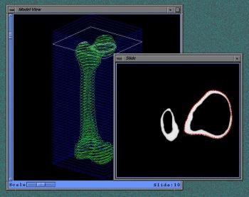
International Journal of Information Technology

process can be repeated until satisfaction is obtained, without touching the patient. In surgical
simulators, a collision test algorithm for detection of the contact between surgical instruments
and the virtual organs is often performed. In the training system KISMET8, contact forces
between the tissue and the instrument end-effector are calculated which can be used to drive a
force-reflecting surgeon interface 3D-simulation support during the design of new surgical
instruments and manipulator.
    A comprehensive software system for interactive visualization, manipulation and
measurement of multi-modality 3-D medical images has been developed, used and evaluated at
the Mayo Clinic for Computer Aided Surgery and Radiation Treatment Planning for more than
a decade.9,10 This software system, called ANALYZE, has provided surgeons and physicians
with powerful and flexible computational support both for pre-operative surgical and treatment
planning and for post-operative evaluation. The software has been applied to a variety of
surgical and medical problems, and used on significant numbers of patients at the Mayo Clinic
and at many other institutions.
    The training systems available on the market are oriented on solving the particular
problems. For example, Limb Trauma Simulator (Musculographics Inc.) models a gunshot
wound to the leg for military surgeons training.11 Software for VR Surgery mainly works on
UNIX on Silicon workstations or is prepared on the powerful workstations. For example,
Voxel-Man Junior is a subproject of the general VOXEL-MAN-project aiming to transfer
substantial functions of exploring and manipulating 3D anatomical models from the workstation
based VOXEL-MAN system to inexpensive PCs.12,13
    In this paper, we address the problem of developing virtual reality tools for training medical
students and for improving skills and efficiency of orthopedic surgeons in internal fixation of
bone fractures. We developed a desktop VR orthopedic surgery training system capable of
running on commonly available PCs including notebooks without compulsory use of VR
hardware.

2. The Problem Statement
During common orthopedic surgery training, students must fix fractures on synthetic plastic
bones using surgical tools and implants. Fig. 1 shows a typical fractured plastic bone. These
synthetic bones differ in quality and cost. Good-quality synthetic bones aren’t just simple plastic
dummies. They are made from the materials with different densities and textures on the surface
and inside to simulate real bones as closely as possible.
    Normally, students assemble the bone in front of them, in the position typical of the
respective real operation. Then, they reduce the fracture and fix it internally using implants that
include hundreds of different plates, screws, nails, and wires. See Fig. 2, for example, where the
fractured femur is fixed with the nail inserted in its canal and with four screws. For the insertion
of implants, students—or surgeons—use different surgical instruments that let them drill holes,
measure for a length, insert the implants, and so forth. The training lab with all this stuff looks
rather like a repair shop. There is no flesh and blood there, or anesthesia and assisting nurses.
Each student works along independently.

Volume 6, No. 1                                   17                                       May 2000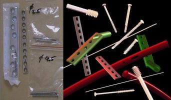
International Journal of Information Technology

                                   Fig. 1. Fractured synthetic bone.

                               Fig. 2. X-ray of the fracture and its fix.

The objectives of this orthopedic training follow:
• To memorize the techniques and respective tools and implants used for fixing fractures, and
   to choose the most relevant technique for each case.
• To learn how to put the implants in place, including exactly where and how deep to drill or
   cut the bone, the proper insertion angle, and so on.
• To acquire “muscle memory ” for the physical efforts applied to the tools and implants to
   avoid damaging the bones.
     Many synthetic bones are spoilt before a student can demonstrate the necessary skills on
this “synthetic patient.” The next step is surgical operations on cadavers. Only after that is the
student allowed to approach a real patient.
    The idea of using a computer for orthopedic surgery training came from surgeons in the
Department of Orthopedics of Singapore General Hospital, who approached the School of
Applied Science at Nanyang Technological University (where the first two authors were
employed at the time). The surgeons were interested, first, in saving the cost of the bones—
good-quality synthetic bones are expensive—and second, in the ability to work with certain
types of bones for which synthetic models might not be available. The latter need also derived

Volume 6, No. 1                                   18                                     May 2000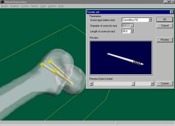
International Journal of Information Technology

from the fact that real Asian bones have their own geometric specifics compared to the
commonly available synthetic bones. Besides that, the surgeons anticipated an attractive
possibility—training the students on virtual models of real fractures obtained from computed
tomography (CT) or magnetic resonance imaging (MRI) data.
    Our feasibility study assured us that at least the first two objectives of the common
orthopedic training could move into the virtual environment relatively easily, with the outside
possibility of complete virtual orthopedic surgery training if special haptic devices were used.
Certainly, this virtual training won’t substitute completely for the compulsory training on
synthetic bones and on cadavers, but it might let the students perform the initial routine work
entirely in the virtual environment, thus saving cost and time.
    To make this virtual training easily accessible, we decided to develop a software system
capable of running on PCs, available in every medical clinic and in homes. This desktop or
“pocket” VR system had to produce acceptable realism even without special VR input devices
such as head-mounted displays (HMDs) or goggles, although using them obviously would
provide better immersion.
   Eventually, we came up with a project aiming to develop an inexpensive alternative to
common orthopedic surgery training that would allow the surgeons the following:
• To keep in the computer every single bone of different sizes and specific features that might
   be difficult and expensive to obtain using synthetic models.
• To create models of the fractured bones from real patients using data from CT and MRI
   images.
• To perform surgical operation planning before undertaking the actual operation.

3. Virtual Orthopedic Surgery Training
First initial reports on this project were published in papers.14-16 In this paper, we provide an
extended report on the project describing the whole pipeline from the CT scanner through the
SGI-workstation to the personal computer where eventually the program works. We also
describe the methods and mathematical models that we used for implementing the ultimate goal
of the project—to allow surgeons to perform virtual surgical operations even at their home
computers without any special expensive hardware devices.
     Achieving immersion in most VR systems usually requires the following:
1.   Realistic 3D geometric models with behavior and constraints.
2.   Real-time simulation including collision detection, sounds, and so forth.
3.   Real-time 3D rendering.
4.   VR rendering and input techniques based on special hardware devices.
    Since we aimed to develop a system capable of running on PCs including notebooks
without compulsory use of VR hardware, we concentrated on the first three requirements at the
current stage of the project. Also, while designing the system, we kept in mind the significant
difference between computer-assisted surgery and training systems. The former assist and guide
the surgeon during the real surgery operation, while the latter simulate only certain aspects of
these operations with a sufficient degree of realism.
    In the virtual environment, the orthopedic surgeon deals with models of bones, surgical
tools, and implants. Fixing fractures requires using certain virtual fixation techniques. To
achieve our goals, we had to find a reasonable balance among the processing time, the size, and
the complexity of the models and techniques.

Volume 6, No. 1                                   19                                     May 2000
International Journal of Information Technology
4. The Data Pipeline
Except for cases where the data for reconstruction of the bones, the tools, and the implants come
from the atlases or third parties, the following pipeline was developed for the project.
Initially, the CT-images are obtained. Images like those depicted in Fig. 3 are to be
delivered to a computer that is responsible for 3D model reconstruction. Normally, these images
come in the proprietary data formats and have to be converted in one of the common image
formats. In our case, for all these purposes, we used SGI workstations though it could be
virtually any Unix workstation or Linux computer. The choice of SGI workstation was done
simply because we planned to use the 3D reconstruction software that was available on it.
Actually, the same processes could be implemented entirely on the personal computer.
After the CT images are copied to the workstation, there are basically two ways to work
with them.
First, they can be automatically processed by the special software that reconstructs 3D
models from the 2D slices. For these purposes, we used the free-ware software NUAGES by
Bernhard Geiger18 that creates 3D polygonal meshes from the 2D contours.
Fig. 3. Example of CT-images. Fig. 4. A 3D reconstruction from the images.
Volume 6, No. 1 20 May 2000International Journal of Information Technology
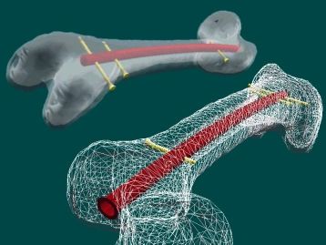
Fig. 5. The interactive 3D modeling tool for extracting and editing contour lines.
Second, for the editing purposes, we developed an interactive 3D tool that builds and edits contour
lines for the CT slices and converts them to the data file later processed by NUAGES. In Fig. 4, an
example of the 3D reconstruction from the data in Fig. 3 is depicted. In Figs. 5 and 6, the 3D editing tool
and the reconstructed models of the femur are presented. The 3D data of the bones obtained later are
converted to the data format used by our program and then transferred to the personal computer.
Fig. 6. The reconstructed femur and the femur neck
5. Objects and Operations in the VE
Analyzing the application area, we understood that most bone fractures appear to occur in
predictable patterns. For example, there are 27 types of femoral fractures (see Fig. 7) and 9
types of pelvic fractures. This encouraged us to create a geometric database of the fractured
bones using geometric models of the broken bones created from standardized geometric models.
These bones were “broken” as needed using interactive modeling tools and other methods.
To fix fractures, surgeons use hundreds of different screws, plates, nails, wires, and locking
bolts. Their geometric models and the models of the tools were also created and stored in the
respective geometric databases (see Fig. 8).
Volume 6, No. 1 21 May 2000International Journal of Information Technology
Since the software had to be implemented on a common personal computer, we had to find
a reasonable balance between the processing time, the size, and the complexity of the model.
Analyzing the goals that must be achieved, probably the straightforward method can be to
implement something like the functionally based spatial spline.19 This spline would interpolate
the bones from the scattered points, and for any trial point would answer the question whether
the point is inside, outside or on the boundary of the bone. Unfortunately, all these methods,
providing a general solution to practically every problem, assume very extensive use of
computer memory, and will not provide the required interactivity. Thus, we decided to combine
different models for different purposes in our representations.
Fig. 7. Several femoral fractures from the geometric database of common fractures
implemented for the project.
Fig. 8. Real and virtual surgical tools and implants from the geometric database implemented
for the project.
Internally, each geometric model in the VE is represented as a polygonal mesh with the
functional description that—like geometric DNA—constitutes its shape. Extra application data
is stored in the attachment to the geometric model. Thus, for example, extra information about
the number, the size, and the locations of the holes for each plate are stored with the polygonal
Volume 6, No. 1 22 May 2000International Journal of Information Technology
mesh and functional description of the plate. For the bones, the essential information is the
location of their medial axes.
Since the final assembly somewhat resembles complex objects in CAD, each object in the
VE and the scenes composed from the objects are stored in the hierarchical geometric database.
For example, each plate with holes is a hierarchy of the solid plate body as a root object with the
addressable holes as its children nodes. When inserting a screw through the plate, first the
surgeon picks up the hole, then the inserted screw becomes yet another child node of this
assembly. If the guide wire is inserted through the plate’s hole, later it will be used for inserting
the cannulated screw. In that case, the coordinate systems of the guide wire and the cannulated
screw will be aligned to provide the guiding effect. After the insertion, the guide wire will be
removed from the scene, and the screw will substitute for it in the hierarchy.
Many common surgical techniques handle internal fixation of bone fractures, such as
fixation with cancellous screws20, fixation with an intra-medullary nail21, dynamic condylar
screw (DCS) implant system technique22, dynamic hip screw (DHS) implant system technique23,
and so on. Analyzing them systematically from a geometric point of view, we managed to find
common and specific parts that let us come up with the generic virtual fixation techniques that
became foundation virtual methods in our project. These methods have been implemented on
the internal level, while the surgeon sees the familiar names and commands at the user interface.
6. Collision Detection
All the geometric and nongeometric data are used together for pseudo-physical collision
detection—crucial for VR modeling. Each particular operation required developing its own
respective methods and models.
For example, when inserting a screw into bone through a plate’s hole, the proximity sensors
of each plate implemented as functional fields engage to attract the screw tip to the nearest hole.
Then, depending on the mode selected, the screw will either automatically align with the hole
axis or the surgeon will have a limited ability to change its orientation, thus simulating the
actual insertion of a screw. The screw being inserted never goes through the solid parts of the
plate. Since the length of each screw and all the sizes of the plate are among the parameters
stored in the geometric database, the screw never penetrates deeper than it should. For the
surgeon, the result looks very realistic.
Another example of handling collision detection involves an implementation of nail
insertion in the femoral canal (Fig. 9a). The femur with its canal has a curvature of a certain
radius. The nail to be inserted (Fig. 9b) is a piece of a large torus with the same curvature as that
of the femur.
Mathematically, the problem of insertion is to find the initial location of the torus so that it
lies in the same plane with the bone’s medial axis and so that its axis is coincident with the axis
of the bone curvature. From the surgeon’s point of view, the skill required in this operation is to
find the proper point and angle of insertion for the nail. These initial parameters let us solve the
geometric problem of the axes’ coincidence and answer the question whether in principle the
nail goes freely all the way through the canal or seriously damages the bone.
These are just two examples of the methods used to solve collision detection problems in
our project. Each particular operation required developing its own respective methods and
techniques.
Volume 6, No. 1 23 May 2000International Journal of Information Technology
(a)
(b)
Fig. 9. Universal femoral nail.
7. Virtual Bone-setter
We called the software implemented in this project Virtual Bone-setter. The program runs on
PCs with the most common architecture though the following features are preferred:
• Powerful CPU not less than 150 MHz.
• Extended RAM 64MB or above.
• Video card supporting 3D polygon rendering and real transparency.
Using a mouse as an input device controlling object relocations may suffice, but for some
users it might not be as realistic as expected. Better immersion results from using relatively
cheap 3D mice or graphics pads instead. The software is implemented with Criterion’s
Renderware, which provides the required interactivity with a reasonable quality of rendering.
Comparing with the de facto standard OpenGL, it provides better speed of rendering though
slightly sacrificing the rendering quality since it uses less number of bits for representing the
colors. The ability of Renderware to model in hierarchical coordinate systems and its support of
many VR devices provides a very natural environment for the geometric modeling in the
project.
Starting the virtual surgery, the surgeon locates the fractured bone in 3D space looking at
the screen. Then, depending on the fracture, the surgeon can issue the following basic
commands:
Volume 6, No. 1 24 May 2000International Journal of Information Technology
• Applying the instruments and putting the implants in place.
To simulate the surgical techniques, the following explicit and implicit commands are used:
“Insert threaded guide wire”, “Insert pin”, “Remove threaded guide wire”, “Remove pin”,
“Insert multiple guide wires”, “Measure for the screw length”, “Insert screw”, “Seat plate”,
“Insert nail”, etc. These operations are applied to the current objects in the scene. The user
is required to locate the application point on the bone, to define the orientation or to follow
the guided orientation, and to apply or to insert the instrument or implant to the proper
depth. In common training where expensive non-reusable synthetic bones are operated, it
usually takes hours of practicing to achieve the required skills. All the models were created
with the maximum possible realism to train the surgeons visually so that they will
remember how all this hardware looks before they enter the training lab. Collision detection
is implemented to ensure proper insertion of the implants (e.g., for the screws being inserted
through the plate holes and for the cannulated screws being inserted by the guiding wires).
The screws rotate about their axes when inserted. The speed of insertion slows down when
the tension is growing. Drilling and cracking sounds are played for the realistic audio
feedback.
• Moving, rotating, and zooming the scene and the objects in the scene.
The whole scene or individual objects can be moved, rotated or zoomed in any direction.
• Looking at the assembly through the X-ray lens.
The surgeon often inserts the wires, nails or pins to the appropriate depth under image
intensification (X-ray). In the virtual environment, it is provided either for the whole scene
or for the particular part of it through the so-called X-ray lens.
• Walking through the bone canal.
It allows the surgeon to look at the seated implants from inside the bone. This effect can not
be achieved during the real surgery and is implemented here for education and control
purposes.
• Reversing process.
If something has been done wrong, the multilevel undo operation can be applied without
any damage to the models.
• Setting the lights and the background.
Different type of lights can be applied for better illumination of the surgical field thus
simulating the actual environment in the operation theatre. These can be omni-directional or
spot-lights.
The program guides the surgeon through the whole surgery, although the surgeon retains the
ability to make independent decisions. Pseudo-physical collision detection is implemented.
Realistic sounds play when the instruments are used. The scene with the fracture being fixed can
be saved at any time and stored in the hierarchical geometric database for further use. If the
surgeon requires an implant not in the database, its model can be created interactively and used
immediately. If the surgeon wishes to study a real fracture rather than those stored in the
fracture database, its geometric model can be reconstructed from CT data.
8. Examples of Virtual Fracture Fixation
Femoral neck fracture fixation with cancellous screws, as depicted in Fig. 10,requires the
following actions:
Volume 6, No. 1 25 May 2000International Journal of Information Technology
Fig. 10. Virtual femoral neck fracture fixation with cancellous screws.
1. The first step includes guide wire insertion to the appropriate depth. The surgeon chooses
“Insert threaded guide wire ”and locates the point of insertion on the bone. The wire appears
on the screen touching the bone at the selected point. The surgeon rotates the wire to the
proper insertion angle and inserts it. The result of the insertion can be checked with the
simulated X-ray imaging.
2. The next step is placing multiple parallel guide wires at various distances from the first
wire. The surgeon selects “Place multiple guide wires ”and defines the respective points on
the bone. Wires are inserted automatically parallel to the first one, thus implementing the
adjustable parallel wire guide device.
3. Then, the surgeon measures for the screw length. The reading indicates the appropriate
screw length. It simulates the result of applying the cannulated screw-measuring device.
4. Next comes inserting the screws. The surgeon selects the cannulated screw of the
appropriate length from the database, picks up the wire, and places the screw over the wire.
After that, other cannulated screws are placed over the respective wires.
5. The last step is to remove and discard the guide wires.
Another example is the inter-trochanteric fracture fixed with a DHS implant system (see
Fig. 11). In this case, the surgeon inserts the guide wire first, next the guide pin, and then
removes the wire. The surgeon measures the pin for a screw length and inserts the DHS screw
with the appropriate length over the pin. After that, the surgeon seats the appropriate DHS plate
over the screw, removes the pin, and fixes the plate with cortex screws through the plate holes.
Finally, the surgeon applies the DHS compressing screw to secure the assembly.
Volume 6, No. 1 26 May 2000International Journal of Information Technology
Fig. 11. Virtual femoral neck fracture fixation with DHS implant system.
Yet another example of the virtual femoral fracture fixation with an intra-medullary nail
appears in Fig. 12. This VR surgery simulates the real surgery illustrated by the x-rays in
Fig. 2 and by the diagram in Fig. 9a. Here, the nail—in fact, a piece of a torus—is to be inserted
in the bone canal and then fixed with the screws. This operation requires hours of training
before the surgeon learns exactly where to insert the nail and what the insertion angle should be.
Fig. 12. Virtual femoral fracture fixation with an intra-medullary nail.
9. Conclusion
The implemented tools let surgeons learn how to fix fractured bones and perform preoperative
planning without wasting expensive synthetic bones. The software works on common PCs and
Volume 6, No. 1 27 May 2000International Journal of Information Technology
simulates surgical techniques, implants, and tools. Using more sophisticated virtual input/output
devices, although not compulsory, increases realism and provides better immersion. Besides
that, our software also may be used for simulating internal operations for some bone diseases,
like slipped capital femoral epiphyses, ankle arthrodeses, sacroiliac joint disruptions, subtalar
arthrodeses, and so forth.
The long-term goals of the project follow:
• Photorealistic representation of the operating room environment.
• Force feedback and tactile response using VR haptic devices.
• Object-management at different levels-of-detail.
• Greater immersion, letting the surgeon study and manipulate virtual objects on the screen or
on a virtual workbench with a higher degree of realism.
These goals assume software development in both directions—on PCs and advanced
software implemented on a virtual workbench. Both lines of software will use the same models
and data format, allowing surgeons to handle simple cases on their home or office computers
and to ring the most important or complex cases to expensive graphics workstations.
10. Acknowledgements
This project was inspired by the Singapore General Hospital Trauma Service, Department of
Orthopedics. The software has been developed at the School of Applied Science, Nanyang
Technological University where the first two authors were employed until April 1999.
Currently, the project continues internationally between Singapore General Hospital, Moscow
Institute of Physics and Technology, and Institute of Computing for Physics and Technology,
Russia. The authors are grateful to all mentioned institutions for the opportunity to work on the
project.
References
1. Muriel D. Ross, 3-D Imaging In Virtual Environment (A Scientific, Clinical And Teaching Tool.
Biocomputation Center NASA Ames Research Center Moffett Field, CA 94035)
http://biocomp.arc.nasa.gov/visible_human/
2. Satava R. M., Virtual Reality Surgical Simulator: The First Steps, in Proc. of the Third Annual
Conference on Virtual Reality, pp.103-105.
http://www.ee.cua.edu/~davies/THESIS/REFERENCES/sata2.htm
3. M. Bro-Nielsen, Mvox, Interactive 2-4D medical image and graphics visualization software, in Proc.
Computer Assisted Radiology (CAR'96), pp. 335-338, 1996
http://www.imm.dtu.dk/documents/users/mvox/
4. M. Bro-Nielsen, P. Larsen and S. Kreiborg, Virtual teeth: A 3D method for editing and visualizing
small structures in CT scans, in Proc. Computer Assisted Radiology (CAR'96), pp. 921-924, 1996
5. Delp S.L. and Loan J.P., A Software System to Develop and Analyze Models of Musculoskeletal
Structures, Computers in Biology and Medicine, vol. 25, pp. 21-34, 1995.
http://www.musculographics.com/compasssurg.htm
6. Dev P., Fellingham L.L., Vassiliadis A., Woolson S.T., White D.N. and Young S.L, 3D Graphics for
Interactive Surgical Simulation and Implant Design, in Proc. and Display of Three-Dimensional Data
II, SPIE vol. 507, San Diego, California, pp. 52-57, 23 - 24 August, 1984.
http://www.ee.cua.edu/~davies/THESIS/REFERENCES/devp84.htm
7. U.G. Kühnapfel et. Al., Endosurgery Simulations with KISMET: A Flexible Tool for Surgical
Instrument Design, Operation Room Planning and VR Technology based Abdominal Surgery
Training, Proceedings VR'95 WORLD Conference, Feb. 21-23 1995.
http://iregt1.iai.fzk.de/KISMET/kis_apps_med.html
Volume 6, No. 1 28 May 2000International Journal of Information Technology
8. Krumm H.G., Kuehnapfel U., Kuhn Ch., Huebner M., Neisius B., Force Feedback for Medical
Virtual Reality Applications Using the KISMET System. Medicine meets Virtual Reality 5, San
Diego, Calif., January 25, 1997.
9. MusculoGraphics' Limb Trauma Simulator (LTS) http://www.musculographics.com/
10. Robb, R.A., Three-Dimensional Biomedical Imaging. CRC Press, Inc., Boca Raton, FL,
vols. I & II, 1985.
11. Robb, R.A., Multidimensional Biomedical Image Display and Analysis in the Biotechnology
Computer Resource at the Mayo Clinic. Machine Vision and Applications, 1:75-96, 1988.
12. B. Pflesser, U. Tiede, and K. H. Höhne, Towards Realistic Visualization for Surgery Rehearsal,
Computer Vision, Virtual Reality and Robotics in Medicine, in Proc. CVRMed '95 (N. Ayache, ed.),
vol. 905 of Lecture Notes in Computer Science, pp. 487-491, Berlin: Springer-Verlag, 1995.
13. K. H. Höhne, ed., VOXEL-MAN Junior: Interactive 3D Anatomy and Radiology in 40 Virtual
Reality Scenes, (Heidelberg, Springer-Verlag Electronic Media, 1997)
14. Sourina O., Sourin A., Virtual Simulation of Orthopedic Surgery Training, in Proc. Joint Workshop
on Computer Graphics with ACM Symposium on Virtual Reality Software and Technology 1998,
VRST’98, Taipei, Taiwan, November 2-5, pp.48-51, 1998.
15. Sourina O., Sourin A., Orthopaedic Surgery Simulation, in Proc. Applied Informatics’99, Innsbruck,
February 15-18, 1999, pp.234-237.
16. O.Sourina, A.Sourin, Virtual Simulation of Orthopaedic Surgery Training, SAS Research Link,
Nanyang Technological University, Singapore ISSN 0218-9534, No5/99, 1999, pp.17-18.
17. Sourin A., Sourina O., Howe Tet Sen, “Virtual Orthopedic Surgery Training”, IEEE Comp. Graphics
and Applications, May/June, 2000, in print.
18. J-D. Boissonnat and B. Geiger, Three dimensional reconstruction of complex shapes based on the
Delaunay triangulation, Technical Report 1697 INRIA 1992. Available at
19. http://www-sop.inria.fr/prisme/personnel/geiger/geiger.html
20. Savchenko V.V., A.A.Pasko, A.I.Sourin, T.L.Kunii, Volume Modelling: Representations and
Advanced Operations”, Proc. of CGI’98, June 22-26, 1998, Hannover, Germany, pp.4-13.
21. Main Catalogue, Original Instruments and Implants of the Association for the Study of Internal
Fixation –AO/ASIF, SYNTHES (U.S.A.), 1690 Russell Road, Paoli, Pa. 19301-1222.
22. Universal Tibial and Femoral Nail System, Original Instruments and Implants of the Association for
the Study of Internal Fixation –AO/ASIF, SYNTHES (U.S.A.), 1690 Russell Road, Paoli, Pa. 19301-
1222.
23. DCS. Dynamic Condylar Screw Implant System, Original Instruments and Implants of the
Association for the Study of Internal Fixation –AO/ASIF, SYNTHES (U.S.A.), 1690 Russell Road,
Paoli, Pa. 19301-1222.
24. DHS. Dynamic Hip Screw Implant System, Original Instruments and Implants of the Association for
the Study of Internal Fixation –AO/ASIF, SYNTHES (U.S.A.), 1690 Russell Road, Paoli, Pa. 19301-
1222.
Volume 6, No. 1 29 May 2000You can also read