VISION THROUGH ELECTRONICS- To make 'India Vision 2020' a Possibility - IJIRSET
←
→
Page content transcription
If your browser does not render page correctly, please read the page content below
ISSN(Online) : 2319-8753
                                                                                                  ISSN (Print) : 2347-6710

        International Journal of Innovative Research in Science,
                      Engineering and Technology
                                    (An ISO 3297: 2007 Certified Organization)

                                        Vol. 5, Issue 12, December 2016

         VISION THROUGH ELECTRONICS-
         To make 'India Vision 2020' a Possibility
                                                       V.Umaiyaal
  IV year Student, Dept of Biomedical Engineering, PSNA-CET, Kothandaraman Nagar, Dindigul, Tamil Nadu, India

ABSTRACT: The dream of using electronic or artificial retinal replacements to treat blindness has long been held. In
the development of prosthetic vision, it is also possible to stimulate the visual pathway at other sites other than the
retina to gain visual perceptions The visual pathway functions as a complex image processor as well as an information
conduit. At higher levels, the visual signals arrive with significant processing completed. Treatment options for the
associated cataract and macular oedema, have been limited,retinal prostheses offer the only treatment option for
patients at the severe end of the disease spectrum at present. There are currently two models of retinal prostheses
available commercially: (i) Argus® II retinal prosthesis system (Second Sight Medical Product, Inc., Sylmar), which
received CE (Conformité Européenne) marking in March 2011 and the Food and Drug Administration (FDA) approval
in February 2013[3] and (ii) the alpha-IMS (Retinal Implant AG, Reutlingen), which obtained CE marking in July
2013.Apart from technological advances in prosthetic vision, development in other biomedical fields has also shed new
hope on restoring vision in patients with end-stage retinal diseases, most notably the cellular therapy. This video
camera is embedded in the inter-ocular bridge of the glasses frame. The video-processing unit (VPU), which converts
the images captured by the video camera into electrical signals. These signals are then passed onto the External Coil for
information relay.Red-free fundus photograph of an Argus®II retinal implant placed on the retinal surface (epiretinally)
over the macular region, within the retinal vessel arcades. There are 60 (10 × 6) microelectrodes in the array. Large
clumps of intra-retinal pigmentation (bone-spicule pigments) and the pale atropic underlying RPE are seen,
characteristic of end-stage RP. An area of four adjacent microelectrodes is marked by a white square.

KEYWORDS: Retinal prosthesis Prosthetic vision Therapy Argus II

                                                I.INTRODUCTION

 India, world’s second largest population, has the distinction of being the home of the world's largest number of blind
people. World Health Organization (WHO) statistics revealed that approximately 63 million people in India are
visually impaired, and of these 8 million people are totally blind. The number of blind persons in India in 2010 was
estimated to be 24.1 million and 31.6 million 2020 [1]. Based on Government of India statistics (2011), one out of
every three blind persons in the world lives in India. Over 15 million people are blind out of which 11.75 million live in
the rural and most backward areas. Over 9.4 million have cataract related blindness. 2.8 million are blind due to
refractive error. 6 million people become blind with low vision every year. 3.2 million Children are blind under the age
of 16 years, only 5% of them receive any education. 12 million people could be saved from become blind. The Union
health ministry has already launched a national programme to control blindness and expects to reach its blindness
elimination target of 0.3% by 2015[11], five years before the WHO deadline of 2020.Although it has reached three by
fourth of its goal it is still the work of Bio medical Engineers to overcome this issue, since all the blind cannot be given
the natural eye we can give them through electronics.

Copyright to IJIRSET                                     DOI:10.15680/IJIRSET.2016.0512131                           20906
ISSN(Online) : 2319-8753
ISSN (Print) : 2347-6710
International Journal of Innovative Research in Science,
Engineering and Technology
(An ISO 3297: 2007 Certified Organization)
Vol. 5, Issue 12, December 2016
II.WORKING OF NATURAL EYE
Fig.1. Human Eye
Light rays enter the eye through the cornea, the clear front “window” of the eye. The cornea’s refractive
power bends the light rays in such a way that they pass freely through the pupil the opening in the center of the iris
through which light enters the eye. The iris works like a shutter in a camera. It has the ability to enlarge and shrink,
depending on how much light is entering the eye[2]. After passing through the iris, the light rays pass thru the eye’s
natural crystalline lens. This clear, flexible structure works like the lens in a camera, shortening and lengthening its
width in order to focus light rays properly. Light rays pass through a dense, transparent gel-like substance, called the
vitreous that fills the globe of the eyeball and helps the eye hold its spherical shape.
In a normal eye, the light rays come to a sharp focusing point on the retina. The retina functions much like the
film in a camera. It is responsible for capturing all of the light rays, processing them into light impulses through
millions of tiny nerve endings, and then sending these light impulses through over a million nerve fibers to the optic
nerve.
The retina receives the image that the cornea focuses through the eye’s internal lens and transforms this image
into electrical impulses that are carried by the optic nerve to the brain. We can tolerate very large scars on our bodies
with no concern except for our vanity. This is not so in the cornea. Even a minor scar or irregularity in the shape can
impair vision. No matter how well the rest of the eye is functioning, if the cornea is scarred, clouded or distorted, vision
will be affected.
III.NEED FOR AN ARTIFICIAL EYE
There are a number of retinal diseases that attack these cells, which can lead to blindness. The most notable of
these diseases are:
1. Retinitis pigmentosa
2. Age-related macular degeneration
Retinitis Pigmentosa (RP) is the name given to a group of hereditary diseases of the retina of the eye. In
macular degeneration, a layer beneath the retina, called the Retinal Pigment Epithelium (RPE), gradually wears out
from its lifelong duties of disposing of retinal waste products. Both of these diseases attack the retina, rendering the
rods and cones inoperative, causing either loss of peripheral vision or total blindness. However, it’s been found that
neither of these retinal diseases affects the ganglion cells or the optic nerve[10]. This means that if scientists can
develop artificial cones and rods, information could still be sent to the brain for interpretation.
Copyright to IJIRSET DOI:10.15680/IJIRSET.2016.0512131 20907ISSN(Online) : 2319-8753
ISSN (Print) : 2347-6710
International Journal of Innovative Research in Science,
Engineering and Technology
(An ISO 3297: 2007 Certified Organization)
Vol. 5, Issue 12, December 2016
IV.BIONIC EYE
Fig.2. Chip placed on the eye
A blind person could be made to see light by stimulating the nerve ganglia behind the retina with an electrical
current. This test proved that the nerves behind the retina still functioned even when the retina had degenerated. Based
on this information, scientists set out to create a device that could translate images and electrical pulses that could
restore vision. Today, such a device is very close to be available to the millions of people who have lost their vision to
retinal disease. In fact, there are at least two silicon microchip devices that are being developed. The concept for both
devices is similar, with each being:
Small enough to be implanted in the eye
Supplied with a continuous source of power
Biocompatible with the surrounding eye tissue
Perhaps the most promising of these two silicon devices is the artificial silicon retina. The ASR is an
extremely tiny device. It has a diameter of just 2 mm (.078 inch) and is thinner than a human hair[8]. In order for an
artificial retina to work it has to be small enough so that doctors can transplant it in the eye without damaging the other
structures within the eye.
Groups of researchers have found that blind people can see spots of light when electrical currents stimulate
cells, following the experimental insertion of an electrode device near or into their retina. Some patients even saw crude
shapes in the form of these light spots[5]. This indicates that despite damage to cells in the retina, electronic techniques
can transmit signals to the next step in the pathway and provide some form of visual sensation. Researchers are
currently developing more sophisticated computer chips with the hope that they will be able to transmit more
meaningful images to the brain.
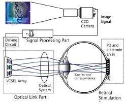
V.WORKING OF BIONIC IMPLANT
A.MARC SYSTEM
The intermediary device is the MARC system. The schematic of the components of the MARC to be
implanted consists of a secondary receiving coil mounted in close proximity to the cornea, a power and signal
transceiver and processing chip, a stimulation-current driver, and a proposed electrode array fabricated on a material
such as silicone rubber, thin silicon, or polyimide with ribbon cables connecting the devices. The biocompatibility of
polyimide is being studied, and its thin, lightweight consistency suggests its possible use as a non-intrusive material for
an electrode array. Titanium tacks or cyanoacrylate glue may be used to hold the electrode array in place
Copyright to IJIRSET DOI:10.15680/IJIRSET.2016.0512131 20908ISSN(Online) : 2319-8753
ISSN (Print) : 2347-6710
International Journal of Innovative Research in Science,
Engineering and Technology
(An ISO 3297: 2007 Certified Organization)
Vol. 5, Issue 12, December 2016
Fig.3. Circuit of Marc system.
The MARC system would consist of two parts which separately reside exterior and interior to the eyeball. Each part is
equipped with both a transmitter and a receiver[6]. The primary coil can be driven with a 0.5-10 MHz carrier signal,
accompanied by a 10 kHz amplitude modulated (AM/ASK) signal which provides data for setting the configuration of
the stimulating electrodes. A DC power supply is obtained by the rectification of the incoming RF signal. The receiver
on the secondary side extracts four bits of data for each pixel from the incoming RF signal and provides filtering,
demodulation, and amplification. The extracted data is interpreted by the electrode signal driver which finally generates
appropriate currents for the stimulating electrodes in terms of magnitude, pulse width, and frequency.
B.WORKING PROCEDURE
An artificial eye provokes visual sensations in the brain by directly stimulating different parts of the optic
nerve.
A bionic eye works by stimulating nerves, which are activated by electrical impulses. In this case the patient
has a small device implanted into the body that can receive radio signals and transmit those signals to nerves. The
Argus II[9] implant consists of an array of electrodes that are attached to the retina and used in conjunction with an
external camera and video processing system to provide a rudimentary form of sight to implanted subjects The Argus II
Copyright to IJIRSET DOI:10.15680/IJIRSET.2016.0512131 20909ISSN(Online) : 2319-8753
ISSN (Print) : 2347-6710
International Journal of Innovative Research in Science,
Engineering and Technology
(An ISO 3297: 2007 Certified Organization)
Vol. 5, Issue 12, December 2016
Fig.4.Working of an artificial eye
Retinal Prosthesis System can provide sight, the detection of light, to people who have gone blind from
degenerative eye diseases. Diseases damage the eyes’ photoreceptors, the cells at the back of the retina that perceive
light patterns and pass them on to the brain in the form of nerve impulses, where the impulse patterns are then
interpreted as images. The Argus II system takes the place of these photoreceptors.
The second incarnation of Second Sight’s retinal prosthesis consists of five main parts:
• Digital Camera - built into a pair of glasses, captures images in real-time sends images to microchip.
• Video processing microchip - built into a handheld unit, processes images into electrical pulses representing patterns
of light and dark; sends pulses to radio transmitter in glasses
• Radio transmitter - wirelessly transmits pulses to receiver implanted above the ear or under the eye
• Radio receiver - receiver sends pulses to the retinal implant by a hair-thin, implanted wire
• Retinal implant - array of 60 electrodes on a chip measuring 1 mm by 1 mm[12].
The entire system runs on a battery pack that is housed with the video processing unit. When the camera
captures an image-of, say, a tree-the image is in the form of light and dark pixels. It sends this image to the video
processor, which converts the tree-shaped pattern of pixels into a series of electrical pulses that represent “light” and
“dark.” The processor sends these pulses to a radio transmitter on the glasses, which then transmits the pulses in radio
form to a receiver implanted underneath the subject’s skin. The receiver is directly connected via a wire to the electrode
array implanted at the back of the eye, and it sends the pulses down the wire. When the pulses reach the retinal implant,
they excite the electrode array[7]. The array acts as the artificial equivalent of the retina’s photoreceptors. The
electrodes are stimulated in accordance with the encoded pattern of light and dark that represents the tree, as the
retina’s photoreceptors would be if they were working (except that the pattern wouldn’t be digitally encoded). The
electrical signals generated by the stimulated electrodes then travel as neural signals to the visual center of the brain by
way of the normal pathways used by healthy eyes -- the optic nerves. In macular degeneration and retinitis pimentos,
the optical neural pathways aren’t damaged. The brain, in turn, interprets these signals as a tree, and tells the subject,
“You’re seeing a tree”. All of this takes some training for subjects to actually see a tree. At first, they see mostly light
and dark spots. But after a while, they learn to interpret what the brain is showing them, and eventually perceive that
pattern of light and dark as a tree. Thus bionic eye helps a blind people to see the objects and recognize them.
VI.DEVICES OTHER THAN ARGUS II
Bionic Vision Australia, collaboration between of researchers working on a bionic eye, has announced that its
prototype implant has completed a two year trial in patients with advanced retinitis pigmentosa. Three patients with
Copyright to IJIRSET DOI:10.15680/IJIRSET.2016.0512131 20910ISSN(Online) : 2319-8753
ISSN (Print) : 2347-6710
International Journal of Innovative Research in Science,
Engineering and Technology
(An ISO 3297: 2007 Certified Organization)
Vol. 5, Issue 12, December 2016
profound vision loss received 24-channel[4] suprachoroidal electrode implants that caused no noticeable serious side
effects. Moreover, though this was not formally part of the study, the patients were able to see more light and able to
distinguish shapes that were invisible to them prior to implantation. The newly gained vision allowed them to improve
how they navigated around objects and how well they were able to spot items on a tabletop.
The 44-electrode prototype is designed to help researchers learn more about how the bionic eye can be
optimized. The device will be fully implantable and include a patient-worn vision processor. Participants will be able to
take the device out of the lab and into the real world. Feedback from patients will allow researchers to develop more
sophisticated vision processing and stimulation techniques.
ADVANTAGES
Helps correct vision
No longer has limited access
American one is FDA approved
Can be easily implanted
Research is not limited by budget.
DISADVANTAGES
Australian one is still being researched
Both eyes has research cost in the millions of dollars
Australian one has to undergo human trials
American one doesn't correct vision to 100%
Si based photo detectors have been tried in earlier attempts. But Si is toxic to the human body and reacts
unfavorably with fluids in the eye.
VII.CONCLUSION AND RESULTS
The result is based on Conditions : System off and both eyes unpatched; system on in ‘scrambled’ mode with eyes
patched and unpatched; and system in standard mode with eyes patched and unpatched
Methods:
• Test I: letter identification
• Test II: letter size reduction
• Test III: word recognition
Copyright to IJIRSET DOI:10.15680/IJIRSET.2016.0512131 20911ISSN(Online) : 2319-8753
ISSN (Print) : 2347-6710
International Journal of Innovative Research in Science,
Engineering and Technology
(An ISO 3297: 2007 Certified Organization)
Vol. 5, Issue 12, December 2016
Restoration of sight for the blind is no more a dream for people today. Bionic Eyes have made this true. Though
there are a number of challenges to be faced before this technology reach the common man, the path has been laid. This
paper has tried to present the concept of Artificial Vision called “Bionic Eyes”. It is just a matter of time, may be 4-5
years that the blind will be able to see through these Bionic Eyes, with thanks to Science and Technology.
REFERENCES
[1]. Ashley Hall,” Diamond shines as basis for bionic eye prototype”, ABC News, December 09, 2010
[2].Humayun MS, de Juan E Jr., Dagnelie G, et al. Visual perception elicited by electrical stimulation of retina in blind humans. Archives of
Ophthalmology; vol 114.
[3].International Journal of Medical Research and Review
[4].Julia Layton, “How does a 'bionic eye' allow blind people to see?” Discovery Communications, LLC.
[5].W.H. Dobelle, ``Artificial Vision for the Blind by Connecting a Television Camera to the Visual Cortex,'' ASAIO Journal (American Society for
Artificial Internal Organs), January - February 2000.
[6]. "The Artificial Silicon Retina Microchip for the Treatment of Vision Loss From Retinitis Pigmentosa" Arch Ophthalmol. 2004;122:460-469.
[7]. Wikipedia Articles on: Retina, Retinitis Pigmentosa, Macular Degeneration, Photoreceptors, Optic Nerve, Photodiode
[8]. Figure Anatomy of human eye available at : http://seedoctoradams.com/the_amazing_human_eye
[9]. FDA Approves First Retinal Implant for Rare Use: Available at: http://www.reuters.com/article/2013/02/14/ussecondsight-fda-
eyeimplantidUSBRE91D1AK20130214.
[10]. Vision 2020 Australia, Clear Focus: The Economic Impact of Vision Loss in Australia in 2009, prepared by Access Economics. 2010.
[11]. George H. van Doorn, Barry L. Richardson, Dianne B. Wuillemin, International Journal of Autonomous and Adaptive Communications
Systems ,Volume 6, Issue 4 ,DOI: 10.1504/IJAACS.2013.056822
[12]. Anthony’s textbook of Anatomy and Physiology -Gary A Thibodeau, Kevin T Patton Image processing for a high-resolution optoelectronic
retinal prosthesis. Asher, A; Segal, WA; Baccus, SA; Yaroslavsky, LP; Palanker, DV; IEEE Transactions on Biomedical Engineering, 54(6): 993-
1004 (2007).
Websites
1. http://www.thescientist.com/?articles.view/articleNo/41052/title/The-Bionic-Eye/
2. https://www.scribd.com/doc/27004363/Bionic-Eye
Copyright to IJIRSET DOI:10.15680/IJIRSET.2016.0512131 20912You can also read