VivoSight OCT Skin Imaging and Measurement System - Advancing Research & Management of
←
→
Page content transcription
If your browser does not render page correctly, please read the page content below
VivoSight OCT
 Skin Imaging and Measurement System

Advancing Research & Management of:
    § Immune and Inflammatory Diseases
    § Non Melanoma Skin Cancer
    § Vascular Conditions
    § Microneedle Research
    § Skin Ageing and Skin Quality

Healthy Skin for Happy Living
VivoSight Optical Coherence Tomography
 An optical analogue of ultrasound using laser scanning

VivoSight scans a
6 x 6 mm2 patch of skin
and produces up to 500
cross-sectional slices
(frames) per scan.

                     Each frame visualizes 1 mm deep
                     creating a 3D image block that
                     can be reviewed frame by frame
                     and at any depth.

 VivoSight resolution defines skin architecture to
       generate accurate measurements

 VivoSight scan of healthy, normal skin (6 mm x 1 mm)

Healthy Skin for Happy Living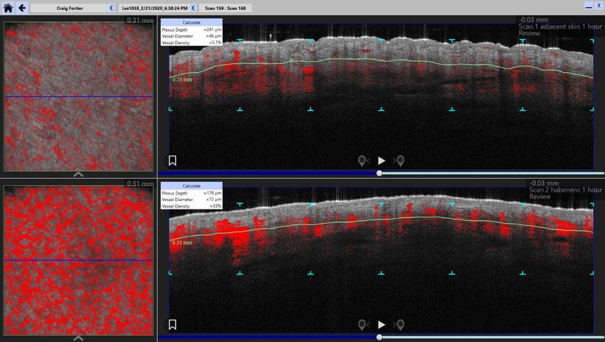
VivoSight Dynamic Scanning Analyzes
             Vascular Morphology

Superficial Vascular Plexus
 Automatically Calculate:
 •   Depth of the superficial plexus
 •   Modal vessel diameter
 •   Density of vessels at this level

Vascular Density by Depth
 Automatically Calculate:
 •   Vessel density by depth in the dermis
 •   Visualize and compare density by
     depth before and after treatments

Normal scan of volar forearm             Same site after exposure to capsaicin

 Healthy Skin for Happy Living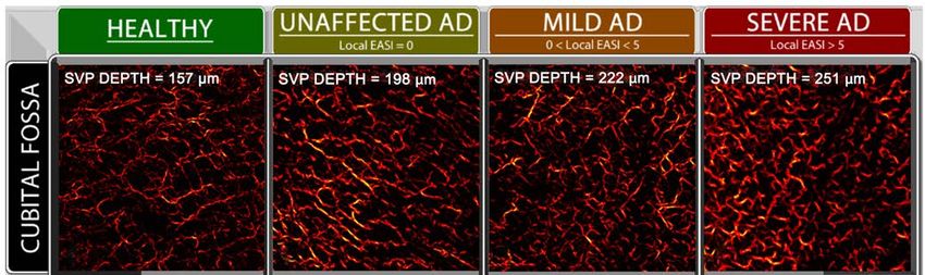
Advance Your Research and Development
                Program with VivoSight

    Accelerate development
    timelines and reduce costs

    §   Visualize & quantify treatment effects

    §   Scan in-vivo and ex-vivo targets

    §   Multiple skin and vascular
        measurements

    §   Rapid scan time < 30 seconds

    §   Automated report generation with
        batch processing

        VivoSight Scan Screen and User Interface
Video camera view of skin surface facilitates
targeting and probe placement

                                                                            Top-down,
                                                                            birds eye view
View 3D surface
                                                                            at any depth
texture and 3D
                                                                            in the 6x6 mm2
vascular network                                                            image stack

Annotation tools to mark up                     Up to 500 cross-sectional slices (frames)
images for easy reference                       can be viewed individually or as a video

   Healthy Skin for Happy Living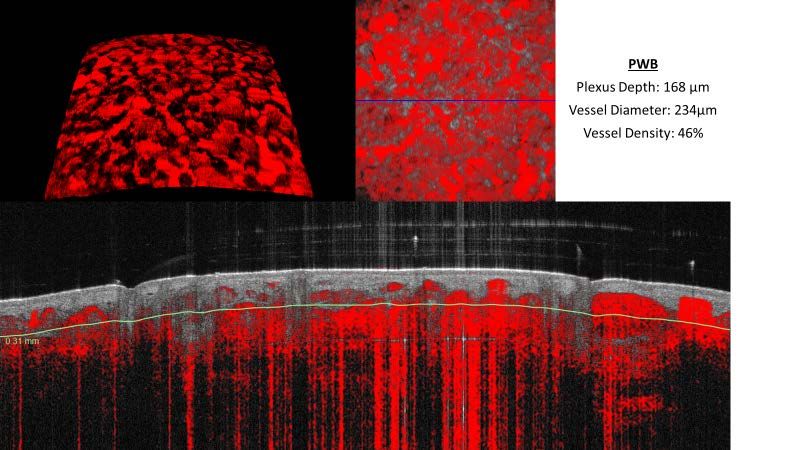
VivoSight OCT
Treating Vascular Lesions More Intelligently

    Do you know the depth and diameter of
    the vessels you are attempting to treat?

Healthy Skin for Happy Living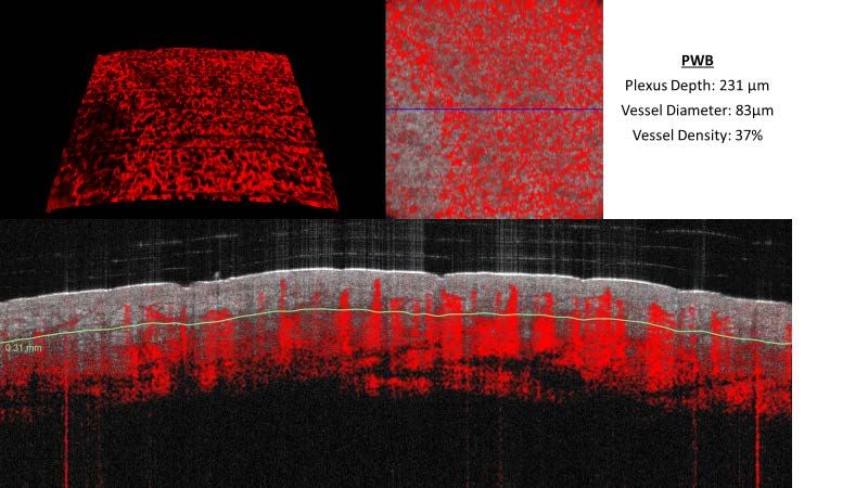
Selective Photothermolysis
Laser treatment of vascular lesions is based on the Theory of Selective Photothermolysis
(Anderson and Parrish, Science 1983) which requires:
•       Wavelength that is preferentially absorbed by the desired target structure

•       Laser pulse width that is less than or equal to the thermal relaxation time of the
        vessel targeted as determined by Vessel Diameter

•       Sufficient radiant exposure, Fluence, to reach a damaging temperature in the target.
        Vessel Depth can impact fluence requirements.

Start with Vessel Measurements for Better Targeting

Vascular Plexus Profile:
•       Depth of the superficial plexus
•       Modal vessel diameter
•       Density of vessels at this level

                                            Vessel depth and diameter
                                            measurements will guide laser
                                            pulse width and fluence settings.

    Changes in Vascular Density:
    •    Quantify the density of vessels by depth in the dermis

                                              Quantify treatment effect by
                                              measuring vessel density
                                              before and after treatments.

    Healthy Skin for Happy Living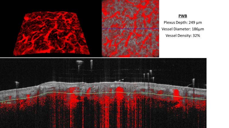
VivoSight Images of PWS and Rosacea
         VivoSight measurements show the high variability
   in vessel morphology, diameter and depth, demonstrating the
     need for variable laser pulse duration and fluence settings.

Healthy Skin for Happy Living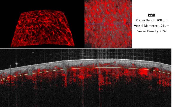
Autoimmune and
       Inflammatory Diseases

      Can you see beyond the skin surface
       to extract objective measures and
          identify sub-clinical disease?

Healthy Skin for Happy Living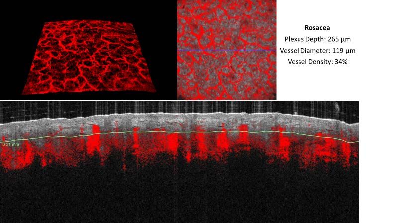
Researchers are Finding Value in Quantification
              and Monitoring of Atopic Dermatitis (AD)

VivoSight Dx capabilities relevant to inflammatory disease include:
•     Epidermal remodeling and thickness measurement
•     Alterations in vascular morphology, depth and density as a measure of inflammation
•     Optical attenuation as a proxy for collagen density
•     Skin surface roughness

Epidermal Alterations 1

OCT image of healthy individual      OCT image captured from an           OCT image captured from an
with no history of AD                uninvolved site on an eczema         involved site on a different eczema
                                     patient, showing slightly extended   patient, showing what appears to
                                     rete-pegs and an undulating DEJ      be inflammatory acanthosis (Long
                                                                          thin epidermal papillae/rete-pegs)

 Vascular Alterations 1
              VivoSight images show morphologic differences and variable
              superficial vascular plexus (SVP) depth for different EASI scores.

    Healthy Skin for Happy Living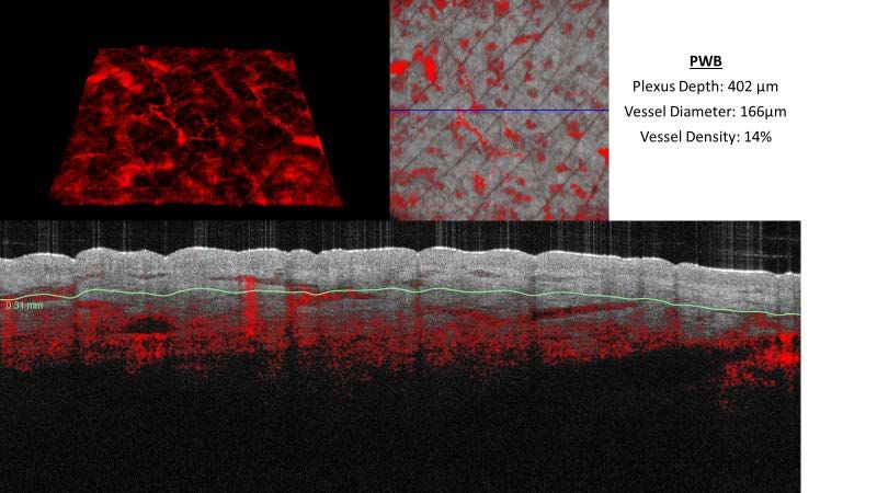
Better Control of Flares Through Monitoring of Sub-clinical AD

Unlike visual scores, dynamic OCT has the potential to quantify sub-clinical AD that allows
for determination of treatment duration or when to start new, proactive therapies

                                                         Visualize and quantify sub-clinical AD
                                                         for advanced, proactive management

  Bieber T. Ann Dermatol. 2010 May;22(2):125-137

Quantification of Pharmaceutical Treatment Effects on AD

Recently, a correlation between clinical improvement of AD and dynamic OCT imaging
metrics has been demonstrated on patients undergoing a systemic therapy

                                                              Significant OCT metrics for
                                                              pharmaceutical treatment effects:
                                                              •    Vascular plexus depth
                                                              •    Epidermal thickness

                               Baseline                       •    Collagen density
                                                              •    Vesicle density

                                                              OCT imaging biomarkers quantify
                                                              pharmaceutical treatment effects
                                                              to more efficiently accelerate drug
                                                              development cycles
                     90 days after dupilumab

Healthy Skin for Happy Living
Microneedle & Transdermal
Applications
Can you image and measure insertion depth,
swelling or degradation of microneedles in-vivo
and see the vascular response and skin changes?
Do you know the depth and diameter of the vessels
you are attempting to treat?
Healthy Skin for Happy LivingVisualize micro-channel creation and monitor the
time course of needle degradation or swelling
VivoSight Dx capabilities to advance your
microneedle and drug delivery research include:
• In-vivo imaging of microneedles in real time
• Measure microneedle dimensions,
penetration depth, dissolution and swelling VivoSight image with vascular overlay.
Microneedle penetrates 800 μm deep [2]
• Measure inflammatory response via
vascular changes
• Understand morphology of device created
skin defects
• Observe kinetics of pore closure and skin
recovery
• Verify reproducibility, consistency of results VivoSight images of hydration profile of
microneedles over a 34 minute period
VivoSight imaging and measurement of
microneedle changes over 60 minutes in-vivo
Proportion of needle affected Air gap between skin and Pore size shown to slightly reduce
increases linearly with time substrate shrinks over 20 min with time as the needle dissolves
Healthy Skin for Happy LivingVascular Dynamics and Skin Changes
VivoSight can monitor the inflammatory response over time after MAP removal
VivoSight scans prior to MAP application and after MAP removal showing
measurable changes in the superficial vascular plexus.
Plexus Depth: 430 µm Plexus Depth: 241 µm
Modal Diameter: 36 µm Modal Diameter: 57 µm
Vessel Density: 9% Vessel Density: 40%
Skin blood flow returns to Modal vessel diameter Plexus depth increase
normal after about 25 min decreasing by about 30% to normal levels
VivoSight produces 3-D surface images, top down and
frame views, to monitor hole closure after MAP removal
Immediately after MAP removal
21 hours after MAP removal
Healthy Skin for Happy LivingNon-Melanoma Skin Cancer
Nodular BCC
Can you see the extent of NMSC lesions or follow
clearance progression non-invasively?
Superficial BCC
Infiltrative BCC
Healthy Skin for Happy LivingSee the whole picture with VivoSight to inform
and optimize treatment planning and monitoring
Aid decision making with more information than can be
obtained through clinical assessment or dermoscopy
Lesion Assessment
Obtain images of sub-surface, sub-clinical tumor tissue
• White asterisks mark hyporeflective tumor nests
• Thin arrows mark dark peripheral borders
• Thick white arrows mark the DEJ
• Black asterisk marks thinning epidermis
Image courtesy of Themstrup [4]
• Thick black arrow marks a hair casting a shadow
Treatment Monitoring
Topical Treatment of BCC Lesion [5] Laser Treatment of BCC Lesion [6]
Before imiquimod therapy 1 week with imiquimod therapy
1 month with imiquimod 4 weeks post therapy cysts have Pre laser treatment Immediate post showing
replaced the BCC islands reduced vascularity
Mohs Surgery
Margin Assessment Prior to Mohs Surgery [7]
Lesion is outlined with Reflective marker casts a Example showing lesion
reflective marker shadow on VivoSight image outside of marker shadow,
perimeter must be expanded
Healthy Skin for Happy LivingThe Full Suite of VivoSight
Skin Measurements
Quantify your treatment’s impact by measuring:
• Surface Roughness
Roughness measures including
peak to trough, average
roughness and 3D image
• Epidermal Thickness,
Variation and Contrast
Average epidermal
thickness over the entire
6 x 6 mm scan area
• Vascular Plexus Profile
Depth of the superficial plexus,
Modal vessel diameter,
Density of vessels at this level
• Changes in Vascular Density
Quantify the density of
vessels by depth in the dermis
• Beam Attenuation
Proxy measure for collagen
content
Healthy Skin for Happy LivingCompanion Products
Maximize Versatility and Performance
Laboratory Fixture
Increased utility for laboratory research
Precision fixture for the VivoSight OCT handheld probe
converts VivoSight into a powerful OCT microscope for
scanning laboratory samples. Fine adjustment in all 3 axes
makes sample analysis easy, convenient and accurate.
Dermatoscope
DermoScan dermatoscope USB port
Combine dermoscopy with VivoSight OCT in a
single workstation. Dermatoscope images are
displayed onscreen.
DermoGenius Ultra Polarized
Software Upgrades
VivoSight Dx software updates two to three times per year
Customers with support contracts receive an annual
software update to their VivoSight free of charge. From
time to time, Michelson may also release new major
software upgrades which will be available for an extra fee.
References
1. Byers RA, Maiti R, Danby SG, Pang EJ, Mitchell B, Carre MJ, Lewis R, Cork MJ, Matcher SJ. Sub-clinical assessment of atopic dermatitis severity using
angiographic optical coherence tomography. Biomedical Optics Express (BOE), Vol. 9, No. 4, 1 Apr 2018.
2. S. Sharma, et al., Rapid, low cost prototyping of transdermal devices for personal healthcare monitoring, Sensing and Bio-Sensing
Resear(2016), http://dx.doi.org/10.1016/j.sbsr.2016.10.004
3. R.F. Donnelly et al. Evaluation of the clinical impact of repeat application of hydrogel-forming microneedle array patches. Drug Delivery and
Translational Research (Feb 2020). https://doi.org/10.1007/s13346-020-00727-2
4. Themstrup L, De Carvalho N, Nielsen SM, Olsen J, Ciardo S, Schuh S, Nørnberg BM, Welzel J, Ulrich M, Pellacani G, Jemec GBE. In vivo differentiation
of common basal cell carcinoma subtypes by microvascular and structural imaging using dynamic optical coherence tomography. Exp Dermatol.
2018;27(2):156–65.
5. C. A. Banzhaf, L. Themstrup, H. C. Ring, M. Mogensen and G. B. E. Jemec. Optical coherence tomography imaging of non-melanoma skin cancer
undergoing imiquimod therapy. Skin Research and Technology 2013; 0: 1–7
6. De Carvalho N, Schuh S, Kindermann N, Kästle R, Holmes J, Welzel J. Optical coherence tomography for margin definition of basal cell carcinoma
before micrographic surgery—recommendations regarding the marking and scanning technique. Skin Res Technol. 2017;00:1–7.
https://doi.org/10.1111/srt.12407R
Healthy Skin for Happy LivingVivoSight Specifications
Morphologic and Angiographic Imaging of Skin
• Uses a low-power 1300nm laser
• Up to 1 mm depth penetration
• Scanning area of 6 x 6 mm
• < 7.5 µm lateral resolution
• < 5.5 µm axial resolution
• Eye-safe
Power
Supply Voltage 100-240V~ 50-60Hz Nom. (Earthed supply)
Maximum Power 250VA
Mains Input Connector IEC 320 C13 Socket 10A
Fuse T 5AL 250V
Weight and dimensions
Dimensions (W x D x H) 0.55 x 0.57 x 1.61 m
(Monitor at maximum height)
Weight 56 kg
For more information please contact:
Michelson Diagnostics
Cell (408) 504-7391
Compliant with European CE Mark. Email: craig.fortier@vivosight.com
FDA cleared for clinical use 510(k) K093520.
VivoSight is a Multi-Beam Optical Coherence Tomography (OCT) system indicated for use in the
two-dimensional, cross-sectional, real-time imaging of external tissues of the human body. This
indicated use allows imaging of tissue microstructure, including skin, to aid trained and competent
clinicians in their assessment of a patient's clinical conditions.
Healthy Skin for Happy Living
1028.DO.113 Issue 1You can also read