X-linked reticulate pigmentary disorder in a 4-year-old boy - Termedia
←
→
Page content transcription
If your browser does not render page correctly, please read the page content below
Letter to the Editor

X-linked reticulate pigmentary disorder in a 4-year-old boy

Yu-Kun Zhao1, Li-Hua Fan2, Jing-Fa Lu3, Ze-Yu Luo4, Zhi-Miao Lin5,6, Hui-Jun Wang5,6,7, Di-Qing Luo1

1
 Department of Dermatology, The East Division of The First Affiliated Hospital, Sun Yat-sen University, Guangzhou, China
2
  Zhangzhou Dermatology Hospital, Zhangzhou, Fujian Province, China
3
 Department of Dermatology, The First Affiliated Hospital of Gannan Medical University, Ganzhou, Jiangxi Province, China
4
  Department of Dermatology, Guangzhou Development District Hospital, Guanghzou, China
5
  Peking University First Hospital, Beijing, China
6
  Beijing Key Laboratory of Molecular Diagnosis on Dermatoses, Beijing, China
7
 National Clinical Research Center for Skin and Immune Diseases, Beijing, China
                                                                                         Adv Dermatol Allergol 2022; XXXIX (2): 410–412
                                                                                           DOI: https://doi.org/10.5114/ada.2022.115893

     A 4-year-old boy from a nonconsanguineous Chinese                                After informed written consent was obtained from the
Han family was referred to the Department of Derma-                              patient’s parents and other family members, DNA was ex-
tology, The East Division of The First Affiliated Hospital,                      tracted from peripheral blood of the patient and his family
Sun Yat-sen University, China, because of dyschromato-                           members according to standard methods. Then exome se-
sis. He was born at full term through an uncomplicated                           quencing of the POLA1 gene was performed and revealed
delivery, and developed photophobia as well as bilateral                         hemizygous mutation of NC_000023.10:g.24744696A>G
lop ears at the age of 3 months. When he was 6 months                            for the patient and heterozygote for his mother and sister
old, he developed hypohidrosis that was over the whole                           (Figures 2 A–C). Then a diagnosis of X-linked reticulate pig-
body except the neck and outer aspects of the thighs, he                         mentary disorder was confirmed.
also had mild fever of about 37.5°C during the summer                                 X-linked reticulate pigmentary disorder [XLPDR;
or after physical activity, leading to erythema. Hereafter,                      MIM301220], first described in a Canadian pedigree [1],
generalized hyperpigmentation with mottled hypopig-                              is an exceptionally rare genodermatosis inherited as X-
mentation developed over his whole body. He had nor-                             linked recessive trait [1–9]. In males, it is always charac-
mal growth and normal mental development, and there                              terized by diffused reticulate dyschromatosis with pho-
were no systemic manifestations as well as associations                          tophobia, dry skin and typical facial appearance featuring
such as pruritus, pain, or burning sensations since his                          upswept frontal hairline and flared eyebrows. Hypohi-
birth. His mother, and grandmother as well as the mater-                         drosis is also a distinctive feature, with predilection for
nal grandmother on mother's side had multiple, asymp-                            trunk and extremities [2, 3] as our patient presented. The
tomatic, brownish macules in linear and whorled pat-                             systemic manifestations include recurrent infections and
terns over the trunk, axillae, groin and extremities after                       autoimmune reactions against various organs including
birth, which relieved after adolescence. His elder sister                        respiratory, gastrointestinal, and neurological systems
had cleft palate in addition to linear hyperpigmentation.                        [2], however, not all patients including the present one
During a 4-year follow-up, the patient’s hypohidrosis                            had such presentations. The reasons for the absence of
and the rise of body temperature had mild improvement                            systemic manifestations remain unknown which needs
although no treatments were ordered, while mottled                               further study. Males always have more severe symptoms
hypopigmentation increased and was accompanied by                                than female carriers. Whereas the females always pres-
more severe hyperpigmentation.                                                   ent patchy hyperpigmentation along the Blaschko’s lines
     Cutaneous examination showed diffuse reticulate                             alone that are similar to stage III incontinentia pigmenti
hyperpigmentation with mottled hypopigmentation over                             and will alleviate and even disappear after puberty, but
the whole body, associated with photophobia, coarse and                          without systemic involvements [2, 4, 5], as did in our pa-
dried skin, upswept frontal hairline and flared eyebrows                         tient’s mother and his sister. Although cutaneous symp-
(Figures 1 A–C). Skin biopsy was refused.                                        toms and peculiar facial appearance are considered to

Address for correspondence: Hui-Jun Wang PhD, Department of Dermatology, Peking University First Hospital, Beijing Key Laboratory
of Molecular Diagnosis on Dermatoses, Beijing 100034, China; National Clinical Research Center fo Skin and Immune Diseases, Beijing 100034,
China, e-mail: drhuijunwang@pku.edu.cn; Prof. Di-Qing Luo, Department of Dermatology, The East Division of The First Affiliated Hospital,
Sun Yat-sen University, Guangzhou 510700, China, phone: +86 20 82493439, fax: +86 20 82398840, e-mail: luodq@mail.sysu.edu.cn
Received: 22.04.2020, accepted: 07.08.2020.

This is an Open Access article distributed under the terms of the Creative Commons Attribution-NonCommercial-ShareAlike 4.0 International (CC BY-NC-SA 4.0).
License (http://creativecommons.org/licenses/by-nc-sa/4.0/)

410                                                                                  Advances in Dermatology and Allergology 2, April/2022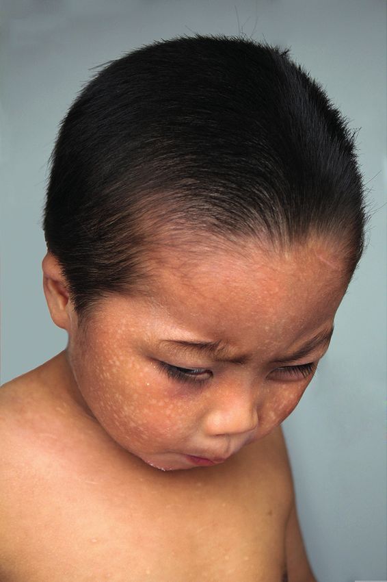
X-linked reticulate pigmentary disorder in a 4-year-old boy
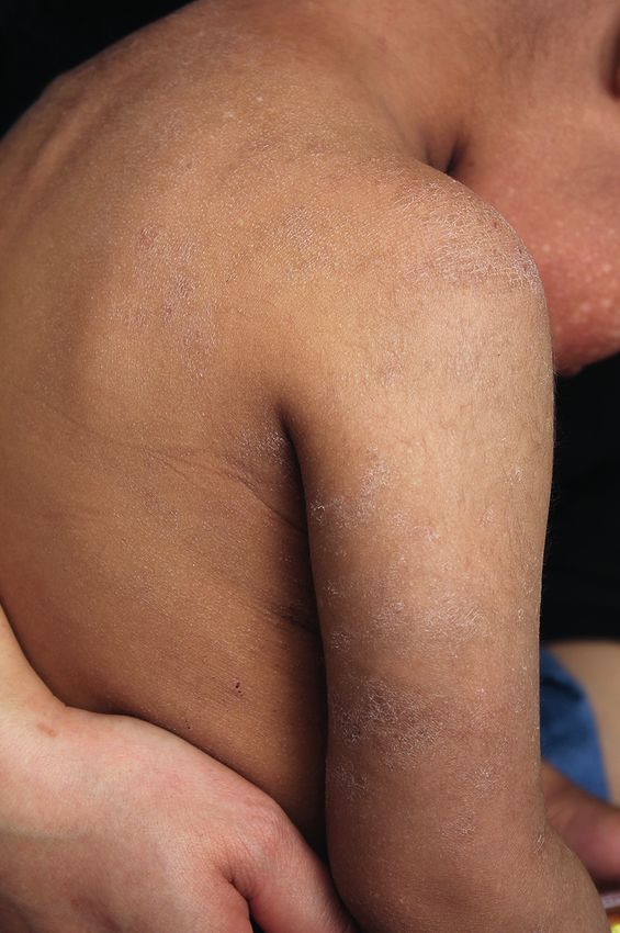
A B C
Figure 1. Diffuse reticulate hyperpigmentation with mottled hypopigmentation on the whole body as well as upswept
frontal hairline, flared eyebrows and photophobia (A, B), and coarse and dried skin (C)
be the distinctive features of XLPDR [3, 4], absence of A NC_000023.10:g.24744696
skin lesions in infancy had also been reported [4]; in rare
instances, the reticulate hyperpigmentation developed
until the patient was 8 years old [6]. The absence of skin
lesions in early stage may lead to a delayed diagnosis
for the patient. Up to date, 25 cases from different fami-
lies including the present one have been reported [1–9].
However, it is not until 2015 that the mutation of c.1375-
354A>G or g.24744696A>G in POLA1 gene was demon- Normal control
strated as a causal factor for XLPDR [7], and all the pa-
tients with genetic demonstration including the present B
one were detected to have the same intronic mutation
in POLA1. Because of the rarity of the disease, it remains
to be elucidated whether XLPDR is caused by this muta-
tion specifically, or this mutation stands for one mutation
hotspot with additional mutations in POLA1 underlying Patient
XLPDR. Nevertheless, it is undoubtedly cost-effective to
screen preferentially for this mutation in cases of XLPDR. C
The exact mechanisms for XLPDR as well as POLA1
remain fully unknown. POLA1 protein, or DNA polymerase
alpha catalytic subunit, is necessary for initiation of DNA
replication and the synthesis of cytosolic RNA:DNA, Mother and sister
which directly modulates interferon-regulatory factors
[3, 7]. The reduction of POLA1 expression may result in Figure 2. Exome sequencing of the POLA1 gene shows
normal control gene (A), hemizygous mutation of
diminished cytosolic RNA:DNA hybrids [3, 7], which are
NC_000023.10:g.24744696A>G in the patient (B) and het-
responsible for the negative regulation of interferon-reg- erozygous mutation in his mother and his sister (C)
ulatory factors, leading to increase of type I interferon [3,
7]. It has been reported recently that XLPDR was associ-
ated with a decreased number and selective cytotoxic- described up to date. The histopathological features of
ity defect of NK cells [3]; this might be why the patients XLPDR are similar in both sexes, including mild hyper-
have autoinflammation and recurrent infection [6, 7]. keratosis, acanthosis, basal hyperpigmentation, and pig-
Interestingly, no recurrent cutaneous infection has been mentary incontinence in the upper dermis [2, 4, 6]. De-
Advances in Dermatology and Allergology 2, April/2022 411
Yu-Kun Zhao, Li-Hua Fan, Jing-Fa Lu, Ze-Yu Luo, Zhi-Miao Lin, Hui-Jun Wang, Di-Qing Luo

position of amyloid-like material in the papillary dermis            9. Fraile G, Norman F, Reguero ME, et al. Cryptogenic multi-
and mild superficial lymphocytic perivascular infiltrate                focal ulcerous stenosing enteritis (CMUSE) in a man with
may also be present [2, 4, 6]. Direct immunofluorescence                a diagnosis of X-linked reticulate pigmentary disorder (PDR).
                                                                        Scand J Gastroenterol 2008; 43: 506-10.
may reveal granular deposits of C3 at the dermoepidermal
junction [6]. However, these histopathological findings
are non-specific for the disease [2, 4]. The mechanisms
for pigmentation also remain elusive. Accumulation of
melanophages and amyloid-like materials had been
found in the upper dermis [1, 2, 4, 5, 6], suggesting that
those might be responsible for the hyperpigmentation.
Of course, further studies are still required to verify this
speculation and clarify the function of POLA1 in skin.
     Differential diagnosis of XLPDR includes incontinentia
pigmenti, primary cutaneous amyloidosis, Rothmund-
Thomson syndrome, Kindler syndrome, congenital dys-
keratosis, and Naegeli–Franceschetti syndrome [1, 4, 5, 8].
Based on the featuring presentations and inherited trait,
it is not hard to make a correct diagnosis, sometimes, se-
quencing of the POLA1 gene is an optimal option. No ther-
apeutics are recommended for the disease at present yet.

Acknowledgments
    The authors sincerely thank the patient and his fam-
ily members participating the present work.
    Yu-Kun Zhao, Li-Hua Fan and Jing-Fa Lu contributed
equally for the present work.

Conflict of interest
      The authors declare no conflict of interest.

   References
 1. Partington MW, Marriott PJ, Prentice RS, et al. Familial cuta-
    neous amyloidosis with systemic manifestations in males.
    Am J Med Genet 1981; 10: 65-75.
 2. Kim BS, Seo SH, Jung HD, et al. X-Linked reticulate pigmen-
    tary disorder in a female patient. Int J Dermatol 2010; 49:
    421-5.
 3. Starokadomskyy P, Wilton KM, Krzewski K, et al. NK cell de-
    fects in X-linked pigmentary reticulate disorder. JCI Insight
    2019; 4: e125688.
 4. Pezzani L, Brena M, Callea M, et al. X-linked reticulate pig-
    mentary disorder with systemic manifestations: a new fam-
    ily and review of the literature. Am J Med Genet A 2013;
    161A: 1414-20.
 5. Anderson RC, Zinn AR, Kim J, Carder KR. X-linked reticulate
    pigmentary disorder with systemic manifestations: report of
    a third family and literature review. Pediatr Dermatol 2005;
    22: 122-6.
 6. Starokadomskyy P, Sifuentes-Dominguez L, Gemelli T, et al.
    Evolution of the skin manifestations of X-linked pigmentary
    reticulate disorder. Br J Dermatol 2017; 177: e200-1.
 7. Starokadomskyy P, Gemelli T, Rios JJ, et al. DNA polymerase-
    alpha regulates the activation of type I interferons through
    cytosolic RNA:DNA synthesis. Nat Immunol 2016; 17: 495-504.
 8. Ades LC, Rogers M, Sillence DO. An X-linked reticulate pig-
    mentary disorder with systemic manifestations: report of
    a second family. Pediatr Dermatol 1993; 10: 344-51.

412                                                                    Advances in Dermatology and Allergology 2, April/2022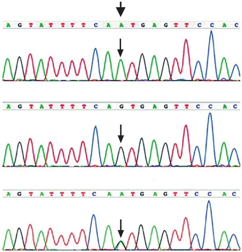
You can also read