Spontaneous ventral hernia through the rectus abdominis muscles: a case report
←
→
Page content transcription
If your browser does not render page correctly, please read the page content below
Spontaneous ventral hernia through the rectus
abdominis muscles: a case report
mitsutsune Benedict washiro ( mitsutsune.washiro@healthcoop-nagano.or.jp )
Naganochuo hospital
Takuya Kitahara
Naganochuo Hospital
Nobuo Yanagisawa
Naganochuo Hospital
Jun Narita
Naganochuo Hospital
Takao Danzuka
Naganochuo Hospital
Masayuki Ohtsuka
Chiba University: Chiba Daigaku
Research Article
Keywords: Ventral, hernia, rectus abdominis muscles, transverse abdominis, fascia, elderly
Posted Date: June 2nd, 2022
DOI: https://doi.org/10.21203/rs.3.rs-1697525/v1
License: This work is licensed under a Creative Commons Attribution 4.0 International License.
Read Full License
Page 1/17Abstract
Background
A hernia is a condition in which tissues or organs are abnormally protruding through a defect
in the surrounding wall. We experienced a rare spontaneous ventral hernia that penetrated
the right transverse abdominal fascia and rectus abdominis muscles.
Case presentation
A 92-year-old woman was admitted to our hospital with major complaints of right abdominal
pain and some quadrant mass. She had no history of trauma and lower abdominal surgery.
Computed tomography revealed that a protrusion of the hernia from the right lower
abdominal wall. The hernia contents were small intestine, which penetrated the transverse
abdominis fascia and rectus abdominis muscles. The herniation has been manually reduced.
Elective surgery was performed 3 days later. The intraoperative findings indicated that the
hernia sac was located around the middle of the rectus abdominis muscles in the right lower
Page 2/17abdominal wall. Hernioplasty was performed using Bard Light PerFix Plug®. The patient fully
recovered and was discharged 8 days after surgery. No recurrence was identified for 42
months during patient follow-up.
Conclusions
We presented a rare case of hernia, which was protruded through the rectus abdominis
muscles. Intrinsic anatomical weakness of the abdominal wall and age-related muscle
atrophy might have caused this rare hernia. This case report provides clinicians useful
information for accurate diagnosis and successful treatments of this type of hernia in the
elderly, and contributes to identifying the anatomical features of some rare ventral hernias.
Background
A hernia is a condition in which tissues or organs are abnormally protruding through a defect
in the surrounding wall. It can occur congenitally or acquired, and the common types of hernia
include inguinal, femoral, ventral, and incisional. Intraperitoneal, Richer and Littre hernias of
Page 3/17the abdominal wall, as well as sciatic obturator and perineal hernias in the pelvis are also
known, but those types of hernias are uncommon.1)
Here, we present a rare case of another type of hernia, a ventral hernia that penetrates the
right transverse abdominal fascia and rectus abdominis muscles. There is no defect around
the rectus abdominis muscles, which appeared to have a normal anatomy. This is the first
case report of the rectus abdominis muscles hernia.
Case Presentation
A 92-year-old woman was admitted to our hospital with major complaints of right lower
abdominal pain and some quadrant mass. Her medical history included oral treatment for
osteoporosis. The patient had a history of pregnancy but no history of trauma and lower
abdominal surgery. She had no psycho-social history. On admission, the blood pressure,
pulse rate and body temperature were 150/90 mmHg, 76 beats per minute and 36.7℃,
respectively. Clinical examination showed a small mass about 2.0×2.0 cm on the right
Page 4/17lower abdomen, with moderate tenderness around the mass. Physical examination did not
reveal bowel obstruction. All laboratory findings were within normal limits, except for a slight
increase of lactate dehydrogenase (261 IU/L). It was observed that the patient had neither
drinking nor smoking. The patient’s family history was noncontributory. Computed
tomography (CT) of her abdomen demonstrated a protrusion of the hernia from the right
abdominal wall (Fig. 1). The hernia contents were small intestine, which penetrated the
transverse abdominis fascia and rectus abdominis muscles. There was a slight dilatation of
the bowel loops. The herniation has been manually reduced. There was no evidence of the
bowel ischemia and elective operation was performed 3 days later. The intraoperative
findings were an atypical ventral hernia. The hernia sac was located around the middle of the
rectus abdominis muscles in the right lower abdominal wall. The defect size was 2.0×2.0 cm
(Fig. 2a). Hernioplasty was done using Bard Light PerFix Plug®. The defect was filled with
Page 5/17Plug and covered by Onlay patch (Fig. 2b). No defect was found on the contralateral site,
and the left space was not repaired by any method. The patient recovered uneventfully and
was discharged on postoperative day 8. No recurrence was identified for 42 months during
patient follow-up.
Discussion
In clinical anatomy of the abdominal wall, the rectus abdominis muscles is protected by the
rectus sheath. Above the arcuate line, the rectus sheath was formed by aponeurosis of
inferior oblique muscles, external oblique muscles and transverse abdominis muscles.
However, below the arcuate line, the sheath was completely lacking. These anatomical
features make the abdominal wall below the arcuate line weaker when compared to other
sites.2)3) In addition, the patient presented here was 92-year-old, and her muscles may have
progressively atrophied, as she grew older. In fact, in retrospect, CT one year before surgery
showed that there was already a narrow space in the bilateral rectus abdominis muscles (Fig.
Page 6/173). We suggest this had developed. Aging and pregnancy could contribute expanding the
space. Although there is a slight high-density area in the subcutaneous adipose tissue on
the right rectus abdominis muscle, detailed interview about patient’s medical history
revealed that she had neither trauma nor surgery. Anatomical weakness of the abdominal
wall and age-related muscle atrophy might have caused herniation from this space in the
present case.4) This type of hernia should be distinguished from Spigelian hernia as a
differential diagnosis. Spigelian hernia is named after Adrian van der Spiegel, who depicted
the semilunar line in1645.5) The semilunar line is defined as the transition of the transverse
abdominis muscle to its aponeurotic tendon.6) Spigelian fascia is located between the
semilunar line and the lateral edge of the rectus abdominis muscle. Spigelian hernia is a
spontaneous abdominal hernia caused by a defect in the Spigelian fascia. The
characteristics of the hernia in our case, as well as similar hernias such as Spigelian hernia,
Page 7/17incisional hernia and spontaneous posterior rectus sheath hernias are reported in Table 1.
We indicate differences about the site of hernia orifice, etiology and the hernia sac position
of each case. Spigelian hernia protrudes from Spigelian fascia and some cases cause
congenitally.7) Spontaneous posterior rectus sheath hernias are a type of interparietal hernias
where the hernia sac lies between the various layers of the abdominal wall muscles and dose
not penetrate into the subcutaneous tissue.1)8) Etiologically, in addition to the similar to other
type of hernias, posterior sheath components of linea alba to be thinner than the anterior
components.9) Colonal Plane CT demonstrates that the position relationship between a
hernial orifice, the rectus abdominis muscles, the semilunar line and the Spigelian
fascia. (Fig.4) We also showed the arcuate line where the inferior epigastric vessels perforate
the rectus abdominis.10) This location was found to be at a mean of 2.1 ±2.3 cm superior to
the level of the anterior superior iliac spines.11) The most common complications of abdominal
Page 8/17wall hernias are bowel obstruction secondary to the hernia, incarceration, and strangulation.1)
CT is the gold standard for diagnostic modality due to its high sensitivity in diagnosing such
complications.1) For hernia treatment, Preoperative accurate diagnosis is very important.
Most abdominal wall hernias are surgically repaired, considering the risk of development of
above mentioned complication, even if asymptomatic.1) Repair of the defect of abdominal
wall entails fascial closure, or fascial suturing reinforced with synthetic mesh in the case of
large defects.12)13) Small hernia defects can be repaired only by laparoscopic
herniorrhaphy.14) Placement of sutures too close or too far from the defect margin and pulling
sutures too tight can result in hernia recurrence.13) However, in the current case, we
performed open hernioplasty using Bard Light PerFix Plug® because of muscle weakness
and fascial attenuation in the elderly. Preoperative knowledge of the type of the herniation,
whether below the arcuate line or not, help preoperative planning to facilitate primary fascial
Page 9/17repair, which can then be supported with on-lay mash, depending on the clinical situation.11)
An excellent anatomic layer-by-layer closure can be achieved using the anterior approach.15)
Conclusions
To the best of our knowledge, this is the first case report of spontaneous trans-rectus
abdominis muscle hernia. This is notable because hernia was located around the middle of
the right rectus abdominis muscles. This hernia is associated with severe abdominal pain
and incarceration. Therefore, prompt diagnosis and surgery are important to achieve a
successful outcome in the elderly patients. Surgeons should be aware of this very rare type
of hernia, and may be able to suspect it preoperatively on imaging studies. Principle of
surgical repair of this type of hernia is using on-lay mesh.
Abbreviations
CT
Computed tomography
Declarations
Ethics approval and consent to participate
Not applicable.
Consent for publication
Written informed consent was obtained from the patient’s daughter for publication of this
Page 10/17case report and accompanying images.
Availability of data and materials
Not applicable.
Competing interests
The authors declare that they have no competing interests.
Funding
The authors did not receive any funding.
Author’s contributions
TK, MW, and NY performed the surgery. JN and TD attended the patient postoperatively.
MO revised the article. All authors have read and approved the final manuscript.
Acknowledgments
The authors thank Mr. Susumu Idegawa and Mrs. Natsuko Obuse for their technical
Page 11/17assistance in uploading images and literature search.
Author’s information
Not applicable.
Endnotes
Not applicable.
References
1) Aguirre DA. Abdominal wall hernias: imaging feature, complication, and diagnostic
pitfalls at multi-detector row CT. RadioGraphics. 2005; 25:1501-20.
2) Losanoff JE. Spontaneous hernia through the posterior rectus abdominis sheath: case
report and review of the published literature 1937-2008. Hernia. 2009; 13:555-8.
3) Johnson TG. Clinical anatomy of the abdominal wall: hernia surgery. OA Anat.
2014; 2:3.
4) Reznichenko A. Case of rare abdominal wall hernia. J Curr Surg. 2014; 4:99-100.
Page 12/175) Spangen L. Spigerian hernia. World J Surg. 1989; 13:573-80.
6) Sachs M. The so-called Spigelian hernia-a rare lateral hernia of the abdominal wall.
Zentralbl Chir. 1998; 123:267-71.
7) Skandalakis PN, Zoras O, Skandalakis JE, Mirilas P. Spigelian hernia: surgical anatomy,
embryology, and technique of repair. Am Surg. 2006; 72:42-48.
8) Felfel M, Khoury ME, Marboeuf Y, Strohl D, Menu Y. Incarcerated hernia through the
posterior rectus sheath. AJR. 2005; 185:1185-6.
9) Korenkov M, Beckers A, Koebke J, Lefering R, Tiling T, Troidl H. Biomechanical and
Morphological types of the linea alba and its possible role in the pathogenesis of
midline incisional hernia. Eur J Surg. 2001; 167:909-14.
10) McVay CB, Anson BJ. Composition of the rectus sheath. Anat Rec. 1940; 77:213-225.
11) Loukas M, Myers C, Shah R, Tubbs RS, Wartmann C, Apaydin N, et al. Arcuate line of
Page 13/17the rectus sheath: clinical approach. Anat Sci Int. 2008; 83:140-144.
12) Onal A. Spigelian hernia associated with strangulation of the small bowel and appendix
Hernia. 2003; 7:156-7.
13) Losanoff JE. Recurrent Spigelian hernia: a rare cause of colonic obstruction. Hernia.
2001; 5:101-4.
14) Bittner JG. Mesh-free laparoscopic Spigelian hernia repair. Am Surg. 2008; 74:713-20.
15) Schumpelick V. Atras of hernia surgery. Philadelphia: BC Decker; 1990.
Tables
Table 1 is in the supplementary files section.
Figures
Figure 1
Preoperative CT. CT investigation revealed protrusion of the small intestine from
the right rectus abdominis muscle.
Figure 2
Page 14/17Intraoperative findings. a hernia sac was located around the middle of the rectus
abdominis muscle. b Hernioplasty was made using Bard Light PerFix Plug®.
Figure 3
Previous CT. a narrow space among the bilateral rectus abdominis muscle was
revealed 1 year before the CT.
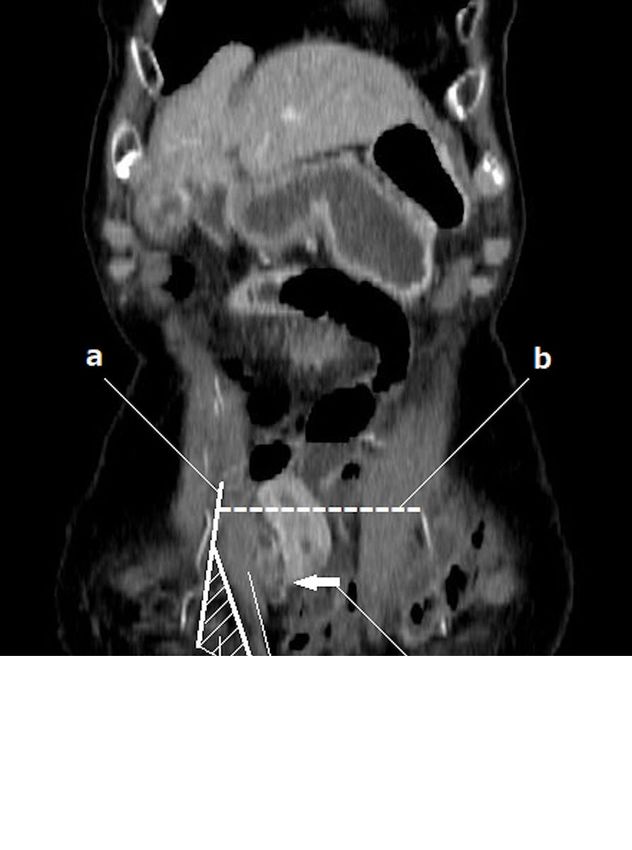
Page 15/17Figure 4
Colonal CT. We demonstrate the location of a The semilunar line, b The arcuate
line, c Spigelian fascia, d Hernia orifice, e Rectus abdominis muscles.
Page 16/17Supplementary Files
This is a list of supplementary files associated with this preprint. Click to download.
Table1.txt.docx
CAREchecklist.pdf.pdf
Page 17/17You can also read