3 Meniscus Transplantation - Dr. Brian Cole
←
→
Page content transcription
If your browser does not render page correctly, please read the page content below
3
Meniscus Transplantation
GRANT H. GARCIA, DAVID R. CHRISTIAN, MICHAEL L.
REDONDO, JOSEPH N. LIU, ADAM B. YANKE AND
BRIAN J. COLE

CHAPTER OUTLINE                                                                 cartilage restoration and found that 71% were completely
Introduction, 17                                                                satisfied with their results. If careful selection is performed,
Preoperative Assessment and Complications, 17                                   combined procedures can offer excellent pain relief and
    Preoperative Imaging, Sizing, and Preservation Techniques, 18               improved function.8,9
    Intraoperative (Slot) Technique and Potential Complications, 19                 Complications generally arise from a number of fac-
    Complications Caused by Neglecting Concomitant                              tors including improper preoperative assessment, poor
       Procedures, 19                                                           indications, missed concomitant pathology, inadequate
Preventing Complications With Concomitant Procedures, 21                        graft preparation, improper surgical technique, and insuf-
   Anterior Cruciate Ligament Reconstruction and Meniscus                       ficient postoperative management. All these factors will be
     Allograft Transplantation, 21                                              addressed in this chapter, as well as evidence-based out-
   High Tibial Osteotomy and Meniscus Allograft                                 comes and preventative measures to reduce complication
     Transplantation, 21                                                        risk and improve results of this high-demand and complex
Postoperative Follow-Up and Complications, 22                                   patient population.
Outcomes, 23
Conclusion, 24
                                                                                Preoperative Assessment and
                                                                                Complications
Introduction                                                                    A good preoperative assessment of patients in this popula-
                                                                                tion is essential because they may have other factors con-
Initial treatment of most meniscus tears involves either exci-                  tributing to pain or dysfunction. A variety of features must
sion or repair to prevent further propagation. The medial                       be considered when evaluating a potential candidate for
meniscus bears 40% of the tibiofemoral load, whereas the                        meniscal allograft transplantation (MAT).10,11 Obtaining a
lateral meniscus bears 70% of the load.1 With a better                          thorough history including previous injuries, comorbidities,
understanding of the meniscal deficient biological environ-                     and treatments is imperative. Furthermore, not all patients
ment, surgeons have transitioned in some case from removal                      with meniscal deficiency are candidates for MAT, and a sur-
to meniscal preservation.2,3 If preservation is not possible, it                geon must understand appropriate indications. All previous
is now understood that transplantation of cadaveric menis-                      operative notes, intraoperative photos, and imaging modali-
cus may lead to prevention of early osteoarthritis changes.4                    ties much be reviewed in detail. Recent operative reports
Most meniscal transplant patients require careful selection.                    and arthroscopy images can be helpful to rule out diffuse
In general, most of these patients are relatively young (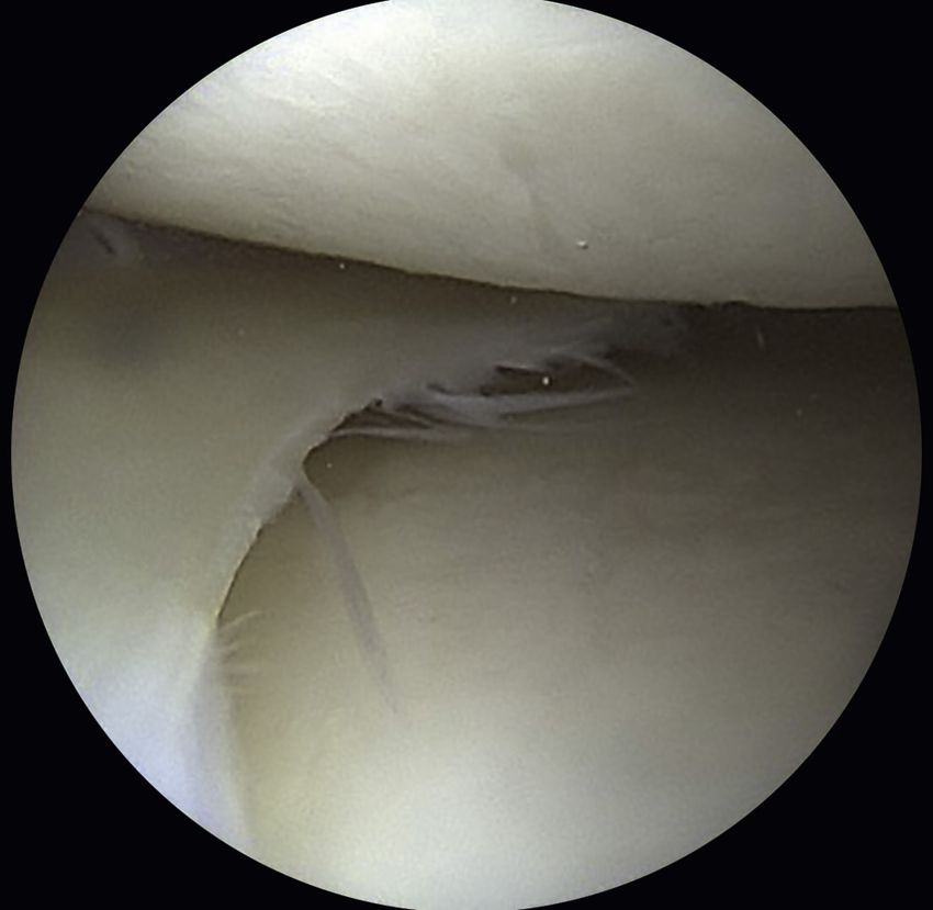
18 PA RT I    The Meniscus

             A                                                                             B
                   •Fig. 3.1 (A) Posteroanterior and (B) lateral radiographs with 10-cm sizing marker used to calculate a
                   patient’s tibial dimensions for appropriate meniscus allograft sizing.

 1)	Patients younger than 50 years old with a chief com-                         malalignment. Magnetic resonance imaging (MRI) is help-
     plaint of pain limiting their desired activities                             ful to assess the extent of previous meniscectomy, the degree
 2)	Body mass index less than 35 kg/m2 and previous total                        of subchondral edema in the involved compartment, and
     meniscectomy with pain localized to the affected com-                        the presence of other articular comorbidities, such as liga-
     partment                                                                     mentous or chondral injury. Bone marrow edema under-
 3)	Normal or correctable coronal and sagittal alignment                         lying the cartilage defect may indicate a source of pain,
 4)	Normal or correctable ligamentous stability                                  and the surgeon should take this into consideration when
 5)	Normal or correctable articular cartilage                                    performing MAT. Correlating this imaging with previous
 6)	Willingness to comply with rehabilitation protocol                           arthroscopic images is crucial.
     Full-thickness chondral defects have traditionally been                          Sizing the meniscal allograft is one of the most important
 considered a contraindication to meniscal transplantation.12                     technical aspects of a successful surgery to prevent postop-
 Several recent reports have demonstrated results of concur-                      erative complications. Dienst et al.15 published a cadaveric
 rent meniscal transplantation and cartilage repair that are                      biomechanical study evaluating the effects of sizing meniscal
 similar to isolated meniscal transplantation.13 As a result,                     allografts on contact stresses in the lateral compartment. An
 we commonly perform concurrent cartilage transplanta-                            oversized graft led to greater contact stresses on the articular
 tion with MAT. Failure to recognize these comorbidities                          surface, whereas undersized grafts led to near-normal con-
 or improper indications can lead to early graft failure, lack                    tact stresses but increased stresses on the allograft itself. They
 of pain reduction, and low patient satisfaction.12 Finally,                      concluded that a size mismatch of 10% or less was needed
 MAT performed as a prophylactic measure in the absence                           for the allograft to function properly.15 Additionally, width
 of appropriate symptoms is highly controversial and not                          should be considered. Huang et al.16 evaluated the cross-
 advocated by the senior author.14 Following these criteria                       sectional parameters of meniscal allografts compared with
 is a critical part of preventing postoperative complications,                    native menisci in cadaveric knees and found increased con-
 graft failure, and poor patient outcomes.                                        tact pressures when allografts did not match native menisci,
                                                                                  with the greatest predictor of differences being the width of
 Preoperative Imaging, Sizing, and                                                the menisci.
 Preservation Techniques                                                              Currently, radiographic measurements, as described by
                                                                                  Pollard et al.,17 are used to appropriately size the meniscus.
 Preoperative imaging and sizing is essential to prevent both                     As a consistent relationship exists between meniscal size and
 intraoperative and postoperative complications. Routinely,                       bony landmarks, most tissue banks currently size the menis-
 we obtain weight-bearing anteroposterior (AP) and postero-                       cus with tibial plateau width and length measurements (Fig.
 anterior 45-degree flexion radiographs, nonweight-bearing                        3.1).18 Sizing using MRI is reserved for cases that are not
 45-degree flexion lateral view, axial view of the patellofemo-                   as well defined because MRI for graft size matching often
 ral joint, and a long-leg mechanical axis view to evaluate                       underestimates meniscus length and width. We recommend

                       Downloaded for Anonymous User (n/a) at Drexel University from ClinicalKey.com by Elsevier on November 30, 2020.
                           For personal use only. No other uses without permission. Copyright ©2020. Elsevier Inc. All rights reserved.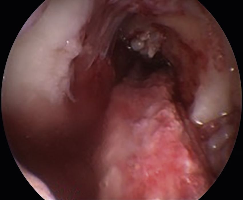
CHAPTER 3         Meniscus Transplantation   19

using radiographs for meniscal sizing to prevent some of                          the tibia. Slot dimensions should be confirmed by placing
these potential complications.                                                    a hooked depth gauge in the reference slot, which also mea-
    In addition to imaging, a diagnostic arthroscopy is use-                      sures the AP length of the tibial plateau. Using a drill guide
ful to obtain a better understanding of the pathology in the                      referencing off the hooked probe, a guide pin is drilled into
patient’s knee and aid in proper surgical planning.19 In all                      the proximal tibia, just distal and parallel to the reference
cases except when a recent (last 6 months) arthroscopy has                        slot. The pin is advanced to, but not through, the posterior
been performed, we perform a diagnostic arthroscopy to                            cortex, being mindful to avoid overpenetration, which could
assess the state of the femoral and tibial cartilage and con-                     injure the posterior neurovascular bundle. If penetration is a
firm a majority of the meniscus is incompetent or missing.                        concern, intraoperative fluoroscopy and/or a posterior lateral
    Once sizing is complete, the appropriate preservation                         or posterior medial portal may be needed to visualize the pin
technique is selected based on the tissue bank. Meniscal                          and protect it from further penetration if this occurs. The
allografts are ideally harvested within 24 hours after death                      pin is subsequently overreamed with an 8-mm cannulated
and frozen. Although other graft preservation methods                             drill bit, again taking care to maintain the posterior cortex
are used, fresh-frozen remains the most commonly used                             and avoid capturing the pin with the drill and advancing
allograft preservation method.20 Graft shrinkage has been                         ahead of the working end. A box cutter is then used to create
reported, which can alter the accuracy of meniscal sizing                         an 8-mm -wide, 10-mm -deep slot which is smoothed and
and compromise the outcome, so careful evaluation of your                         refined with a rasp to ensure that the bone bridge will slide
tissue bank protocols are essential because specific tech-                        smoothly into the slot (Fig. 3.2). In general, following this
niques can vary between tissue banks. Cryopreservation was                        succinct set of steps prevents the frequent complications that
found to induce significant apoptotic cell death in menis-                        can occur with tibial slot preparation.
cal tissue, with a reported cell viability of only 10-40%.19                          Meniscal root anatomy is also imperative to understand,
Furthermore, the storage of the graft itself after harvest and                    not only in the setting of an isolated MAT, but also for com-
before transplantation must be scrutinized because a high                         bination procedures. For the lateral meniscus, a bone bridge
number of freeze-thaw cycles may be detrimental to graft                          technique is used for a MAT. In contrast, the medial menis-
function.21 Our ideal technique is a meniscus harvest within                      cal roots are further apart from each other; thus medial
24 hours after death and preservation using the fresh-frozen                      MAT can be safely performed with either a bone bridge or
technique because in many cases this gives the best viability                     bone plugs.12
and allows for proper surgical planning.                                              There are other fixation techniques, and debate continues
                                                                                  regarding the optimal procedure. Meniscal allografts can be
Intraoperative (Slot) Technique and Potential                                     secured by suture fixation or bony fixation (Fig. 3.2 E–F).
Complications                                                                     Furthermore, bony fixation includes separate bone plugs on
                                                                                  the anterior and posterior horns, or other variations such
Numerous complications can occur during the procedure,                            as keyhole, trough, dove-tail, and bridge-in-slot. In gen-
and an in-depth understanding of surgical anatomy is neces-                       eral, anatomic bony fixation is the gold standard for most
sary. In addition, the type of complication that can arise is                     surgeons, although several surgeons have reported excellent
based on specific techniques and if concomitant procedures                        results with soft tissue-only MAT.
are used.                                                                             When performing a bridge-in-slot technique, bone
    For a MAT slot technique, surgical landmarks include                          bridge fracture is a potential complication. If this occurs, a
the patella, patellar tendon, tibial plateau, and fibular head.                   small K-wire is used to reapproximate the bridge for inser-
During the posterolateral approach or a lateral MAT, struc-                       tion, or conversion to a bone plug technique is used. To
tures most at risk during the procedure include the peroneal                      prevent this from occurring, bone posterior to the posterior
nerve and lateral collateral ligament. For the posteromedial                      meniscal horn attachment is removed, but bone anterior to
approach, the saphenous nerve is most at risk. Also, needle                       the anterior meniscal horn is kept intact to maintain graft
passage during the meniscocapsular repair can injure the                          integrity during graft passage.
posterior neurovascular bundle. One more devastating com-
plication is tibial vein or arterial perforation. This can occur                  Complications Caused by Neglecting
with preparation of the slot, and is most likely to occur with                    Concomitant Procedures
lateral MAT given the anatomy of the arteries and vein.12
    The potentially worst and most preventable complica-                          As discussed earlier, a thorough preoperative evaluation
tions occur with preparation of the tibial slot. We initially                     of these potential patients is essential. Neglecting axial
start with slot preparation using electrocautery to center                        malalignment and concomitant intraarticular injuries can
the anterior and posterior horn attachment sites with a line.                     be detrimental to patient outcomes. Lee et al. found that
This also prevents veering too close to the anterior cruciate                     axial malalignment can exert abnormal pressure on the
ligament (ACL) insertion or too close to the main articu-                         newly placed graft, which can lead to loosening, overload,
lating portion of the plateau. Using this line as a guide, a                      degeneration, and failure.22–24 Also, an osteotomy can inde-
4-mm burr is used to create a superficial reference slot in the                   pendently reduce compartment loads, providing additional
tibial plateau, making sure to follow the posterior slope of                      pain relief.

                      Downloaded for Anonymous User (n/a) at Drexel University from ClinicalKey.com by Elsevier on November 30, 2020.
                          For personal use only. No other uses without permission. Copyright ©2020. Elsevier Inc. All rights reserved.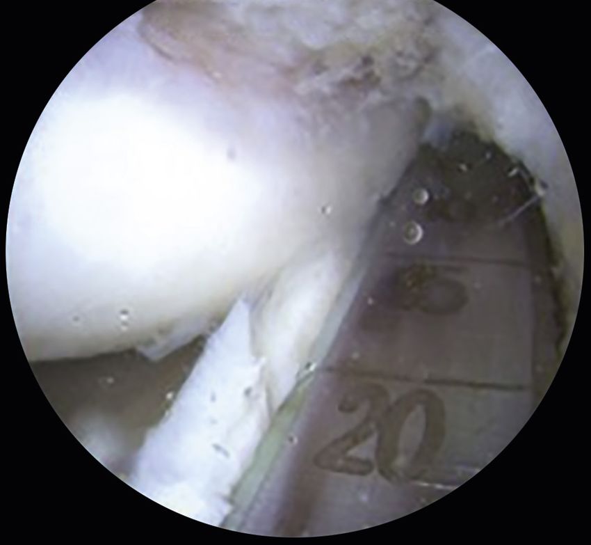
Left knee meniscal trans... Left knee meniscal trans...
A B
Left knee meniscal trans... Left knee meniscal trans...
C D
Left knee meniscal trans...
E F
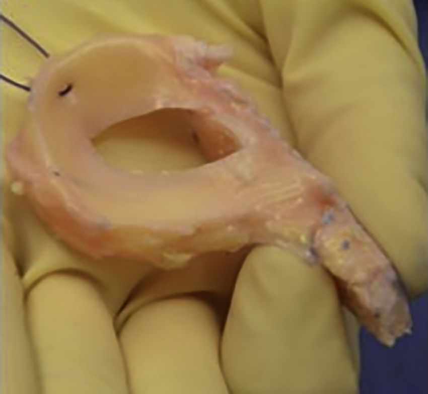
• Fig. 3.2 Intraoperative images demonstrating the medial meniscus allograft transplant bridge-in-slot
technique. (A) An arthroscopic burr is used to create a reference line for slot placement. (B) Once the guide
pin is in place and the slot is reamed, (C) a box cutter is used to create the slot at the desired 8-mm width
and 10-mm depth. (D) A rasp is then used to refine the slot to ensure the bone bridge will be easily placed.
(E) The meniscus allograft is prepared on the back table with the meniscus roots attached to the bone
bridge. A polydioxanone suture is placed in the posterior rim of the meniscus to aid graft insertion. (F) The
meniscus is then sutured to the capsule to aid fixation.
Downloaded for Anonymous User (n/a) at Drexel University from ClinicalKey.com by Elsevier on November 30, 2020.
For personal use only. No other uses without permission. Copyright ©2020. Elsevier Inc. All rights reserved.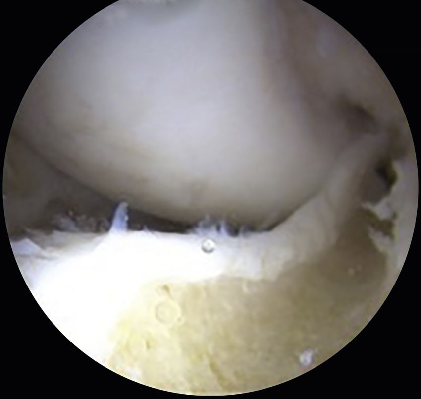
CHAPTER 3         Meniscus Transplantation   21

    A                                                                           B

    C                                                                           D
                  •  Fig. 3.3 Intraoperative arthroscopic images demonstrating concomitant left knee (A) anterior cruciate
                  ligament (ACL) rupture and (B) meniscal deficient medial compartment. Surgical treatment included a
                  combined (C) ACL reconstruction and (D) medial meniscus allograft transplant.

   The ACL-deficient patient needing MAT may benefit                             bone-plug technique described for medial meniscal trans-
from concomitant ACL reconstruction (ACLR) because                               plantation.12 Usually standard femoral and tibial tunnels are
long-term follow-up after this procedure is good, as opposed                     drilled and prepared before meniscal allograft insertion.26
to outcomes in patients with untreated knees.25 Also, the                        The use of allograft decreases associated donor-site morbid-
posterior horn of the medial meniscus is an important sec-                       ity, and tibial tunnel drilling requires special care because this
ondary stabilizer to anterior translation and may prevent                        tunnel often contacts the bone trough for the meniscus.26
secondary stretch of the ACL-reconstructed knee.22,23 These                      Again, you can also leave the ACL drill in when making the
are all potential complications that can be prevented if con-                    trough to protect from “tunnel convergence.” The key is to
comitant or staged procedures, as per the surgeon’s comfort,                     understand the bony anatomy and start with the ACL tibial
are performed with a MAT.                                                        tunnel. These have good results if done properly, with Wirth
                                                                                 et al.27 reporting excellent results in a series of 23 patients
Preventing Complications With                                                    followed up with 14 years postprocedure (Fig. 3.3).
Concomitant Procedures
                                                                                 High Tibial Osteotomy and Meniscus Allograft
Anterior Cruciate Ligament Reconstruction                                        Transplantation
and Meniscus Allograft Transplantation
                                                                                 The key here is to perform all aspects of meniscus trans-
When doing a combined procedure, we use a modification of                        plantation first. The surgeon must perform his or her open-
the bridge-in-slot technique using two smaller bone blocks                       ing wedge osteotomy such that line of osteotomy passes at
rather than one long bridge. The technique is similar to the                     least 1.5 cm below the bottom of the tibial slot. Careful

                      Downloaded for Anonymous User (n/a) at Drexel University from ClinicalKey.com by Elsevier on November 30, 2020.
                          For personal use only. No other uses without permission. Copyright ©2020. Elsevier Inc. All rights reserved.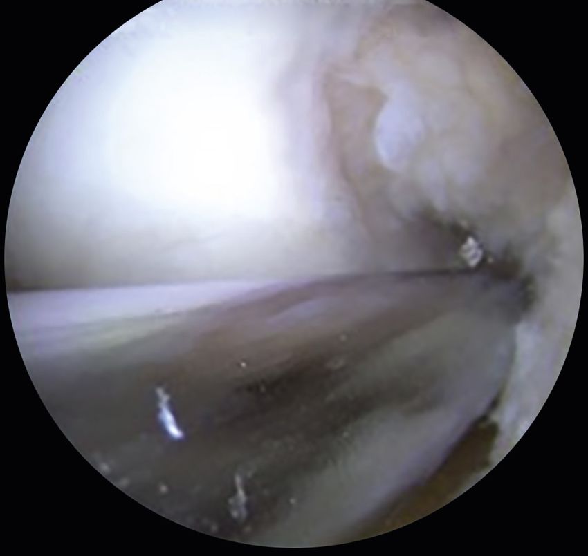
22 PA RT I     The Meniscus

                     A                                                             B

                     C                                                             D
                    • Fig. 3.4  T2 weighted (A) coronal and (B) sagittal magnetic resonance imaging (MRI) scans demonstrating
                    lateral meniscus allograft failure via graft tear in a 17-year-old female 4.5 years after meniscus transplant.
                    T2 weighted (C) coronal and (D) sagittal MRI images demonstrating failure of lateral meniscus allograft
                    transplant via graft extrusion, lateral meniscus allograft tear, and significant subchondral edema of the
                    lateral tibial plateau in a 29-year-old male 7 months posttransplant.

  wedging of the open osteotomy prevents the crack from                                 Most complications following MAT are similar to those
  propagating proximally into the meniscal slot, rather than                        that can follow standard meniscus repair. These include
  laterally toward the fibular head. Verdonk et al.28 found that                    infection, neurovascular damage, stiffness, failure of healing,
  the combination of medial MAT and high tibial osteotomy                           hardware irritation, reoperation, and retear (Fig. 3.4AB). If
  demonstrated better improvements in terms of pain, hos-                           the transplanted allograft is retorn, treatment is similar to
  pital for special surgery score, and knee injury and osteoar-                     that used for a native meniscus, and includes meniscectomy
  thritis outcome score (KOOS) score compared with isolated                         or repair, when indicated. In rare cases, revision MAT can
  medial MAT. Also, Saltzman et al.29 found that the com-                           be performed.30
  bined high tibial osteotomy and MAT group had better                                  Of note, reoperation does not indicate failure because a
  Lysholm, KOOS pain, and KOOS quality of life scores.                              majority of reoperations are for debridement, and patients
                                                                                    experience excellent outcomes following post-MAT
                                                                                    arthroscopic debridement (Fig. 3.5).12 Arthrofibrosis is
  Postoperative Follow-Up and                                                       less common, with 4% of patients requiring manipulation
  Complications                                                                     under anesthesia at 12 years postoperatively.31 Additionally,
                                                                                    the risk of graft complications appears greatest with irra-
  In general, meticulous preoperative evaluation, patient                           diated or lyophilized grafts, grade III to IV osteoarthritic
  selection, and surgical technique are all aimed at preventing                     changes that are not transplanted, soft tissue fixation, and
  long-term complications and improving patient outcomes.                           uncorrected malalignment or instability.12
  Of all the problems discussed, postoperative complications                            One commonly discussed complication is graft
  are the most common.                                                              shrinkage, which has been observed in many studies on

                         Downloaded for Anonymous User (n/a) at Drexel University from ClinicalKey.com by Elsevier on November 30, 2020.
                             For personal use only. No other uses without permission. Copyright ©2020. Elsevier Inc. All rights reserved.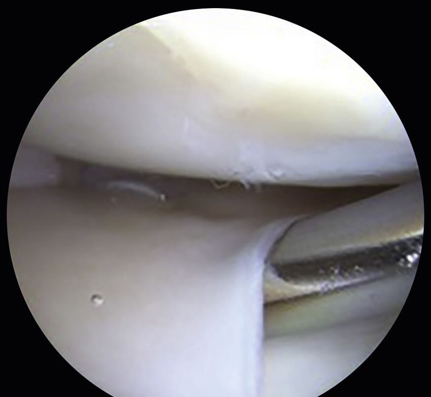
CHAPTER 3         Meniscus Transplantation   23

                                                                                       Most studies view meniscal allograft extrusion as stable
                                                                                   over time, with no clear evidence of related cartilage degen-
                                                                                   eration.38–41 Many studies also show that the degree of
                                                                                   extrusion of MAT fixed with suture-only techniques or poor
                                                                                   bone plug technique is greater than when bony fixation is
                                                                                   used.42,43 Anatomic factors may also play a role in meniscal
                                                                                   body extrusion, including joint laxity, cartilage wear, and
                                                                                   the presence of osteophytes.43
                                                                                       The difficulty with interpreting MAT failure in pub-
                                                                                   lished studies is understanding the criteria the authors use
                                                                                   to define it. Certainly, failure rates will change depend-
                                                                                   ing on the criteria applied, such as reoperation, revision
                                                                                   MAT, conversion to arthroplasty, MRI evidence of graft
                                                                                   extrusion, and/or poor outcomes scores on validated knee
                                                                                   outcome assessment tools. Most studies define failure
                                                                                   as conversion to total knee arthroplasty. Two systematic
                                                                                   reviews demonstrated an overall failure rate that ranged
                                                                                   from 0% to 35%, yet all studies assessed in these two
                                                                                   reviews found the mean failure rate of MAT to be approxi-
                                                                                   mately 10% nearly 5 years after surgery.44,45 Longer-term
• Fig. 3.5  Diagnostic arthroscopy 2.5 years after left medial meniscus            outcomes reporting Kaplan–Meier survival analysis with
allograft transplant in a patient experiencing mild pain and stiffness,
                                                                                   total knee arthroplasty as an endpoint showed 83% sur-
demonstrating an intact graft well healed to the joint capsule.
                                                                                   vival at 10 years in addition to 32% and 38% survival
                                                                                   at 20-year follow-up for medial and lateral meniscus
                                                                                   allografts, respectively.46
second-look arthroscopy and follow-up MRI studies.                                     Another complication can be progression of osteoar-
Although common, it is of unknown clinical significance.                           thritic changes. Verdonk et al.28 found that 52% of a cohort
Graft rupture or tear is also a significant risk in the short                      of patients followed for a mean of 12 years did not show any
term. A meniscal tear of the allograft is one of the most                          change in joint space width. Vundelinckx et al.31 reported
common complications seen. They often occur at the                                 on 49 patients after 15 years of follow-up and found that
capsular junction or the posterior horn, likely secondary                          58% had no increased osteoarthritic changes, whereas 24%
to high contact stresses observed in the allograft, as well                        exhibited an increase by one degree.
as from the suture material.24 Tear rates as high as 36%                               Unaddressed malalignment can lead to progressive com-
at 5-year follow-up have been observed, although other                             plications and failure. Van Arkel et al.47 reported a 13%
studies have reported lower tear rates of 12% at 12-year                           MAT failure rate, and found that failure was primarily
follow-up.31,32                                                                    caused by malalignment resulting in impaired revasculariza-
   Graft extrusion is a commonly discussed complication                            tion of the graft.47 An additional study demonstrated 20
associated with MAT (Fig. 3.4 C–D). Meniscal extrusion                             clinical successes and three failures requiring graft removal.
is defined by displacement of the meniscus beyond the                              The authors considered the failures to be secondary to
tibial plateau margins.33 Although many define 3 mm as                             uncorrected alignment.26
the threshold value for physiological meniscal extrusion                               In some patients, the source of failure is correlated with
in the native joint, it is unclear what amount is tolerated                        progression of oseoarthritis, missed grade III or IV changes,
in meniscal transplant knees and for how long.34 Menis-                            malalignment, suture-only fixation, or graft tears. Some
cal extrusion is more commonly found after MAT than in                             of these failures may be preventable, and thorough step-
normal knees, and has emerged as a potential complica-                             wise preoperative evaluation may decrease the incidence of
tion of MAT, theoretically leading to worse clinical out-                          failure.
comes in the long term.35 Lee and coworkers published the
results of 43 meniscal transplants at a mean of 5.3 years                          Outcomes
postoperatively. All patients had an MRI 1 year postop-
eratively: 26 grafts were read as nonextruded, whereas 17                          Despite the type and number of complications that can
were extruded, although no difference in Lysholm scores                            occur, MAT yields good to excellent results in nearly 85%
was seen between the two groups.36 Extrusion is seen as a                          of patients (Fig. 3.6 and 3.7).6,7,13,25,48
potential complication of MAT; however, the correlation                               Frank et al. reported an overall survival rate of 95% at
with various clinical, radiological, or arthroscopic out-                          5 years.12 Although 33% of patients underwent a second-
comes is not entirely clear, and further research must aim                         ary surgery, most of these procedures were for arthroscopic
at clarifying its significance.37                                                  debridement, and the patients had excellent outcomes

                        Downloaded for Anonymous User (n/a) at Drexel University from ClinicalKey.com by Elsevier on November 30, 2020.
                            For personal use only. No other uses without permission. Copyright ©2020. Elsevier Inc. All rights reserved.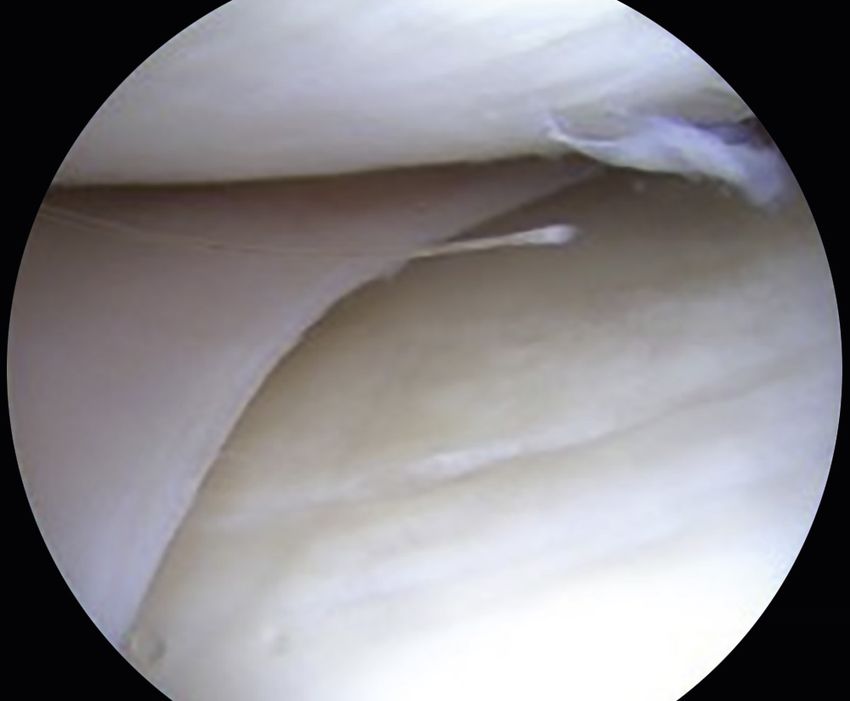
24 PA RT I     The Meniscus

                     A                                                             B

                     C                                                             D
                    • Fig. 3.6T2 weighted preoperative (A) coronal and (B) sagittal magnetic resonance imaging (MRI) scans
                    demonstrating a meniscus-deficient medial compartment of the left knee. Postoperative T2-weighted (C)
                    coronal and (D) sagittal MRI images 2.5 years after surgery demonstrating a well-healed left medial menis-
                    cus transplant.

  2 years after their index surgery. Chalmers et al.49 reported a                   Conclusion
  77% return to play rate at an average of 17 months follow-
  ing surgery in high-level athletes, with significant improve-                     Complications are inherent in a procedure as complex as
  ments in all outcome scales at a follow-up of 3.3 years.                          MAT. The types and frequency of complications vary based
      Verdonk et al.35 reported a survival rate of 74.2%                            on preoperative evaluation, sizing, graft preservation,
  for medial allografts and 69.8% for lateral allografts at                         intraoperative technique, and postoperative follow-up. For
  10-year follow-up. Saltzman et al.29 reported their MAT                           each step in the process, we have outlined potential com-
  results in 22 patients with a minimum follow-up of 7                              plications and what preventative measures can be taken
  years: all patients significantly improved the quality of                         to reduce their occurrence. Also, recognizing factors that
  life, Lysholm, International Knee Documentation Com-                              improve outcomes and reduce early failure, such as appro-
  mittee, and KOOS scores, with excellent satisfaction                              priate concomitant procedures, is essential to mastering
  scores and an overall success rate of 88%. El Attar and                           this procedure. This chapter not only evaluates potential
  colleagues conducted a metaanalysis of 44 separate clini-                         complications throughout the whole process from preop-
  cal studies of over 1000 MATs with a mean patient age of                          erative evaluation to postoperative management, but also
  34.8 years. The investigators concluded that “MAT cn be                           offers expert advice on how to prevent them. The goal of
  considered as safe and reliable for the treatment of refrac-                      MAT is to offer a young, active patient population the
  tory postmeniscectomy symptoms in selected patients”                              potential to return to relatively normal activities pain-free
  (Table 3.1).44                                                                    and with good long-term outcomes.

                         Downloaded for Anonymous User (n/a) at Drexel University from ClinicalKey.com by Elsevier on November 30, 2020.
                             For personal use only. No other uses without permission. Copyright ©2020. Elsevier Inc. All rights reserved.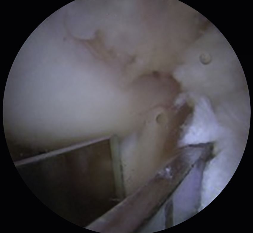
CHAPTER 3         Meniscus Transplantation   25

                       A                                                             B

                       C                                                             D
                      • Fig. 3.7  T2 weighted preoperative (A) coronal and B) sagittal magnetic resonance imaging (MRI) scans
                      demonstrating a meniscus-deficient lateral compartment of the left knee. Postoperative T2 weighted (C)
                      coronal and (D) sagittal MRI images demonstrating a well-healed left lateral meniscus transplant 1 year
                      after lateral meniscus allograft transplant of the left knee.

TABLE
  3.1   Meniscus Allograft Transplant Clinical Results

  Authors                            Follow-Up Period                                                 Clinical Outcome
  Stone et al.6                      Range, 2–7 years                                                 89.1% successful
  Rue et al.7                        Mean, 3.1 years (range, 1.9–5.6)                                 93.5% successful
  Farr13                             Mean, 4.5 years                                                  87.9% successful
  Sekiya25                           Mean, 3.3 years (range, 2–6 years)                               96% improved activity and function
  Marcacci et al.49                  Mean, 40.4 months, (range, 36–66 months)                         94% successful
  Verdonk38                          10-year survival                                                 74.2% medial survival, 69.8% lateral survival
  Saltzman et al.29                  Mean, 8.5 years                                                  88% successful
  El Attar44                         Mean, 4.6 years (range, 8 months–20 years)                       10.6% failure rate
  Chalmers et al.49                  Mean, 3.3 years (range, 1.9–5.7 years)                           77% returned to high-level sporting activity

                           Downloaded for Anonymous User (n/a) at Drexel University from ClinicalKey.com by Elsevier on November 30, 2020.
                               For personal use only. No other uses without permission. Copyright ©2020. Elsevier Inc. All rights reserved.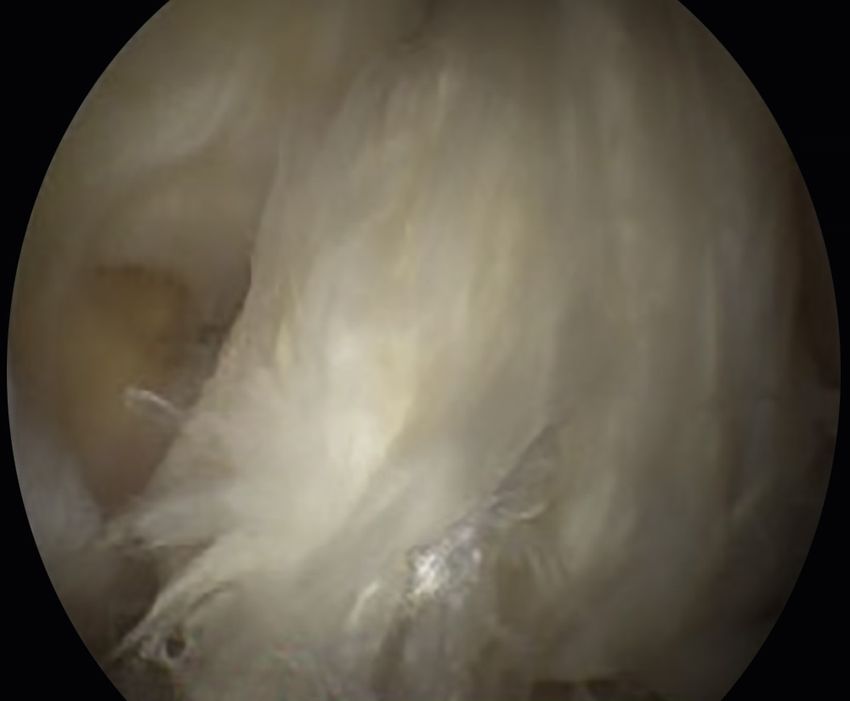
26 PA RT I       The Meniscus

  References                                                                          20.	McDermott ID. What tissue bankers should know about the
                                                                                           use of allograft meniscus in orthopaedics. Cell Tissue Bank.
   1.	Walker PS, Erkman MJ. The role of the menisci in force transmis-                    2010;11(1):75–85.
       sion across the knee. Clin Orthop Relat Res. 1975;109:184–192.                 21.	Lewis PB, Williams JM, Hallab N, Virdi A, Yanke A, Cole BJ.
   2.	Baratz ME, Fu FH, Mengato R. Meniscal tears: the effect of                          Multiple freeze-thaw cycled meniscal allograft tissue: A bio-
       meniscectomy and of repair on intraarticular contact areas and                      mechanical, biochemical, and histologic analysis. J Orthop Res.
       stress in the human knee. A preliminary report. Am J Sports Med.                    2008;26(1):49–55.
       1986;14(4):270–275.                                                            22.	Shelbourne KD, Gray T. Results of anterior cruciate ligament
   3.	Allen PR, Denham RA, Swan AV. Late degenerative changes                             reconstruction based on meniscus and articular cartilage status at
       after meniscectomy. Factors affecting the knee after operation. J                   the time of surgery. Five- to fifteen- year evaluations. Am J Sports
       Bone Joint Surg Br. 1984;66(5):666–671.                                             Med. 2000;28(4):446–452.
   4.	Noyes FR, Barber-Westin SD, Rankin M. Meniscal transplanta-                    23.	Shoemaker SC, Markolf KL. The role of the meniscus in the ante-
       tion in symptomatic patients less than fifty years old. J Bone Jt                   rior-posterior stability of the loaded anterior cruciate-deficient
       Surg - Ser A. 2004;86(7):1392–1404.                                                 knee: effects of partial versus total excision. J Bone Jt Surg - Am.
   5.	Verdonk R, Volpi P, Verdonk P, et al. Indications and limits of                     1986;68(1):71–79.
       meniscal allografts. Injury. 2013;44(suppl 1):S21–S27.                         24.	Yoon JR, Kim TS, Wang JH, Yun HH, Lim H, Yang JH. Impor-
   6.	Stone KR, Walgenbach AW, Turek TJ, Freyer A, Hill MD.                               tance of independent measurement of width and length of lateral
       Meniscus allograft survival in patients with moderate to severe                     meniscus during preoperative sizing for meniscal allograft trans-
       unicompartmental arthritis: a 2- to 7-year follow-up. Arthrosc J                    plantation. Am J Sports Med. 2011;39(7):1541–1547.
       Arthrosc Relat Surg. 2006;22(5):469–478.                                       25.	Sekiya JK, West RV, Groff YJ, Irrgang JJ, Fu FH, Harner CD.
   7.	Rue JPH, Yanke AB, Busam ML, McNickle AG, Cole BJ. Pro-                             Clinical outcomes following isolated lateral meniscal allograft
       spective evaluation of concurrent meniscus transplantation and                      transplantation. Arthroscopy. 2006;22(7):771–780.
       articular cartilage repair: minimum 2-year follow-up. Am J Sports              26.	Sekiya JK, Giffin JR, Irrgang JJ, Fu FH, Harner CD. Clinical
       Med. 2008;36(9):1770–1778.                                                          outcomes after combined meniscal allograft transplantation
   8.	Cole BJ, Carter TR, Rodeo SA. Allograft meniscal transplanta-                       and anterior cruciate ligament reconstruction. Am J Sports Med.
       tion: background, techniques and results. J Bone Jt Surg - Ser A.                   2003;31(6):896–906.
       2002;84:1236–1250.                                                             27.	Wirth CJ, Peters G, Milachowski KA, Weismeier KG, Kohn D.
   9.	Cole BJ, Dennis MG, Lee SJ, et al. Prospective evaluation of                        Long-term results of meniscal allograft transplantation. Am J
       allograft meniscus transplantation: a minimum 2-year follow-up.                     Sport Med. 2002;30(2):174–181.
       Am J Sports Med. 2006;34(6):919–927.                                           28.	Verdonk PCM, Verstraete KL, Almqvist KF, et al. Meniscal
  10.	Verdonk R, Almqvist K, Verdonk P. Meniscal allografts: indi-                        allograft transplantation: long-term clinical results with radiolog-
       cations and results. In: Doral MN, Karlsson J, eds. Sports Inju-                    ical and magnetic resonance imaging correlations. Knee Surgery,
       ries: Prevention, Diagnosis, Treatment and Rehabilitation. 2nd ed.                  Sport Traumatol Arthrosc. 2006;14(8):694–706.
       Springer: Verlag Berlin Heidelberg; 2015:1183–1190.                            29.	Saltzman BM, Bajaj S, Salata M, et al. Prospective long-term
  11.	Verdonk R, Madry H, Shabshin N, et al. The role of meniscal tis-                    evaluation of meniscal allograft transplantation procedure:
       sue in joint protection in early osteoarthritis. Knee Surgery, Sport                a minimum of 7-year follow-up. J Knee Surg. 2012;25(2):
       Traumatol Arthrosc. 2016;24(6):1763–1774.                                           165–175.
  12.	Frank RM, Cole BJ. Meniscus transplantation. Curr Rev Muscu-                   30.	McCormick F, Harris JD, Abrams GD, et al. Survival and reop-
       loskelet Med. 2015;8(4):443–450.                                                    eration rates after meniscal allograft transplantation: analysis of
  13.	Farr J, Rawal A, Marberry KM. Concomitant meniscal allograft                        failures for 172 consecutive transplants at a minimum 2-year
       transplantation and autologous chondrocyte implantation: mini-                      follow-up. Am J Sports Med. 2014;42(4):892–897.
       mum 2-year follow-up. Am J Sports Med. 2007;35(9):1459–1466.                   31.	Vundelinckx B, Vanlauwe J, Bellemans J. Long-term subjec-
  14.	Jiang D, Yu JK. Immediate versus delayed meniscus allograft                         tive, clinical, and radiographic outcome evaluation of menis-
       transplantation: response. Am J Sports Med. 2015;43(5):NP9–                         cal allograft transplantation in the knee. Am J Sports Med.
       NP10.                                                                               2014;42(7):1592–1599.
  15.	Dienst M, Greis PE, Ellis BJ, Bachus KN, Burks RT. Effect of                   32.	Rath E, Richmond JC, Yassir W, Albright JD, Gundogan F.
       lateral meniscal allograft sizing on contact mechanics of the lat-                  Meniscal allograft transplantation. Two- to eight-year results. Am
       eral tibial plateau: an experimental study in human cadaveric                       J Sports Med. 2001;29(4):410–414.
       knee joints. Am J Sports Med. 2007;35(1):34–42.                                33.	Wang Y, Wluka AE, Pelletier JP, et al. Meniscal extrusion pre-
  16.	Huang A, Hull ML, Howell SM, Haut Donahue T. Identifica-                            dicts increases in subchondral bone marrow lesions and bone
       tion of cross-sectional parameters of lateral meniscal allografts                   cysts and expansion of subchondral bone in osteoarthritic knees.
       that predict tibial contact pressure in human cadaveric knees. J                    Rheumatology. 2010;49(5):997–1004.
       Biomech Eng. 2002;124(5):481–489.                                              34.	Costa CR, Morrison WB, Carrino JA. Medial meniscus extrusion
  17.	Pollard ME, Kang Q, Berg EE. Radiographic sizing for meniscal                       on knee MRI: is extent associated with severity of degeneration or
       transplantation. Arthrosc J Arthrosc Relat Surg. 1995;11(6):684–                    type of tear? Am J Roentgenol. 2004;183(1):17–23.
       687.                                                                           35.	Verdonk PCM, Demurie A, Almqvist KF, Veys EM, Verbruggen
  18.	Lubowitz JH, Verdonk PCM, Reid JB, Verdonk R. Meniscus                              G, Verdonk R. Transplantation of viable meniscal allograft. JBJS
       allograft transplantation: a current concepts review. Knee Surg                     Essent Surg Tech. 2006;88(1 suppl 1):109–118.
       Sports Traumatol Arthrosc. 2007;15(5):476–492.                                 36.	Lee BS, Kim JM, Kim KA, Bin SI. Patient-related risk factors for
  19.	Sekiya JK, Ellingson CI. Meniscal allograft transplantation. J Am                   the extrusion of lateral meniscal allograft transplants. Arthroscopy.
       Acad Orthop Surg. 2006;14(3):164–174.                                               2015;31(4):699–706.

                           Downloaded for Anonymous User (n/a) at Drexel University from ClinicalKey.com by Elsevier on November 30, 2020.
                               For personal use only. No other uses without permission. Copyright ©2020. Elsevier Inc. All rights reserved.
CHAPTER 3 Meniscus Transplantation 27
37. Spencer SJ, Saithna A, Carmont MR, Dhillon MS, Thompson P, 43. Verdonk P, Depaepe Y, Desmyter S, et al. Normal and trans-
Spalding T. Meniscal scaffolds: early experience and review of the planted lateral knee menisci: evaluation of extrusion using mag-
literature. Knee. 2012;19(6):760–765. netic resonance imaging and ultrasound. Knee Surgery, Sport
38. Lee DH, Kim TH, Lee SH, Kim CW, Kim JM, Bin Sl. Evaluation Traumatol Arthrosc. 2004;12(5):411–419.
of meniscus allograft transplantation with serial magnetic reso- 44. El Attar M, Dhollander A, Verdonk R, Almqvist KF, Verdonk P.
nance imaging during the first postoperative year: focus on graft Twenty-six years of meniscal allograft transplantation: Is it still
extrusion. Arthrosc - J Arthrosc Relat Surg. 2008;24(10):1115– experimental? A meta-analysis of 44 trials. Knee Surg, Sport Trau-
1121. matol Arthrosc. 2011;19(2):147–157.
39. Jang SH, Kim JG, Ha JG, Shim JC. Reducing the size of the 45. Smith NA, MacKay N, Costa M, Spalding T. Meniscal
meniscal allograft decreases the percentage of extrusion after allograft transplantation in a symptomatic meniscal deficient
meniscal allograft transplantation. Arthrosc - J Arthrosc Relat Surg. knee: a systematic review. Knee Surg, Sport Traumatol Arthrosc.
2011;27(7):914–922. 2014;23(1):270–279.
40. Lee DH, Kim SB, Kim TH, Cha EJ, Bin SI. Midterm out- 46. Verdonk P, Beaufils P, Bellemans J, et al. Successful treatment of
comes after meniscal allograft transplantation: Comparison of painful irreparable partial meniscal defects with a polyurethane
cases with extrusion versus without extrusion. Am J Sports Med. scaffold: two-year safety and clinical outcomes. Am J Sports Med.
2010;38(2):247–254. 2012;40(4):844–853.
41. Lee DH, Lee CR, Jeon JH, Kim KA, Bin SI. Graft extrusion 47. van Arkel ER, Goei R, de Ploeg I, de Boer HH. Meniscal
in both the coronal and sagittal planes is greater after medial allografts: evaluation with magnetic resonance imaging and cor-
compared with lateral meniscus allograft transplantation relation with arthroscopy. Arthroscopy. 2000;16(5):517–521.
but is unrelated to early clinical outcomes. Am J Sports Med. 48. Verstraete KL, Verdonk R, Lootens T, Verstraete P, De Rooy J,
2015;43(1):213–219. Kunnen M. Current status and imaging of allograft meniscal
42. Abat F, Gelber PE, Erquicia JI, Pelfort X, Gonzalez-Lucena G, transplantation. Eur J Radiol. 1997;26(1):16–22.
Monllau JC. Suture-only fixation technique leads to a higher 49. Chalmers PN, Karas V, Sherman SL, Cole BJ. Return to high-
degree of extrusion than bony fixation in meniscal allograft trans- level sport after meniscal allograft transplantation. Arthrosc - J
plantation. Am J Sports Med. 2012;40(7):1591–1596. Arthrosc Relat Surg. 2013;29(3):539–544.
Downloaded for Anonymous User (n/a) at Drexel University from ClinicalKey.com by Elsevier on November 30, 2020.
For personal use only. No other uses without permission. Copyright ©2020. Elsevier Inc. All rights reserved.You can also read